Fundamentals of Clinical Ophthalmology - part 9 ppsx
Bạn đang xem bản rút gọn của tài liệu. Xem và tải ngay bản đầy đủ của tài liệu tại đây (327.14 KB, 20 trang )
(Figure 14.2) may recover better with the
release of entrapped tissues within a day or
two of injury.
Management
The assessment and treatment of systemic,
facial and cranial injury takes precedence over
the repair of orbital fractures. The patient with
acute orbital fracture involving the paranasal
sinuses should be instructed not to blow his/her
nose for 10 days and, in view of the
sight-threatening nature of acute orbital
cellulitis, a short course of systemic antibiotics
should be considered. Oral anti-inflammatory
medications may be given after injury
to accelerate the resolution of orbital
inflammation and oedema.
Orbital floor repair
If surgical repair is indicated, then the
orbital floor is readily approached through a
lower eyelid swinging flap (Chapter 11) or a
subciliary skin-muscle blepharoplasty flap
(Chapter 8). Using one of these routes, the
orbital rim is exposed and the periosteum
incised about 5mm outside the rim, to leave a
margin of periosteum for adequate closure
in front of any orbital floor implant. The
periosteum is raised into the orbit, across the
orbital floor until the site of fracture is located
and then the periosteum around the sides
of the fracture site is raised to define the
extent of tissue incarceration; particular care
must be taken laterally, as this area is liable to
major haemorrhage from the infraorbital
neurovascular bundle in the area of the
inferior orbital fissure. There should be a
clinically evident improvement in the forced
duction test after the incarcerated orbital
tissues are released completely from the
fracture site and the whole of the fracture edge
should be visible; typically there is a ledge of
normal orbital floor at the posterior edge of
the fracture site. Although often not possible,
the sinus mucosa should be kept intact to
avoid formation of sino-orbital fistula.
Once the orbital contents have been
completely freed from the fracture site, an
implant may be shaped and positioned across
the defect in the orbital floor and medial wall
(Figure 14.3). Where the repair is for release
of entrapped tissues (rather than volume
enhancement), it is essential to place the rear
of the implant on the intact fragment of
orbital floor at the orbital apex, behind the
point of emergence of the infraorbital nerve
from the inferior orbital fissure. Bulky
implants should be avoided within 1cm of the
orbital apex, as thick materials may bear upon
the optic nerve or ophthalmic artery and lead
to blindness, and any materials should be
inserted gently and not forced into place.
Likewise, when placing the material it is very
important to avoid snagging of the orbital fat
with the back edge of the implant, or motility
disorders will result. The most useful implant
materials include porous polyethylene and
silicone sheeting, although silicone is
PLASTIC and ORBITAL SURGERY
152
Figure 14.2 Gross restriction of up (a) and down
(b) gaze after a “hairline” blowout fracture of the
orbital floor with entrapment of fascia around inferior
retus muscle.
(a)
(b)
inadvisable where there has been a breach of
sinus mucosa; whilst still widely used, bone
grafts have the disadvantage of reabsorption
and donor-site morbidity. Microplate fixation
may be necessary where there has been
extensive damage to the orbital walls or
fracture of the orbital rim, although treatment
of such facial fractures is outside the realm of
the ophthalmic surgeon.
The anterior edge of the orbital periosteum
is closed with a 5/0 absorbable suture, the
lower eyelid approach repaired in layers with a
6/0 absorbable suture and the eyelid placed on
upward traction with a 4/0 nylon suture. The
site is padded with a firm elastic dressing.
Fractures of the medial orbital wall can be
readily repaired during orbital floor repair,
with extension of the flexible implant upwards
alongside the medial defect. For isolated
fractures of the medial wall, however, it is
possible to use either the extended post-
caruncular incision, directed postero-medially
onto the orbital wall, or the aesthetically less
desirable Lynch incision, through the skin of
the nasal part of the upper eyelid and medial
to the inner canthus.
The patient should be nursed head-up after
surgery and it is important that any severe or
increasing pain is reported. Where pain is
severe or increasing, the vision in the affected
eye and the state of the orbit should be
checked; a very tense orbit with markedly
decreased vision, a relative afferent pupillary
defect and loss of eye movements, suggests
accumulation of orbital haemorrhage and this
may lead to irreversible visual loss. If this
emergency appears to be developing, the
operative site should be reopened at the
“bedside”, without delay, and any accumulation
of blood allowed to drain.
The patient should refrain from nose
blowing for 10 days after orbital floor repair
and should be prescribed a course of
systemic antibiotics and anti-inflammatory
medications. It is possible that eye movement
exercises performed several times daily, with
forced ductions to the extremes of range, may
increase the recovery of tissue compliance and
speed the resolution of post operative
diplopia; if, however, diplopia persists at
several months after repair then squint surgery
may be of value when the Hess charts are stable.
Complications
Post operative haemorrhage, with threat to
vision, is the most feared complication and
should be recognised and treated promptly.
153
ORBITAL TRAUMA
Figure 14.3 Implant material placed across an
orbital floor fracture, approached through a lower
eyelid swinging flap: (a) orbital floor pre- and (b) post
insertion of implant.
(b)
(a)
The risk of this major complication may be
reduced by recognition of the various arterial
branches that cross between the orbit and the
walls (the branches to the infraorbital nerve
and the anterior ethmoidal vessels being the
most troublesome in this context) and
appropriate coagulation of the vessels.
Increased infraorbital nerve hypoaesthesia
is fairly common and generally recovers.
Transient alteration in muscle balance is
almost inevitable, this typically settling over a
week or two, but capture of the released orbital
tissues by an edge of the implant should be
avoided as it may permanently worsen motility.
Infection, more common with entrance into
the sinus cavity, may occur soon after surgery
(Figure 14.4) and necessitates removal of the
implant with later repair after the infection has
settled on systemic therapy. Late infection may
occur where maxillary sinusitis spreads through
a thin interface into the site of orbital repair.
Migration of the implant is less common
with integrating implants, such as porous
polyethylene, and frank extrusion from the
operative site (Figure 14.5) is almost unknown
with avoidance of direct incision over the
inferior orbital rim – an unsightly surgical
approach used widely in the past. Formation of
a pneumatocoele around non-integrating
implants such as silicone (Figure 14.6), lined
by respiratory epithelium that has migrated
through an sino-orbital fistula, may be avoided
by using integrating implants where there is a
defect into the sinuses at the time of surgery;
where a pneumatocoele forms, it can later be
excised and the defect repaired, if necessary,
with an integrating implant material.
Surgical approaches through the lower
eyelid may rarely lead to a cicatricial retraction
of the lower lid, with secondary entropion or
ectropion.
Damage to the lacrimal drainage system,
either during exposure of a fracture site or due
to bearing of the implant on the nasolacrimal
duct, may lead to epiphora that may require
dacryocystorhinostomy. Likewise, surgery on
the medial orbital wall carries a very minor
risk of cerebro-spinal fluid leak or intracranial
damage.
PLASTIC and ORBITAL SURGERY
154
Figure 14.4 Patient referred with acute infection of
an orbital floor implant.
Figure 14.5 Late extrusion of a silicone implant
through a direct incision over the orbital rim.
Figure 14.6 Air-filled cavity (in communication
with the maxillary sinus) that has formed around a
silicone sheet implant for repair of an orbital fracture.
Fractures of the orbital roof,
zygoma and mid-face
Fractures of the orbital roof are uncommon
and usually accompany major head injury,
larger fractures often being comminuted and
involving the frontal sinuses, the cribriform
plate or intracranial injury; the ophthalmologist
is, therefore, unlikely to be in charge of the
primary management of these cases. Similarly,
midfacial fractures are treated by maxillo–facial
surgeons and the ophthalmologist’s role is in
the assessment of visual function, treatment of
the ocular injury and in the late management of
associated soft tissue injury and diplopia.
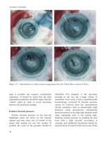
Assessment
Orbital roof injury should be suspected where
head trauma is accompanied by a large upper
eyelid haematoma, hypoglobus, restricted up
gaze and sensory loss over the forehead
(Figure 14.7). Late manifestations include
deformity of the orbital rim underlying the
brow and failure of descent of the upper eyelid
during down gaze due to adhesions between
the fracture site and the levator muscle.
Tripod fracture of the zygoma, with
disarticulation from the neighbouring frontal
bone and maxilla, tends to occur with a major
blow to the cheek and is manifest by a
flattening of the prominence of the cheek
(although this may be masked by overlying
haematoma), by palpable discontinuity of the
orbital rim, by tenderness with upward
pressure below the zygomatic arch, and by an
ipsilateral buccal haematoma.
Le Fort fractures involve the maxilla and
extend posteriorly through the pterygoid
plates. The orbit is involved in types II and III
Le Fort fractures, both extending across the
medial part of the orbit at the level of
the cribriform plate, but the type II fracture
(the commonest) passes infero-laterally to the
level of the inferior orbital fissure, whereas the
type III fracture extends laterally higher in the
orbit, through the zygomatico-temporal suture
line. It is unlikely that the ophthalmologist will
be required to identify such fractures, which
are characterised by dental malocclusion.
When one of these fractures is identified,
adequate CT imaging should be performed to
include an area clear of the clinical site of
injury; damage at the optic canal should be
identified prior to surgery, with particular care
being taken to avoid damage to the nerve or its
circulation by disturbance of bone fragments
near the orbital apex or canal. Treatment
of these fractures is by open reduction,
microplate fixation and dental stabilisation.
155
ORBITAL TRAUMA
Figure 14.7 Child presenting with a delayed onset
of severe compressive optic neuropathy due to a large
subperiosteal haematoma along the orbital roof; the
child had sustained a blunt orbital injury a week
before, with fracture of the orbital roof.
Management
Small fractures of the orbital roof are
managed conservatively if they cause no
functional deficit and only minimal
irregularity of the orbital rim and brow. Small
bone fragments that interfere with the
function of the levator or superior rectus
muscles should be repositioned or removed,
either through the open wound at the time of
primary repair, or through an incision in the
upper eyelid skin crease. Larger bone
fragments require reduction and microplate
fixation, although most such surgery is beyond
the realm of the ophthalmic surgeon and
involves a multi-disciplinary approach.
Adherence of the levator muscle or upper
eyelid scars to fractures of the orbital rim or
roof may cause lagophthalmos and exposure
keratitis.This may be treated by exploration of
the orbital roof through an upper eyelid incision,
division of any adhesions and placement of a
dermis-fat graft sutured inside the orbital rim,
to the periosteum of the orbital roof.
Complications
Both the injury itself and the surgery for the
repair of these complex fractures may be
associated with supraorbital nerve injury, loss
of other ocular motor innervation due to
damage near the superior orbital fissure,
orbital emphysema and pneumocephalus, a
subperiosteal haematoma with a secondary
compressive optic neuropathy (Figure 14.7)
and associated intracranial injuries. Late
complications include persistent ptosis, due
either to mechanical damage or denervation,
lagophthalmos due to scarring and retraction
of the upper eyelid or levator muscle and
chronic or recurrent sinusitis, particularly that
of the frontal sinus.
Intraorbital foreign bodies
The site of entry of an orbital foreign body
may be self-sealing and easily overlooked. High-
speed foreign bodies are more likely to penetrate
the globe, whereas low-speed ones (such as
twigs) are more likely to spare the globe. Failure
to remove an unsterile foreign body is likely to
result in an intraorbital or intracranial abscess,
or an externally draining sinus (Figure 14.8).
The prime investigation for localisation is
thin-slice axial and direct coronal CT scan
(Figure 14.9) and MRI should be considered –
but only after excluding the presence of
intraorbital ferro-magnetic materials – where
wood and other materials of vegetable origin
are thought to be present.
Treatment
Removal of an orbital foreign body is
indicated when there is thought to be
reversible visual impairment, persistent pain,
diplopia, inflammation or infection, or when
the object is palpable in the anterior part of the
orbit. Unless the foreign body is visible under
the conjunctiva, surgery should be under
general anaesthesia as location of the materials
can be difficult. When a foreign body is inert
and posterior within the orbit (Figure 14.10),
it can be left in place and the risk of surgical
damage to the orbital contents avoided.
All organic matter must be removed, as this
typically incites a vigorous inflammatory
response and is liable to infection. Non-
metallic inorganic materials, such as glass,
stone or plastics, may generally be left and
observed and non-reactive metals, such as
stainless steel, steel or aluminium, are well
tolerated. Copper-containing metals, including
PLASTIC and ORBITAL SURGERY
156
Figure 14.8 Wooden foreign body in the inferior
part of the orbit and the pterygopalatine fossa: (a)
coronal and (b) axial view.
(a)
(b)
brass, should be removed as they cause marked
suppurative inflammation. Intraorbital lead
can be left, as it does not appear to cause
systemic toxicity and intraorbital iron does not
have the toxicity of intraocular iron.
Injury to orbital soft tissues
Damage to extraocular muscles
Avulsion of extraocular muscles is rare and
usually results from a penetrating orbital
injury with a “hooking” force, seen
occasionally with deliberate attempts at
enucleation during assault (Figure 14.11). CT
scan allows an assessment of the state of the
musculature, although repair is often difficult
due to oedema, haemorrhage and retraction of
the muscle into the orbit. When enucleation
has been achieved, orbital oedema may be
extreme and bacterial contamination likely; in
these circumstances primary implantation of a
ball is likely to fail and this should be deferred
until both the oedema and the risk of infection
has settled.
Explosive injuries result in ragged wounds
with widespread intraorbital debris, and
should be treated by extensive cleaning of
tissues, debridement where necessary and
repair of the globe and eyelids where possible.
Where there has been a major loss of eyelid
tissues, the principles of reconstruction are
similar to those for eyelid repair after excision
of tumours (Chapter 5), although this may
need to be deferred until the acute oedema
has improved; whilst awaiting reconstruction,
the repaired globe should be kept moist with
regular lubricant/antibiotic ointments and a
“moisture chamber” such as, for example, a
cling-film application over a Cartella shield.
Optic nerve injury
Injury to the optic nerve can either be
direct, due to penetrating orbital foreign
bodies or avulsion, or indirect as part of a
major head injury, with fractures around the
orbital apex – where bone fragments may
impinge on the nerve – or actually involving
the optic canal.
Optic nerve damage anterior to the
entrance of the central retinal artery causes
visual loss with retinal artery occlusion,
whereas optic nerve avulsion (Figure 14.11)
produces extensive peripapillary haemorrhage
and a later fibroglial reaction.
The commonest site for injury to the posterior
part of the optic nerve is in the bony canal
(Figure 14.12) and, more rarely, in the
intracranial nerve or chiasm. Optic neuropathy
may occur with or without fracture of the canal
and recent CT studies suggest that sphenoid
fractures are more common than previously
157
ORBITAL TRAUMA
Figure 14.9 Inferior orbital foreign body with
associated brain abscess.
Figure 14.10 Airgun pellet deep within orbit, thus
not requiring removal.
thought. It is believed that the energy of impact
and shearing forces due to deceleration are
transmitted to the area of the optic canal,
resulting in axonal damage and tearing of the
pial vessels supplying the intracanalicular optic
nerve; oedema of the injured tissues and post-
traumatic vasospasm will both exacerbate neural
ischaemic damage and this forms a rational
basis for the use of high-dose corticosteroids
after such injuries. The role of optic canal
decompression, however, remains in doubt.
Treatment of indirect optic neuropathy may
be empirically based on the results for spinal
cord injury: a loading dosage of 30mg/kg
Methyl-prednisolone within 8 hours of injury
is followed by an infusion of about 5mg/kg/hr
for 24 hours after injury. A tailing dosage of
prednisolone or dexamethasone may then be
continued for a few days.
Surgery may be considered for removal of
bone fragments, where these are thought to be
causing a compressive optic neuropathy, and
for drainage of intraorbital haematomas.
Although visual improvement has been
reported after drainage of haematomas from
within the optic nerve sheath, there is no
evidence that this procedure actually alters the
natural course of the condition.
Decompression of the optic canal, by removal
of the lateral wall of the sphenoid sinus where it
overlies the optic canal, has not been shown to
improve visual recovery after injury to the
intracanalicular optic nerve; it may, however,
have a role where vision deteriorates in the face
of adequate medical therapy. Decompression
may be achieved through a trans-cranial or a
trans-ethmoidectomy approach and may be
usefully incorporated as part of an open repair
PLASTIC and ORBITAL SURGERY
158
Figure 14.11 (a) Major avulsion of the globes and
eyelids during assault with a claw-hammer, (b) the
scleral defect is evident at the site of optic nerve
avulsion.
(a)
(b)
Figure 14.12 Fracture of the right optic nerve canal
with severe optic neuropathy.
(a)
(b)
of cranio-facial injuries. Because of the
proximity of the internal carotid artery to the
operative site, an otorhinolaryngologist or
neurosurgeon familiar with the regional
anatomy best performs the procedure.
Subperiosteal haematoma
A subperiosteal haematoma of the orbit
may follow blunt trauma, is usually superiorly
within the orbit and the presentation – with a
slowly progressive displacement of the globe –
may lead to delayed diagnosis (Figure 14.7).
The haematoma should be drained through a
transcutaneous approach and a vacuum drain
left in place until the bleeding settles;
compressive optic neuropathy, whilst rare,
dictates urgent intervention.
Surgical trauma to the orbit
The orbital contents may occasionally be
damaged due to inadvertent entry into the
orbit during endoscopic sinus surgery and
may result in devastating complications, such
as severe motility restriction or blindness
(Figure 14.13). Direct damage to the orbital
fat, muscles and, more rarely, optic nerve, may
occur, especially during power-assisted
debridement of diseased sinus tissues. The
most important point in the management of
inadvertent orbital entry is recognition and
immediate cessation of further surgery; in
particular the orbit should be observed for
signs of traction on the orbital tissues and for
small movements of the globe.
Injury to the ethmoidal arteries may result
in orbital haemorrhage with compressive optic
neuropathy and this may require anterior
orbitotomy, drainage of the haematoma and
diathermy of damaged vessels.
Orbital haemorrhage more commonly
follows orbital surgery or after retrobulbar or
peribulbar injections for intraocular and
periocular surgery; it may also occur with
blepharoplasty, when it is thought to arise from
traction damage to small deep orbital vessels.
A venous bleed is of slower onset and will
usually self tamponade with vision frequently
recovering. Firm orbital pressure may assist
tamponade and a lateral cantholysis after 5–10
minutes may assist reduction in intraorbital
pressure after tamponade has occurred.
A rapid development of proptosis is likely to
be arterial bleeding and should be dealt with
by very firm orbital pressure applied for about
8–10 minutes, but being released for about 15
seconds every 2 minutes to allow ocular
perfusion. If the orbital pressure rises to a very
high level, with loss of eye movements and
vision not attributable to local anaesthesia,
then the orbit should be drained through a skin
incision in the affected quadrant; once the skin
is opened, a closed pair of blunt-ended scissors
should be gently advanced about 3cm into the
orbital fat of the affected quadrant and the
blades gently opened to spread the tissues and
encourage drainage of blood and tissue fluid.
This manoeuvre is generally sufficient to
release the orbital tamponade, with restoration
of vision, and a drain should be placed until
the bleeding has stopped.
Further reading
Anderson RL, Panje WR, Gross CE. Optic nerve blindness
following blunt forehead trauma. Ophthalmology 1982;
89:445–55.
Baker RS, Epstein AD. Ocular motor abnormalities from
head trauma. Surv Ophthalmol 1991; 35:245–67.
Biesman BS, Hornblass A, Lisman R, Kazlas M. Diplopia
after surgical repair of orbital floor fractures. Ophthal Plast
Reconstr Surg 1996; 1:9–16.
159
ORBITAL TRAUMA
Figure 14.13 Blindness and gross right exotropia
after avulsion of the right medial rectus, inferior
oblique and optic nerve during endoscopic sinus
surgery.
Bracken MB, Shepard MJ, Collins WF et al. A randomised,
controlled trial of methyl prednisolone or naloxone in the
treatment of acute spinal cord injury. Results of the
Second National Acute Spinal Cord Injury Study. N Eng
J Med 1990; 322:1405–11.
Crompton MR. Visual lesions in closed head injury. Brain
1970; 93:785–92.
Dutton JJ. Management of blowout fractures of the orbital
floor. Editorial. Surv Ophthalmol 1990; 35:279–80.
Goldberg RA, Marmor MF, Shorr N, Christenbury JD.
Blindness following blepharoplasty: two case reports, and
a discussion of management. Ophthalmic Surg 1990; 21:85–9.
Goldberg RA, Steinsapir KD. Extracranial optic canal
decompression: indications and technique. Ophthal Plast
Reconstr Surg 1996; 12:163–70.
Gross CE, DeKock JR, Panje WR, et al. Evidence for orbital
deformation that may contribute to monocular blindness
following minor frontal head trauma. J Neurosurg 1998;
55:963–6.
Guy J, Sherwood M, Day AL. Surgical treatment of
progressive visual loss in traumatic optic neuropathy.
Report of two cases. J Neurosurg 1989; 70;799–801.
Harris GJ, Garcia GH, Logani SC, Murphy ML. Correlation
of preoperative computed tomography and post operative
ocular motility in orbital blowout fractures. Ophthal Plast
Reconstr Surg 2000; 16:179–87.
Rose GE, Collin JRO. Dermofat grafts to the extraconal
orbital space. Br J Ophthalmol 1992; 76:408–11.
Smith B, ReganWF Jr. Blowout fracture of the orbit:
mechanism and correction of internal orbital fractures.
Am J Ophthalmol 1957; 44:733–9.
Steinsapir KD, Goldberg RA. Traumatic optic neuropathy.
Surv Ophthalmol 1994; 38:487–518.
Streitman MJ, Otto RA, Sakal CS. Anatomic considerations
in complications of endoscopic and intranasal sinus
surgery. Ann Otol Rhinol Laryngol 1994; 103:105–9.
PLASTIC and ORBITAL SURGERY
160
161
Watering eyes result from excessive tear
production (hypersecretion), reduced drainage
or a combination of the two. A good history
and thorough assessment (Chapter 10) are
essential to determine the nature of the
underlying problem and decide on its
management.
Dacryocystorhinostomy
indications
Dacryocystorhinostomy (DCR) involves
removal of the bone lying between the
lacrimal sac and the nose, with anastomosis
between the lacrimal sac and nasal mucosa;
the lacrimal sac, with the internal opening
of the common canaliculus, is incorporated
into the lateral wall of the nose and provides
a direct route for tears to reach the nose.
The usual indication for DCR is complete or
partial obstruction of the nasolacrimal duct:
such obstruction can cause skin excoriation,
visual impairment, social embarrassment,
chronic ocular discharge and acute or chronic
dacryocystitis. Less common indications for
DCR include lacrimal calculi, facial nerve
palsy, gustatory lacrimation (crocodile tears),
and lacrimal sac trauma. In the presence of
lacrimal sac mucocoele, DCR is mandatory
prior to intraocular surgery because of the risk
of post operative endophthalmitis.
Patients with acute dacryocystitis require
treatment with systemic antibiotics prior to
undertaking DCR.
15 Basic external lacrimal surgery
Cornelius René
Anaesthesia
Open lacrimal surgery can be performed
under general or local anaesthesia. Local
anaesthesia with sedation provides excellent
intraoperative haemostasis, but may be
associated with somewhat prolonged post
operative nasal oozing. Some patients and
surgeons, however, prefer general anaesthesia
with controlled intraoperative hypotension;
with newer short-acting anaesthetic drugs,
daycase surgery under general anaesthesia is
readily achievable in most cases.
Local anaesthesia is particularly useful for
elderly or debilitated patients who are unfit for
general anaesthesia. The anterior nasal space
is sprayed with 4% lignocaine and packed with
1·2m of 12·5mm ribbon gauze thoroughly
moistened with 2ml of a 10% cocaine
solution, this producing very effective
intranasal anaesthesia and mucosal
vasoconstriction. Using angled nasal forceps,
short loops of the ribbon gauze are firmly
packed far anteriorly and superiorly within the
nasal space – high against the lateral wall of
the nose and the anterior aspect of the middle
turbinate, at the site of the proposed
rhinostomy. Although not essential, the
headlight and nasal speculum may aid correct
placement of the nasal pack. A regional block
of the anterior ethmoidal branch of the
nasociliary nerve is given by infiltration of
2–3ml of 0·5% bupivacaine with 1:200,000
adrenaline along the medial wall of the orbit,
immediately above the medial canthal tendon
PLASTIC and ORBITAL SURGERY
162
and below the trochlea. Additional anaesthesia
and vasoconstriction at the site of incision is
achieved by skin infiltration with 2–3ml of
the same local anaesthetic preparation. To
achieve maximal vasoconstriction, the local
anaesthetic should be administered at least 10
minutes before surgery commences and may
usefully be given just prior to scrubbing, skin
preparation and sterile draping; topical ocular
anaesthesia, such as 0.5% amethocaine
eyedrops, is also required at the time of
surgery.
Vasoconstriction and haemostasis
During local anaesthesia, nasal packing
with cocaine generally provides sufficient
vasoconstriction of the nasal mucosa, although
during surgery it can be supplemented by the
intramucosal or submucosal injection of a
local anaesthetic (such as 2% lidocaine) with
1:200,000 adrenaline. With this technique,
intraoperative bleeding tends to be minimal
and any oozing into the nasal space may be
readily aspirated with a 12G bronchial
aspiration catheter placed within the mid-
nasal space throughout the procedure. Where
general anaesthesia is used, vasoconstriction
of the nasal mucosa is equally important and,
after preparation of the sterile field, is most
conveniently achieved by placing three cotton-
tipped applicators, moistened with 1:1000
adrenaline, high in the antero-superior nasal
space. Infiltration of local anaesthetic with
adrenaline at the site of the skin incision
further contributes to vasoconstriction and
haemostasis, but is not essential in general
anaesthetic cases.
Apart from vasoconstriction, several factors
help to reduce or control perioperative
haemorrhage. Controlled hypotension during
general anaesthesia greatly facilitates the
surgery and significant bleeding is unusual
with a systolic blood pressure below
90mmHg. A head up tilt also reduces cephalic
venous pressure and is helpful during both
general and local anaesthetic cases. The
continuous use of a sucker in the non-
dominant hand is a mainstay to aiding
viewing, and to displacing tissues and
protecting them from other instruments. The
appropriate use of bipolar diathermy, careful
handling and retraction of the tissues, respect
for the surgical planes, and widespread
suturing of mucosal flaps, also help to control
haemorrhage during open lacrimal surgery.
The judicious use of bone wax may, rarely, be
necessary to stop persistent haemorrhage
from bone.
Surgical technique
A 12–15mm straight skin incision
(8–10mm in children), starting just above the
medial canthus and extending inferiorly, is
made just medial and parallel to the angular
vein (Figure 15.1). A straight incision in the
thick paranasal skin tends to heal rapidly with
an imperceptible scar, whereas more posterior
incisions in the concavity of the thinner eyelid
skin sometimes heal with a contracted,
bridging scar (Figure 15.2). The incision
should involve only skin and should not be
carried straight down to the bone, as marked
haemorrhage is common with the latter
approach, due to disruption of the orbicularis
muscle and the angular vessels.
The skin is bluntly dissected from the
underlying orbicularis oculi muscle, using
blunt-tipped scissors directed posteriorly, and
the pretarsal and preseptal parts of the
orbicularis muscle separated along the line of
the fibres down to the bone of the lacrimal
crest, using scissors in a spreading motion just
lateral to the angular vein. A squint hook is
used to retract the preseptal orbicularis and
angular vessels medially and any bleeding
vessels are carefully cauterised.
A Rollet’s rougine is used in an oblique,
spreading mode to incise the periosteum on
the anterior lacrimal crest, starting at the
inferior edge of the medial canthal tendon and
163
BASIC EXTERNAL LACRIMAL SURGERY
extending to the origin of the nasolacrimal
duct. Using the sharp cutting edge of the
Rollet’s rougine, the medial canthal tendon is
transected close to its insertion, the
periosteum divided up to the top of the
lacrimal sac fossa and the paranasal
periosteum reflected as far anteriorly as
possible. The sucker is used to displace the
lacrimal sac laterally, with its periosteal
covering, as the periosteum is stripped
backwards as far as the posterior lacrimal
crest. At this stage 2/0 silk traction sutures
may be passed through the anterior periosteal
edge, with encirclement of the orbicularis
muscle and angular vessels, and the sutures
clipped under tension to the surgical drapes.
Similar sutures are used to encircle the
orbicularis laterally, the increased surgical
exposure and haemostasis being particularly
useful for the less-experienced surgeon
(Figure 15.3). With local anaesthesia, traction
sutures are not possible and a small, self-
retaining retractor is a useful alternative.
Having exposed the whole lacrimal sac
fossa, the pack or cotton-tipped applicators
are removed from the nasal space and the thin
bone at the suture between the lacrimal bone
and the frontal process of the maxilla is
breached with a Traquaire’s periosteal
elevator. In cases where the bone is extremely
thick, a curved haemostat may be required to
make the initial break or, failing that, a
hammer and chisel may be used to thin the
anterior lacrimal crest until the bone can be
breached. Some surgeons prefer to use a burr
or trephine.
Angular vein
Naso-lacrimal
duct
Medial canthal
tendon
Figure 15.1 Incision for external dacryocysto-
rhinostomy (bold line), just medial to the angular
vein; the position relative to the medial canthal tendon
and lacrimal system (dotted line) is indicated.
Figure 15.2 Bowed scar due to contracture in a
posteriorly-placed dacryocystorhinostomy incision.
Figure 15.3 Traction sutures are useful for
increasing exposure of the operative site, especially for
the surgeon in training.
PLASTIC and ORBITAL SURGERY
164
A large rhinostomy is fashioned using bone
punches, trephine or a burr. When using
punches, it is easiest to first enlarge the
opening to at least 1cm in front of the anterior
lacrimal crest, as far superiorly as possible; the
Traquaire’s periosteal elevator being used in a
sweeping motion, between bites, to separate
the underlying nasal mucosa from the bone.
The mucosa is more adherent and more
friable anteriorly, where greater care is
required. Bone removal from the side of the
nose, anterior to the frontal process of the
maxilla, is directed inferiorly to about the level
of the orbital floor – thereby creating an
L-shaped rhinostomy (Figure 15.4); in so
doing, the thick bone of the anterior lacrimal
crest is significantly weakened and removed
relatively easily with a downward-cutting bone
punch.The thin bone lying between the upper
part of the nasolacrimal duct and the nasal
mucosa, the hamular process of the lacrimal
bone, is then removed using a Jensen bone
nibbler. Attention is now directed superiorly,
where further bone should be carefully
removed to extend the rhinostomy to the skull
base; this is essential to ensure that the
internal opening of the common canaliculus is
not obstructed by bone or scarring after
surgery. Excessive tearing forces should be
avoided during bone removal from the upper
part of the rhinostomy, as this may very rarely
result in a hairline fracture of the cribriform
plate and cerebrospinal fluid leak. The
rhinostomy should extend posteriorly into the
anterior ethmoid air cells, where it is generally
necessary to remove small flakes of ethmoid
bone to allow an adequate mucosal
anastomosis.
Having completed the rhinostomy, a “00”
Bowman lacrimal probe is passed via the
inferior canaliculus into the lacrimal sac.
The probe tents the medial wall of the sac
(Figure 15.5), which is then incised using a
No. 11 style blade, directed away from the
internal opening of the common canaliculus
to avoid damaging it. The sac must be widely
opened from the fundus down into the
nasolacrimal duct using Westcott scissors or
Werb’s angled scissors and a common error is
to open the relatively thick overlying fascia
only, leaving the very thin sac mucosa intact.
The lumen of the sac should be examined and
the free patency of the internal opening of
the common canaliculus confirmed; if the
opening is obstructed by a membrane, the
obstruction should be carefully excised, using
Westcott scissors or a No. 11 blade, and
silicone intubation placed.
Being careful to avoid damage to the nasal
septum, the nasal mucosa is incised with a
Lacrimal
sac
Figure 15.4 Creating the rhinostomy: the arrows
indicate the easiest direction of bone removal, relative
to the lacrimal sac (dotted line).
Lacrimal
sac
Canalicular
probe
Figure 15.5 Creating mucosal flaps: the canalicular
probe tents the medial wall of the lacrimal sac and the
dotted lines indicate incisions in the lacrimal sac and
nasal mucosa.
165
BASIC EXTERNAL LACRIMAL SURGERY
No. 11 blade to create a larger anterior flap
(about two-thirds of the antero-posterior
extent) and a smaller posterior flap and, after
cauterisation wherever possible, relieving
incisions are made at the superior and inferior
bone edges to mobilise both flaps. Incising the
nasal mucosa often results in some bleeding
which, under general anaesthesia, can be
controlled by passing the sucker up the nose
and positioning it just behind the posterior
flap – thus acting similarly to the second,
intranasal “sump” drain placed during surgery
under local anaesthesia. A 6/0 absorbable
suture on an 8mm diameter half-circle needle
is passed through the middle of the free edge
of the anterior nasal flap and the ends secured
with a bulldog clip, with the needle left
attached. This is slung across the nasal bridge
to retract the anterior flap medially whilst
suturing the posterior flaps (Figure 15.6).
The posterior mucosal flaps are
approximated and sutured using a similar 6/0
absorbable suture, the needle being reverse-
mounted in an angled, non-locking needle
holder in such a way that its entire length is
used – this facilitating maximum suture
rotation at quite a distance below the small
incision. Either a locked continuous suture, or
three or four interrupted sutures, is placed
from the sac to the nasal mucosa. Keeping the
lacrimal probe in the sac helps to identify and
protect the internal opening while the
mucosal flaps are being united, and this can
later be replaced by intubation where there is
common canalicular disease or a markedly
inflamed lacrimal sac. The absorbable suture
retracting the anterior nasal flap is then used
to unite the anterior mucosal flaps and three
or four sutures are usually necessary to secure
a good anastomosis (Figure 15.7). The cut
medial canthal tendon is sutured to the medial
periosteum using the same suture, and closure
of the orbicularis is not usually necessary if the
fibres were bluntly separated in the plane
between the palpebral and orbital parts. Skin
closure is achieved with 6/0 nylon interrupted
or continuous mattress sutures, antibiotic
ointment instilled into the conjunctival sac,
and a pressure dressing applied to the
incision. Post operative haemorrhage is
infrequent with primary anastomosis of the
mucosal edges, and packing of the nasal space
is not required routinely, but an adrenaline-
moistened pack may be placed if there is
persistent brisk haemorrhage after surgery is
completed; such a pack should be left
undisturbed for five days.
Post operative management
In the immediate post operative period the
patient is nursed semi-erect on bed rest to
Anterior nasal
mucosal flap
Lacrimal probe
through common
canaliculus
Traction
suture
Figure 15.6 Anastomosis of the posterior mucosal
flaps, with the anterior nasal mucosal flap held aside
by a traction suture resting across the nasal bridge.
Anterior flap
of sac mucosa
Anterior nasal mucosal flap
Figure 15.7 Closure of the anterior mucosal flaps.
PLASTIC and ORBITAL SURGERY
166
reduce the nasal venous congestion that can
contribute to nasal oozing and, for similar
reasons, hot drinks should be avoided for 24
hours. A topical combined antibiotic and anti-
inflammatory medication is prescribed for a
few weeks and, unless systemic antibiotics
have been given during surgery, a short course
of oral antibiotics is recommended to reduce
the incidence of post operative infection. The
pressure dressing is removed on the first post
operative day and nose blowing discouraged for
the first week, to reduce the risk of secondary
epistaxis or subcutaneous emphysema. Skin
sutures are removed at about one week
after surgery and the intubation at about four
weeks after surgery, by which time
epithelialisation of the surgical fistula has been
completed.
Complications
Serious complications due to external
lacrimal surgery are extremely rare, but there
are several minor complications (Box 15.1).
In cases of severe continued haemorrhage,
nasal packing may be required either with ribbon
gauze moistened with a mixture of 1:1000
adrenaline and antibiotics, with an absorbable
haemostatic sponge, or with a commercially
available expanding nasal tampon.
Minor leak of cerebrospinal fluid may,
extremely rarely, result from an inadvertent
fracture of the cribriform plate. Once identified,
the site of leakage may be plugged with a slip of
orbicularis oculi muscle obtained from the
surgical field, and post operative systemic
antibiotics administered. Although most cases
resolve without further complication, close
post operative monitoring is required for
continued CSF rhinorrhoea or meningitis and
neurosurgical advice is recommended.
Orbital fat prolapse may occur if the lateral
wall of the lacrimal sac or the orbital
periosteum is breached while incising the
lacrimal sac or performing a membranectomy.
To avoid the risk of orbital haemorrhage,
traction on the orbital fat should be avoided in
such cases, but a small fat prolapse does not
require any specific treatment.
Whilst early post operative nasal oozing is
common and requires no treatment except
upright positioning of the patient and
avoidance of hot beverages, continued brisk
primary haemorrhage is very rare. If simple
measures, such as pinching of the nasal bridge
or icepacks applied to the nasal bridge, do not
control brisk primary haemorrhage, then the
nose should be packed with 12·5mm ribbon
gauze moistened in 1:1000 adrenaline, the pack
being left undisturbed for five to seven days and
a systemic antibiotic given for that period.
Prophylactic systemic antibiotics reduce the
risk of post operative infection (Figure 15.8)
and probably reduce the risk of surgical
failure. A single dose of a systemic antibiotic is
as effective as a post operative course, but
antibiotics should be continued after surgery
where there has been significant preoperative
infection, placement of a nasal tamponade, or
significant primary or secondary epistaxis.
Use of nasal packs also increases the risk of
post operative infection.
Box 15.1 Complications of external
lacrimal surgery
Peri-operative
• Canalicular damage
• Haemorrhage
• Cerebrospinal fluid leak
• Inadvertent orbital entry
Post operative
• Haemorrhage
• Wound infection
• Wound necrosis
• Preseptal/orbital cellulitis
• Hypertrophic scar
• Lacrimal tube prolapse
• Medial migration (cheesewiring) of
lacrimal tubes
167
BASIC EXTERNAL LACRIMAL SURGERY
Although the incision line after almost all
external DCR is imperceptible by six months,
the incision may rarely heal with excessive scar
contracture, especially with posteriorly sited
incisions (Figure 15.2). Subcutaneous sutures
may also increase the tendency to
hypertrophic scar formation. Prominent scars
may become less noticeable as they mature,
especially if massaged or if pressure is applied
by, for example, the wearing of glasses on the
scar. Very rarely revision of an operative scar
is required for unacceptably tight scars,
and generally involves a double Z-plasty
technique. Wound necrosis is occasionally
seen at the incision margins after prior
radiotherapy or in patients with Wegener’s
granulomatosis (Figure 15.9).
Summary
External DCR surgery is a safe and effective
procedure for managing troublesome epiphora.
With attention to patient preparation,
meticulous surgical technique, an appreciation
of the surgical anatomy and careful tissue
handling, the surgery should not be unduly
difficult and success rates are unmatched by
other techniques. For patients without
canalicular or lid abnormalities, the “volume”
symptoms (due to retention of fluid within the
lacrimal sac) can be cured in everybody,
whereas “flow” symptoms (due to the tear line
height) are affected by canalicular conductance
and these symptoms will be improved in at
least 95% of cases. Failure is usually due to
inadequate primary rhinostomy, failure to
create a primary epithelial anastomosis,
excessive fibrosis at the rhinostomy site (due
to secondary intention healing), stenosis of the
canalicular system, or lid abnormalities.
Further reading
Dresner SC, Klussman KG, Meyer DR, Linberg JV.
Outpatient dacryocystorhinostomy. Ophthalmic Surg 1991;
22:222–4.
Ezra EJ, Restori M, Mannor GE, Rose GE. Ultrasonic
assessment of rhinostomy size following external
dacryocystorhinostomy. Br J Ophthalmology 1998;
82:786–9.
Hanna IT, Powrie S, Rose GE. Open lacrimal surgery: a
comparison of admission outcome and complications after
planned daycase or inpatient management. Br J
Ophthalmol 1998; 82:392–6.
Hartikainen J, Grenman R, Pukka P, Seppa H. Prospective
randomized comparison of external dacryocystorhinostomy
and endonasal laser dacryocystorhinostomy. Ophthalmology
1998; 105:1106–13.
Jordan DR. Avoiding blood loss in outpatient
dacryocystorhinostomy. Ophthal Plast Reconstr Surg 1991;
7:261–6.
Jordan DR, Miller D, Anderson RL. Wound necrosis
following dacryocystorhinostomy in patients with
Wegener’s granulomatosis. Ophthalmic Surg 1987;
18:800–3.
Linberg JV. Lacrimal surgery; contemporary issues in
ophthalmology volume 5. New York: Churchill Livingstone,
1988.
McNab AA. Diagnosis and investigation of lacrimal disease.
In McNab AA. Manual of orbital and lacrimal surgery (2nd
ed.) Oxford: Butterworth Heinemann, 1988.
Neuhaus RW, Baylis HI. Cerebrospinal fluid leakage after
dacryocystorhinostomy. Ophthalmology 1983; 90:1091–5.
Tarbet KJ, Custer PL. External dacryocystorhinostomy.
Surgical success, patient satisfaction, and economic cost.
Ophthalmology 1995; 102:1065–70.
Walland MJ, Rose GE. Soft tissue infections after open
lacrimal surgery. Ophthalmology 1994; 101:608–11.
Figure 15.8 Early post operative infective cellulitis
around the dacryocystorhinostomy site.
Figure 15.9 Post operative skin necrosis at the
dacryocystorhinostomy incision in a patient with
previous radiotherapy.
168
Endonasal dacryocystorhinostomy is performed
entirely within the nose, either by direct
visualisation or by using a rigid Hopkins
endoscope as the light source and to magnify
the structures on the lateral nasal wall. Surgical
instruments or laser (or a combination) are used
to create an anastomosis between the lacrimal
sac and the nasal space (Figure 16.1).
It differs from external DCR (Chapter 15)
in that there is no external incision, there are
no sutured mucosal flaps and there is usually
temporary silicone intubation.
Caldwell, in 1893, first described the use of
an electric drill to open the lacrimal sac and
nasolacrimal duct into the nasal space from
an intranasal approach and West subsequently
described another endonasal approach, a so-
called “window resection of the nasolacrimal
duct”. Recognising that the sac-duct junction
appeared to be the commonest site of
lacrimal outflow obstruction, West later
16 Laser-assisted and endonasal
lacrimal surgery
Jane M Olver
extended the resection to include the lacrimal
sac and this remains the principle of modern
endonasal lacrimal drainage surgery. Between
1950 and 1990 most lacrimal surgery
was performed using an external approach
by ophthalmologists, with only few
otolaryngologists continuing to practise
endonasal surgery. The increased usage of
rigid nasal endoscopy and laser surgery has,
however, helped to popularise modern
endonasal dacryocystorhinostomy, with the
results of endonasal endoscopic DCR ranging
from 63–99% (Tables 16.1–16.2).
Indications
Endonasal endoscopic DCR is moderately
quick, easily performed under local
anaesthesia, and bilateral surgery may be
Inferior
turbinate
Septum
Middle
turbinate
Site of endonasal
anastomosis
Figure 16.1 Right lacrimal system, with the site of
the anastomosis outlined.
Table 16.1 Reported results for endonasal endoscopic
surgical dacryocystorhinostomy.
Author Bone Number Success
instruments of cases
Whittet Drill, hammer, 19 94·7%
chisel, rongeurs
Weidenbecher Chisel, drill, 54 86–95%
backbiting forceps
Sprekelsen Drill, rongeur 152 85·5–96%
Yung Rongeur 81 93%
Zilelioglu Drill 23 78·3%
Zilelioglu Drill and Mitomycin 14 78·5%
C application
Moore Chisel, rongeur 34 83%
readily performed with relatively little
perioperative bleeding. The procedure is,
therefore, particularly useful in elderly or frail
patients, and in patients who do not wish to
accept the low risk of a visible cutaneous scar
after external DCR, albeit accepting the
higher failure rate for endonasal surgery.
Most cases of primary acquired stenosis,
“functional block” or complete obstruction
of the nasolacrimal duct are suitable for
endonasal DCR, although patients with
secondary obstruction may be treated if nasal
access is adequate and there is no major nasal
disruption. Major mid-facial fractures, being
associated with gross disruption of anatomy,
are not suitable for this approach and patients
with inflammatory nasal diseases associated
with scarring (such as sarcoidosis or Wegener’s
granulomatosis) should probably not undergo
endonasal surgery, as the technique is
dependent on secondary intention healing.
Whilst membranous block of the common
canaliculus may be treated during endonasal
dacryocystorhinostomy, more extensive
common canalicular block or obstruction of
the individual canaliculi is better treated by an
external technique (Chapter 17).
Endonasal surgery does not allow a full
inspection of the lacrimal sac mucosa and
external surgery should, therefore, be used if
there is suspicion of a tumour, or other
problem, within the lacrimal sac. Other
relative contraindications to endonasal DCR
include a very narrowed nasal space, a small
sac and failed previous DCR with extensive
fibrosis at the rhinostomy.
Endonasal endoscopic
dacryocystorhinostomy
Local anaesthesia is achieved as previously
described (Chapter 15), but with particular
attention paid to establishing vasoconstriction
of the nasal mucosa (Figure 16.2); it is often
necessary to directly infiltrate the submucosal
space, anterior to the middle turbinate, using
lidocaine 2% with 1:80,000 adrenaline.
During the procedure localised haemorrhage
may be treated with 1:1000 adrenaline
solution applied to the bleeding points with
neurosurgical patties.
169
LASER-ASSISTED and ENDONASAL LACRIMAL SURGERY
Table 16.2 Reported results for endonasal laser-assisted
dacryocystorhinostomy.
Author Bone Number Success
instruments of cases
Gonnering CO
2
(5) or 5 + 15 indeterminate
KTP (15) laser
Woog Holmium: YAG 40 82%
laser with or
without surgical
instruments
(25 cases where
only laser used ) (72%)
Boush Argon and surgical 46 70%
instruments
Sadiq Laser 28 79%
(22 cases where
no tubes used) (59%)
Hartikainen CO
2
or Nd:YAG 32 63%
Szubin Argon or Holmium: 31 97%
YAG with surgical
instruments
Camera Holmium:YAG 48 89·6%
laser only
Laser with 123 99·2%
Mitomycin C
Middle
meatus
Septum
Nasal
floor
Figure 16.2 Right nasal space; topical anaesthesia and
vasoconstrictive medications (both spray and packing)
should be applied to these areas before surgery.
Endonasal dacryocystorhinostomy may be
performed solely with surgical instrument-
ation, or with laser-assistance.
Endoscopic surgical
dacryocystorhinostomy
After punctal dilation, a 21-gauge vitrectomy
light pipe is inserted (unilluminated) into the
lacrimal sac, along either canaliculus, and
directed infero-medially at an angle of about
40° to the vertical. When illuminated, the light
is usually visible on the lateral nasal wall, close
to the middle turbinate (Figure 16.3a and
16.3b) and typically anterior to the uncinate
process, behind the lacrimal ridge; it may
be visible just inside the middle meatus, but
sometimes the middle turbinate has to be
pushed medially to create adequate operating
space. The nasal mucosa overlying the light
source is injected with further local
anaesthetic.
A flap of nasal mucosa overlying the light
source is raised using a Freer elevator (Figure
16.4a and 16.4b) and excised using Blakesley
forceps (Figure 16.5a), or the mucosa
curetted away with a J-curette, and the thin
lacrimal bone pierced and elevated with the
Freer elevator and removed with Blakesley
forceps (Figure 16.5b). The heavy bone of the
frontal process of the maxilla, lying anterior to
the area of lacrimal bone removal, should be
removed with a Kerrison rongeurs, fine chisel
or drill – care being taken not to damage the
mucosa of the underlying lacrimal sac or
nasolacrimal duct. The light pipe may be used
to tent the lacrimal mucosa and feel for
residual overlying bone fragments.
The medial wall of the nasolacrimal duct
and sac, tented over the light source, is readily
opened with an angled keratome and any
debris, such as mucopus or dacryoliths, may
then be evacuated (Figure 16.6a and 16.6b).
Transcanalicular silicone intubation is passed,
care being taken not to damage the nasal
mucosa whilst withdrawing the bodkins from
the nose, and the ends of the intubation either
knotted or clipped together within the nasal
space (Figure 16.7a and 16.7b).
Endoscopic laser-assisted
dacryocystorhinostomy
Two lasers are in common use for
endoscopic laser-assisted dacryocystorhino-
stomy: the Holmium:YAG (2100nm pulsed)
laser is used at 6–8 W for mucosa and 10 W for
bone, although the shallow (0·4mm) tissue
penetration of this laser is inadequate for
removal of the frontal process of the maxilla.
The potassium-titanyl-phosphate (KTP) laser,
a 532nm superpulsed 15 W laser, is effective
for the removal of thicker bone due to good
tissue penetration (up to 4mm) and is very
well absorbed by haemoglobin, generating
excellent haemostasis during surgery.
After local anaesthesia has been induced,
the transcanalicular illumination is set-up as
for solely surgical endonasal dacryocyst-
orhinostomy. Using a non-contact probe, the
laser is used to ablate the nasal mucosa
overlying the area of transillumination
(Figure 16.8) and, if the laser is of adequate
power, the underlying lacrimal bone; all
laser-assisted endonasal surgery requires
continuous intraoperative aspiration of the
smoke plume. It may be necessary to
manually remove chips of charred bone from
the operative site, and it is often much quicker
to remove thick bone (such as the frontal
process of the maxilla) using rongeurs. When
an adequate rhinostomy has been fashioned,
the lacrimal sac and upper duct should be
opened with a keratome and silicone
intubation placed.
Post operative management and
complications
A topical steroid-antibiotic combination
should be prescribed and the nose inspected
PLASTIC and ORBITAL SURGERY
170
171
LASER-ASSISTED and ENDONASAL LACRIMAL SURGERY
LR
Light
MT
S
E
16.3a
16.3b
F
16.4a
16.4b
B
Lacrimal
bone
Maxilla
bone
Mucosal
edge
16.5a
16.5b
Sac
mucosa
K
16.6a
16.6b