Care of Musculoskeletal Problems in the Outpatient Setting - part 5 pot
Bạn đang xem bản rút gọn của tài liệu. Xem và tải ngay bản đầy đủ của tài liệu tại đây (732.36 KB, 35 trang )
interphalangeal (DIP) and proximal interphalangeal (PIP) joint dislocation
and ligamentous tears as well as fractures of various bones. Ask about
osteoarthritis (OA) and rheumatoid arthritis (RA) in other joints of the body
as both diseases can produce some characteristic hand problems that need to
be recognized and treated. Trauma no matter how insignificant can produce
significant injury to an arthritic joint.
2. Focused Physical Examination
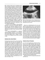
Start by observing for deformities, swelling, and discoloration. Do this on
both the palmar and the dorsal surfaces of the hands and fingers. The usual
position of the hand is a flexed position as the flexors of the metacarpals and
fingers are stronger than the extensors (Figure 8.1). Observe for any deviation
from the usual anatomic position. Next, ask the patient to extend and flex the
metacarpal phalangeal (MCP) joints (Figures 8.2 and 8.3), the PIP joints
(Figures 8.2 and 8.4), and the DIP joints (Figures 8.2 and 8.5).
3. Case
3.1. History
A 30-year-old male construction worker comes to your office after being hit on
the dorsum (top) of his hand with a piece of machinery 1 day ago. He noted
immediate swelling and some tenderness on the dorsal side of hand. He
138 E.J. Shahady
MCP Joint
PIP
Joint
DIP
Joint
FIGURE 8.1. Flexion as the dominant hand position.
placed some ice on it and the discomfort subsided. He was able to work a few
more hours but was then sent home because of the pain. The next day he
went to work but was unable to use the hand without pain. The examination
reveals some swelling over the dorsum of the hand and early ecchymosis.
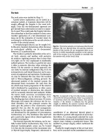
Most tenderness is over the shaft of the third metacarpal. You ask him to flex
his fingers into his palm and note that the middle finger is in an unusual posi-
tion (Figure 8.6). The fingers should all point in a similar direction as noted
in Figure 8.7.
8. Hand Problems 139
Dorsal side
MCP
PIP
DIP
Volar or Palmar side
FIGURE 8.2. Extension of the MCP, PIP, and DIP joints.
FIGURE 8.3. Flexion of MCP joint.
140 E.J. Shahady
PIP
FIGURE 8.4. PIP flexion.
FIGURE 8.5. Flexion of the DIP joint.
8. Hand Problems 141
FIGURE 8.6. Abnormal finger alignment.
FIGURE 8.7. Normal finger alignment.
3.2. Thinking Process
The unusual position of the finger in the flexed palm indicates malrotation of
the bone. If a fracture of the metacarpal shaft has occurred, flexion at the
MCP joint will produce malrotation of that metacarpal and the unusual posi-
tion noted in Figure 8.6. Fractures of the proximal and middle phalanx can
produce similar malrotation with flexion at the PIP or DIP joint. The mus-
cles of the hand and fingers function in perfect balance and fractures of the
shaft of the metacarpals and/or phalanx can cause malrotation and a defor-
mity. The key points are (1) a fracture of the shaft is most likely present and
(2) this type of fracture will require more than a cast.
X-rays revealed a spiral fracture of the shaft of the third metacarpal.
Computerized tomography (CT) and magnetic resonance imaging (MRI)
ordering is usually not needed to make this diagnosis. This patient was
referred to an orthopedic surgeon because the malrotation may require oper-
ative management.
4. Metacarpal Fractures
Fractures of the metacarpals can occur at the base, shaft, and neck. The man-
agement of metacarpal fractures depends upon the location on the
metacarpal of the fracture and which metacarpal is fractured. The fourth and
fifth metacarpals (ring and little finger) can tolerate more angulation than the
others and can often be managed conservatively. Fractures of the neck of the
fifth metacarpal are called boxer’s fracture because they commonly occur
when the fist strikes an object. Metacarpal fractures of the index and middle
fingers tolerate less angulation and may require operative management.
Thumb metacarpal fractures are more problematic and usually require ortho-
pedic evaluation. Primary care clinicians can manage many of these fractures
if they fully understand how to immobilize and protect the fractures.
Additional reading, training, and experience are required to understand these
principles. Many orthopedists are happy to help you understand these prin-
ciples and respond to your questions when you are not sure. This allows the
orthopedist to concentrate on the fractures that require more complex evalu-
ation and treatment.
5. Metacarpal Phalangeal Joint Dislocations
The MCP joint, because of its architecture, allows more freedom of move-
ment than the interphalangeal joints. The surrounding soft tissues are there-
fore more critical in maintaining the stability of the joint. Dislocations of the
MCP joint are not that common and dorsal dislocation (top of the knuckle)
142 E.J. Shahady
is the rule. Volar dislocations are rare and often require operative treatment.
Dorsal dislocations usually respond to reduction. Some simple rules help
with understanding how to reduce the dislocation. First, provide adequate
anesthesia with 1% or 2% lidocaine infusion into the joint. This not only
reduces pain and spasm but will assure that any volar tissue torn during the
dislocation will appropriately move to allow an easier reduction. Next, have
the patient flex the wrist and digits. This relaxes the dominant flexor system.
Reduction is now accomplished by hyperextending the MCP joint while
pulling the proximal phalanx forward, maintaining the tension of the pull
and flexing the MCP joint. Figure 8.8 demonstrates a simple dorsal disloca-
tion with the volar plate tissue in an appropriate place and directions on how
to relocate the dislocation. Figure 8.9 demonstrates a complex dislocation
with the volar plate tissue blocking the relocation. The ability to flex and
extend the joint actively and passively indicates a successful relocation.
Inability to do this indicates the possibility of a complex dislocation and the
patient should be referred. X-rays should be taken to assure that reduction is
complete. Splinting in full flexion for 1 week followed by buddy taping (tap-
ping one finger to the one adjacent to it) for two additional weeks to prevent
hyperextension is indicated. The MCP joint is more stable in the flexed posi-
tion and this position is known as the safe position because of its stability.
More than 1 week in this position may lead to stiffness and an increased need
for rehabilitation. Return to activity is permitted as long as the joint can be
protected from hyperextension.
8. Hand Problems 143
Try to push base
of phalanx volar
while pulling
finger distal
Volar plate still
draped over
metacarpal head
while base of
phalanx sits dorsal
Simple MCP
dislocation
FIGURE 8.8. Simple MCP dislocation and relocation. (Reproduced from Richmond J,
Shahady E, eds. Sports Medicine for Primary Care. Cambridge, MA: Blackwell Science;
1996:354, with permission.)
6. Case
6.1. History
A healthy 35-year-old woman comes to your office complaining of left
thumb pain. She was involved in a minor automobile accident 2 days before
the visit. She was on the passenger side and braced herself on the dashboard.
She did not seek medical attention initially but does now because of left
thumb pain and difficulty in maintaining her grasp. Her examination reveals
tenderness and swelling over the ulnar side of the MCP. The ulnar side points
toward the ulna in contrast to the radial side of the joint, which points
toward the radius (Figure 8.10). Stressing the thumb MCP joint into abduc-
tion (Figure 8.10) reveals laxity or increased opening on the left compared
with the right. No good end point at the end of abduction is felt on the left.
6.2. Thinking Process
The car stopped moving but her body did not. The position of her hand and
thumb made the MCP joint the focal point of this change in velocity. The
thumb was stressed and forcefully abducted at the MCP joint. This force
most likely injured the UCL of her thumb MCP joint when she braced her-
self against the dashboard. For the thumb this is a common type of injury. It
occurs in sports that predispose to thumb abduction stress like football and
skiing. The best name for the injury is UCL injury but names like skier’s or
gamekeepers’ thumb are commonly used. The term gamekeepers’ thumb
comes from an injury suffered by gamekeepers in England when they twisted
144 E.J. Shahady
PREVENT THIS
Complex MCP
dislocation
Volar plate has become
completely displaced
dorsal to metacarpal
head and is now
irreducibly interposed
FIGURE 8.9. Complex MCP dislocation and relocation. (Reproduced from Richmond J,
Shahady E, eds. Sports Medicine for Primary Care. Cambridge, MA: Blackwell Science;
1996:354, with permission.)
the neck of the game they caught. This maneuver would injure the UCL lig-
ament and cause a chronic instability. Skiing accidents associated with the ski
pole can produce a forceful radial and palmar abduction of the thumb and
subsequent disruption of the UCL. This injury can lead to significant dis-
ability because of the importance of the thumb. If a fracture is present and/or
the UCL has migrated into the joint space a more complex injury is likely.
The physical examination helps access the extent of the damage. Perform
abduction stress (Figure 8.10) in both neutral and flexed positions. The flexed
position is usually more stable and weakness in this position indicates a more
serious problem.
Some patients will resist attempts to assess for instability because of the
pain. Lidocaine can be infused into the joint to allow for a more complete
examination. Once adequate anesthesia is accomplished, the examination can
be easily performed. It is important to perform thumb abduction in both the
neutral and the flexed positions to assess for stability in both positions.
A plain film X-ray is indicated in most cases especially if instability in flex-
ion is demonstrated. Fractures may be present and the fracture fragment or
tissue may become lodged in the joint. This fragment or tissue must be
removed surgically for proper healing to occur. The X-ray in this patient
revealed no fracture or indication of tissue in the joint. Magnetic resonance
imaging may be indicated to access the amount of tissue that has been dis-
placed into the joint. Save the ordering of the MRI for the orthopedic or
hand surgeon.
8. Hand Problems 145
UCL
FIGURE 8.10. Test for UCL integrity.
6.3. Treatment
Most patients seen in the primary care setting can be treated nonoperatively.
Minimal injury to the ligament is indicated by tenderness to palpation and
pain with abduction but no instability. Treat this injury by taping the thumb
to the index finger for a period of 3 weeks. This type of injury is commonly
seen in football and basketball players who probably have only strained the
UCL. Instability in the neutral position but not the flexed position and a neg-
ative X-ray can be treated with a thumb spica cast or splint. The initial splint
is placed for 3 weeks. An additional splint that allows wrist flexion but limits
thumb extension and abduction should be used for an additional 2 weeks. This
patient was treated like this and did well. Exercises to regain lost strength and
range of motion (ROM) are indicated as part of the treatment. Patients who
are athletes can return to play within 1 week of the injury. A rubberized cast
can be constructed. Participation in organized sports with a rubberized
cast depends on the rules of your local athletic association. Injuries that are
associated with thumb flexion weakness, fractures, or suspicion of bone or
tissue in the joint should have an orthopedic consultation.
7. Bennett’s Fracture
About 25% of metacarpal fractures involve the base of the thumb. These are
common when someone falls and the thumb takes the brunt of the force in
breaking the fall. The names Bennett and Rolando are attached to commin-
uted (more than one piece of bone) fractures of the thumb base. The strength
of the thumb abductors produces a deformity that most of the time will
require a surgical solution. All thumb metacarpal fractures are best treated
by an orthopedic surgeon.
8. Case
8.1. History
A 25-year-old female softball player comes to your office after being hit in the
hand by a softball 2 h previously. She says her middle finger was pushed back
by the ball and came out of place. The coach tried to push it back in place
but was not successful. Your examination reveals the middle phalanx is dis-
placed dorsally above the proximal phalanx at the PIP joint. The patient is
unable to extend or flex the finger. You reassure the patient and do a digital
block with lidocaine to obtain adequate anesthesia. Once the pain has disap-
peared, you grasp the finger firmly over the middle phalanx and gently hyper-
extend the joint while at the same time applying longitudinal traction and
then flexion similar to the MCP joint dislocation. The middle phalanx nicely
146 E.J. Shahady
came back into place. The patient was now able to flex and extend the finger
at the PIP joint. A postreduction X-ray revealed no fracture and good align-
ment of the middle and proximal phalanx at the PIP joint. A splint to limit
extension of the PIP joint only was placed for 1 week and this was followed
by 2 weeks of buddy taping the third finger to the index finger. There was
some residual swelling and minor discomfort for the next 2 months but the
patient completely recovered. She went back to playing softball after the
splint was removed.
8.2. Thinking Process
Proximal interphalangeal joint dislocation is the most common joint disloca-
tion in the hand. Almost all of them occur in a dorsal direction. They are
usually easily reduced and many are reduced on the field by the coach,
trainer, or the athlete. Thus, the name “coaches finger.” The sooner it is
reduced the easier the reduction. On-the-field reduction is ideal. An X-ray is
not needed before reduction unless the reduction cannot be accomplished
with the usual means. This may indicate that either bone or tissue is in the
joint space, limiting the ability to reduce the dislocation. The key to prevent-
ing complications with dislocations is the postreduction care. An X-ray is
always indicated after the reduction to assure that the reduced bones (middle
phalanx and proximal phalanx) are now aligned/congruent. The radiologist
will be attentive to this if the request is marked “after reduction.” If it is not
aligned/congruent, an orthopedic consultation is indicated. The other com-
plication that should concern you is the boutonnière deformity. This results
from a disruption of the extensor mechanism over the PIP joint (Figure
8.11B). This deformity is not noted immediately but weakness or absence of
extension will be noted immediately. Discussion of this injury occurs in
Section 9.
The vast majority of the time, PIP dislocations relocate easily and after a
reassuring X-ray and demonstrating the extensor mechanism is intact, buddy
taping is all the treatment that is needed.
Injury can also occur to the collateral ligaments of the PIP joint with the
dislocation. The ligaments known as the radial and UCLs of the PIP joint
should be tested for stability. Test for stability with the finger flexed (bent to
90° at the PIP joint) and also when it is extended (completely straight at 0°).
Place both an ulnar- and a radial-directed force on the joint to see if it opens.
Any opening would be abnormal. This is similar to testing the medial and lat-
eral collateral ligaments of the knee. Significant opening indicates a need for
longer splinting and letting the patient know that this enhances the chances
of chronic deformity and arthritic changes. An orthopedic or hand surgeon
should evaluate significant laxity.
Plain film radiographs are usually all that is needed. Additional imaging
may be needed for more complex fracture dislocations but this will usually be
ordered by the orthopedic or hand surgeon.
8. Hand Problems 147
8.3. Treatment
As previously mentioned, the dislocations without significant fractures or lig-
amentous injury will require 2 to 3 weeks of buddy tapping only. Significant
fractures should be managed by the orthopedists and will require dorsal
extension splints that are gradually straightened over a 4-week period.
Remember to evaluate the ability to extend the joint before splinting in flex-
ion. If a tear in the extensor mechanism exists, treating flexion will increase
the chances of the boutonnière deformity. If a splint is used it should only
involve the PIP joint. Limiting the movement of the DIP and MCP joints will
create stiffness in these joints and increased recovery time.
9. Boutonnière Deformity
This deformity although not common is usually preventable. Unfortunately, it
is not recognized early enough to prevent it. It is up to the primary care prac-
titioner to recognize and treat this injury in its early stages. The mechanism of
the deformity is a disruption of the central extensor slip that inserts on the mid-
dle phalanx. This injury makes it impossible to extend the finger at the PIP
joint (middle knuckle). The head of the proximal phalanx migrates (button-
holes) through the torn extensor mechanism and the two lateral bands of the
extensor slip now migrate down or volar. This now turns the extensors into
148 E.J. Shahady
Extensors
Boutonnière Deformity
A
B
FIGURE 8.11. (A) Mechanism of the deformity in a boutonnière injury. (B) Clinical
appearance of the boutonnière deformity. (Reproduced from Shahady E, Petrizzi M,
eds. Sports Medicine for Coaches and Trainers. Chapel Hill, NC: University of North
Carolina Press; 1991:83, with permission.)
flexors of the PIP joint (Figure 8.11A). The joint can still be passively moved
into extension but only with assistance. This also leads to hyperextension of the
DIP joint. In most cases it takes several weeks for the classic deformity (Figure
8.11B) to develop, leading to high number of missed or delayed diagnosis.
A well-conceived examination early in the injury will help with prevention of
this deformity. Dislocations and jammed fingers are predisposing injuries.
Evaluate any patient with either one of these injuries for weakness or inability
to extend the finger at the PIP joint. Start the evaluation by assessing for the
point of maximum tenderness. Extensor injury will be most tender on the dor-
sal surface (top) of the PIP joint (Figure 8.12) in contrast to the volar plate
injury that is most tender on the volar surface (bottom) of the PIP joint (Figure
8.12). If maximum tenderness is elicited over the dorsal surface of the joint, the
chances of an extensor injury are likely. The next step is to access the ability of
the patient to actively extend the PIP joint. Pain may limit extension. Infusing
the joint with a local anesthetic will assess the influence of pain on the ability
to extend. Do not be fooled by the ability of the patient or yourself to place the
finger in extension and its ability to remain extended. As soon as the patient
flexes the finger it will remain that way until passively extended again.
Treatment in the initial phase of the injury is a splint that immobilizes the
PIP joint in full extension for 4 to 5 weeks followed by nighttime splinting for
another 2 weeks. Do not involve the MCP and DIP joints in the splint. This
will result in unnecessary stiffness in these two joints. Instruct the patient
to flex and extend the MCP and DIP joint while the PIP joint is splinted to
prevent stiffness.
8. Hand Problems 149
Dorsal
Volar
FIGURE 8.12. Dorsal volar surfaces of the PIP joint.
If the diagnosis is delayed and the classic deformity of fixed PIP flexion
and DIP hyperextension is present, refer the patient to an orthopedic sur-
geon. Surgical reconstruction is reserved for the patients that fail nonopera-
tive measures.
10. Proximal Interphalangeal Volar Plate Injury
This injury is common and results from hyperextension of the PIP joint
by a ball or another object that causes hyperextension. This is commonly
called the jammed or stowed finger. The initial injury does not usually
cause significant concern or discomfort but within 24 h, the joint is swollen
and tender to motion. Tenderness is noted on the volar side (Figure 8.12)
and hyperextending the joint will reveal laxity. Collateral ligament injury
often accompanies volar plate injuries. Be sure to test these ligaments for
stability. Collateral ligament stability examination is discussed under PIP
dislocation additional examinations. Order X-rays if there is significant
instability or the patient is a child with a growth plate that has not closed.
Small avulsion fractures are common and do not require different treat-
ment. An orthopedic surgeon should evaluate fractures that involve over
30% of the articular surface.
Almost all jammed fingers seen in the primary care setting will have mini-
mal instability and not require an X-ray. Splinting the PIP joint in slight flex-
ion for 1 to 2 weeks followed by 2 weeks of buddy taping is sufficient. For the
mild injuries, buddy taping is all that is needed. It is not unusual for the joint
to remain swollen for a prolonged period of time because of continued use
and reinjury. Because the injury is usually mild, the patient finds it difficult to
stop the activity that caused the injury. Rather than harass the patient, just
provide the maximum protection against hyperextension while the patient
continues participating in the activity.
11. Collateral Ligament Injury
Partial tears of the radial and lateral collateral ligaments are most common
at the PIP joint but also are noted at the DIP and MCP joints. With jammed
and dislocated MCP, PIP, and MCP joints, these ligaments should be tested
for stability. Testing should be done in full extension and 30° of flexion. The
great majority of the time partial tears and minimum instability is present.
These injuries are treated with 2 to 4 weeks of buddy taping. Marked insta-
bility secondary to complete tears is also usually treated with buddy taping
but orthopedic surgeon advice is suggested because of the deformity that is
inevitable. Remember to explain to the patients that swelling and stiffness can
persist for several weeks.
150 E.J. Shahady
12. Distal Interphalangeal Joint
12.1. Extensor Injury (Mallet Finger)
Another common finger injury it is also sometimes called baseball finger or
drop finger (Figure 8.13). Get a group of 50- to 70-year-old men in a room
and look at their hands and you are likely to find someone with a drop fin-
ger. In the past both patients and physicians neglected this type of injury.
Many young boys and recently girls injure their fingers when a ball strikes the
DIP and forces the joint into flexion while they were trying to extend the fin-
ger. This causes a rupture of the extensor mechanism. The young man or
woman will complain of pain and swelling on the tip of the finger. Testing for
the ability to extend the finger at the DIP joint is important but sometimes
difficult because of the pain.
Often, the patient does not seek medical attention until the finger “drops.”
The initial swelling made the deformity difficult to appreciate and once the
swelling decreases patients then realize they have a more problematic lesion.
Treatment is usually by a splint that provides 5° to 10° of hyperextension for
6 to 8 weeks. Commercially available splints can be purchased. Patients will want
to remove the splint to clean it and wash their hands. Tell them how important it
is to keep the distal phalanx extended when the splint is removed. Some practi-
tioners will have the patients return for a clean replacement splint to their offices
so they can demonstrate how to maintain extension while performing the cleans-
ing. No matter how late the patient presents for treatment, always try to splint the
finger. It may not help after 2 to 3 months but nothing is lost in the effort.
8. Hand Problems 151
FIGURE 8.13. Drop or mallet finger.
X-rays should be obtained to see if a fracture is present and how much of
the articular surface is involved in the fracture. If 50% or greater of the artic-
ular surface of the joint is involved consult an orthopedic surgeon. The treat-
ment will still probably be a splint but let the orthopedist make this decision.
Surgery has little place in the treatment of this type of extensor tear unless
there is significant instability.
13. Case
13.1. History and Exam
A right-handed high school football player comes to your office 3 days after
injuring his right ring finger. The injury occurred when he was trying to
tackle another player by grabbing his shirt. The other player was able to pull
away from him and he noticed immediate pain in his ring finger. He did not
feel the injury was significant so he did not seek attention immediately. That
evening he noted difficulty grasping objects, and increased swelling and
pain. He made a trip to the emergency department. An X-ray was obtained
that was considered normal. He was informed that he had a strained finger
and should follow up with you as his primary care clinician. The discomfort
and disability have increased and his grasp strength has decreased.
Examination reveals tenderness on the volar aspect of the DIP joint and
swelling and tenderness over the volar surface at the MCP joint and a feel-
ing of a lump on the volar surface of the MCP joint. Extension of all the
joints is normal and flexion is normal at the MCP and PIP joints. He was
not able to flex the ring finger DIP joint against resistance or passively. Test
DIP flexion with the patient’s finger in extension at the DIP joint and the
examiner’s finger over the DIP joint on the volar surface. This limits flexion
only to the DIP joint and the deep flexor. The patient is asked to flex (bend)
the finger as demonstrated in Figure 8.14.
13.2. Thinking Process
This is the classical presentation for “Jersey finger,” an avulsion of the
flexor digitorum profundus tendon or deep flexor tendon. This tendon
attaches to the volar surface of the DIP joint and functions to flex the DIP
joint. The flexors are more dominating than the extensors in the hand so a
tear of a flexor tendon results in retraction of that tendon. The retraction
can be minimal or go all the way to the palm. If there is a large piece of
bone on the end of the tendon the retraction will be minimal. In this case
the retraction was more extensive. The retracted tendon is the lump pal-
pated at the volar surface of the MCP joint. This is the most frequent and
most worrisome of the tendon ruptures because the loss of usual blood
supply and quick occurrence of contractures make reinsertion more diffi-
152 E.J. Shahady
cult. Quick diagnosis and surgical treatment produces the best results. After
10 days reinsertion is not as successful.
X-rays are not needed to make the diagnosis. In fact, they may confuse the
situation. The diagnosis is purely clinical and once the primary care practi-
tioner discovers loss of deep flexion the next move is calling the hand sur-
geon. The surgeon may want some imaging studies but leave the decision to
the surgeon.
13.3. Treatment
The best option is early recognition and repair. Ten days is considered the
magic window, anything after that is less likely to be successful. Later repair
may not be successful because of the lack of adequate blood supply and ten-
don contracture. If you are faced with a delayed diagnosis, it is still worth-
while asking the patient to discuss it with a hand surgeon. There may be
procedures that will allow some increased function. If the loss of function is
acceptable to the patient there may not be any indication for surgery but at
least the patient is fully informed of options.
8. Hand Problems 153
FIGURE 8.14. Test for the deep flexor.
14. Medical Problems
14.1. Osteoarthritis
Osteoarthritis (OA) of the hand is common with aging. Most of the time it
is asymptomatic and the patient only seeks care because of appearance and
concern about cause and further deformity. Seventy-five percent of individu-
als over age 65 have some form of OA. It is symptomatic in 26% of women
and 13% of men. The most common area of involvement is the DIP joint
(Heberden’s nodes), followed by the base of the thumb carpal metacarpal
(CMC) joint and the PIP joint (Bouchard’s nodes). The MCP joints are not
commonly involved with OA.
Both Heberden’s and Bouchard’s nodes are bony prominences on the dor-
sal surfaces of the DIP and PIP joints. They are not usually tender unless
there has been trauma to the area. Patients will commonly ask what they are
and want reassurance that they are not an indication of something serious.
Although X-rays are not needed to make the diagnosis, they will show osteo-
phytes formation, joint space narrowing, sclerosis, and occasionally subluxa-
tions. Erosions and cysts are seldom seen in OA and if present another
diagnosis like gout should to be considered.
The examination in base of the thumb OA will reveal palpable bony promi-
nences over the CMC joint secondary to osteophyte formation and occa-
sionally radial deviation of the joint secondary to subluxation. The base of
the thumb CMC joint has four articulations. Osteoarthritis involves only one
of the articulations of the trapeziometacarpal joint and occasionally the
trapezioscaphoid joint. X-rays should be taken to make the diagnosis of OA
in this location. Isolated degenerative changes in the trapezioscaphoid joint
suggest other causes of arthritis like rheumatoid arthritis.
Treatment of hand OA in the asymptomatic patients consists of explaining
the process and excellent prognosis. Patients should be encouraged to stay
active and use their hands but protect them against trauma. Symptomatic
patients also require appropriate explanation of the process and good prog-
nosis. Exercises and continued protected movement is important and occu-
pational and physical therapy may be appropriate. Tylenol in doses up to 4 g
a day is helpful. Cautious use of appropriate nonsteroidal anti-inflammatory
agents (NSAIDs) will also help (see Chapter 1). If exercises and medications
do not relieve symptoms, consider injecting the joint with steroids and/or
splinting and protecting joints.
For the patients with more severe disease, consultation with an occupa-
tional therapist (OT) and/or physical therapist (PT) may provide other sug-
gestions to preserve function and decrease discomfort. Surgery may be
helpful to some patients and consultation with an orthopedic or hand sur-
geon may be indicated in the patients with more severe disease.
Other keys to making the diagnosis of OA of the hand include arthritis of
the other joints commonly afflicted by OA like the neck, back, hip, and knee,
154 E.J. Shahady
and the classic symptoms of little to no pain at rest, increased pain during
and immediately after movement, and minimal morning stiffness. Examina-
tion usually reveals bony prominences over the joints, and crepitations with
joint movement. Routine laboratory tests are negative and only serve to rule
out other diseases. X-rays are diagnostic but not prognostic. Do not let an
X-ray make the decision about the diagnosis. Osteoarthritic radiographic
changes are common in any patient after age 50. Symptoms are not caused
by the bone pathology but the surrounding soft tissue, ligaments, and carti-
lage damage.
14.1.1. Erosive Inflammatory Osteoarthritis
Osteoarthritis is not usually associated with acute inflammatory episodes like
swelling, painful joints, erythema, warmth, and limitation of motion. But,
occasionally it can be and this is called erosive inflammatory OA. Some inves-
tigators feel this is a separate entity but most feel it is an extension of nonin-
flammatory OA. This entity is most common in postmenopausal women and
involves primarily their hands and not the other joints commonly involved
with OA. The DIP and thumb base are the most common joints involved and
can be quite painful. Most clinicians when first faced with this entity think it
is infectious or due to RA. Knowing this entity exists and including it in the
differential diagnosis is helpful to both the patient and the physician.
X-rays reveal erosions. Rheumatoid factor and serum uric acid should be
obtained to rule out these entities. If the patient has all the signs and symp-
toms of OA and nothing else to suggest another disease process treat the
patient symptomatically. The joints need to be protected and pain relief
should be sought with NSAIDs and, if needed, short-term narcotics. Joint
injection with lidocaine and steroids can also help. As with noninflammatory
OA splints, consultations with PT, OT, and an orthopedic surgeon may be of
benefit.
14.1.2. Rheumatoid Arthritis
Swelling and tenderness of one or two PIP joints in the absence of any signs
of OA like Heberden’s or Bouchard’s nodes is suspicious for RA. The age of
the patient is also a tip. It is unusual for patients to have significant OA before
their 50s. Arthritic hand symptoms before age 50 should alert you to the diag-
nosis of RA. Erosive inflammatory OA, gout, and psoriatic arthritis are part
of the differential diagnosis. Once they are ruled out a preliminary diagnosis
of RA can be made. Medications that prevent loss of function in RA if given
in the first 4 to 6 months of the disease are now available. Early diagnosis
decreases the disability that accompanies RA.
Additional tips to aid in the diagnosis of RA, as noted in Chapter 7, may
be ulnar deviation of the wrist, swelling and pain of the MCP joints, and
forefoot pain secondary to involvement of the metatarsophalangeal (MTP)
joint.
8. Hand Problems 155
Morning stiffness lasting more than 45 min and severe fatigue may also be
present. Women in their first year postpartum are at increased risk of
autoimmune disease, so suggestive symptoms in this group should be given
appropriate attention. Laboratory and radiographic confirmation is nice to
have but only 50% of patients will have a positive rheumatoid factor early in
the disease and radiographic signs of bony erosions are not usually present
during the first year.
In the past, symptomatic treatment to make the patient comfortable was
acceptable. Relief with steroids was significant but the destruction of tissue
continued. Diseases-modifying antirheumatic drugs (DMARDs) are now
available. These drugs prevent joint destruction. Joint destruction can begin
as early as 4 months after the onset of symptoms and early drug treatment
preserves function. The drugs are expensive and have significant side effects.
If the primary care clinicians suspect RA a consultation with a physician
who understands how to administer DMARDs is indicated. Primary care
clinicians should familiarize themselves with all the side effects and interac-
tions of all these drugs as these patients will still rely on their primary care
clinician to provide other aspects of their care.
Suggested Readings
Sorock GS. Acute traumatic occupational hand injuries: type, location, and severity.
J Occup Environ Med. 2002;44(4):345–351
Daniels JM. Hand and wrist injuries: Part II. Emergent evaluation. Am Fam
Physician. 2004;69(8):1949–1956.
156 E.J. Shahady
9
Neck Problems
EDWARD J. SHAHADY
Neck pain is a common complaint in the general population. Forty to seventy
percent of adult patients will experience a least one significant episode of
neck pain during their lifetime. Most of the time it is mild and not a source
of concern for the patient. Although not as common as back pain, the preva-
lence is great enough in primary care to warrant attention, particularly in the
geriatric population.
Multiple diseases can cause neck pain. Medical problems like angina or
myocardial infarction refer pain to the neck, meningitis produces neck
stiffness, and migraine headache can be associated with neck pain.
Whiplash injuries after an auto accident, osteoarthritis (OA), and herni-
ated cervical disk are probably the most common causes of neck pain in
primary care. A good working knowledge of the epidemiology, anatomy,
associated symptoms, and examination reduce confusion and enhance the
diagnostic and therapeutic process when evaluating the patient with neck
pain.
Remember the neck supports a 20- to 25-lb weight (the head) that is con-
stantly moving around and places stress on the cervical spine and sur-
rounding musculature. This stress will never be eliminated but it can be
reduced.
Caring for neck problems is easier if a few simple organizational steps are
followed:
1. Step 1 is to realize that 95% of patients seen in the office with neck com-
plaints can be classified into the categories of problems noted in Table 9.1.
2. Step 2 is to take a focused history that segments the categories into acute
trauma, degenerative disease, and medical disease. This process reduces the
number to a manageable list to initiate further investigation.
3. Step 3 is to perform an examination that focuses on the most likely causes
suggested by the history. With a focused history and examination you now
have a likely diagnosis. Your knowledge of the usual history and examina-
tion associated with the most common problems has facilitated this process.
4. Step 4 is ordering confirmatory studies if needed (many times they are
not).
159
5. Step 5 is to start treatment. (This may include appropriate consulta-
tion.) Five percent of the time, the diagnosis will not be so obvious.
However, not being one of the 95% helps the examiner now think of
the 5%.
Some clinicians feel that ruling out the rare is our first responsibility based
on the old theory “I got burned once and it will not happen again.”
Unfortunately, this theory leads to unneeded testing, loss of treatment
time, and missed diagnosis of common problems. Unless the situation is life-
threatening or a red flag is present, ruling out the common is the most effec-
tive way to care for neck pain. If the common is not present, additional
confirmatory studies and/or a consultation will be required to discover the
rare causes.
1. Focused History
Inquiring about general health and constitutional symptoms should start
the process. Questions about chills, fever, and malaise will help rule out the
diagnosis of meningitis. Pain with exertion is suggestive of angina. Angina
pain may start in or radiate to the neck, face, and arms. Myocardial infarc-
tion pain is usually accompanied by sweating and may radiate or start in
the neck and face. Osteoarthritis of the neck is more common in older
patients. If you suspect OA of the neck, attempt to elicit other symptoms
of OA in the hands, hips, and knees. A history of acute blunt trauma
should make you suspicious for a fracture. If the patient was involved in a
car accident, think about whiplash. Burning radiating pain down to the
160 E.J. Shahady
TABLE 9.1. Common neck problems.
Neck pain of no specific cause (cervical strain)
Acute trauma
●
Fractures
●
Whiplash
Degenerative disease
●
Herniated disk
●
Spondylosis
Medical problems
●
Cardiac disease
●
Other types of arthritis
Other
●
Torticollis
9. Neck Problems 161
arms and hands suggest nerve root compression and a herniated cervical
disk. Since cervical root, compression may cause shoulder pain without
neck pain it is important to ask questions to rule out rotator cuff prob-
lems. Rotator cuff disease produces pain with shoulder abduction and cer-
vical root compression usually does not. Patients with rotator cuff
problems have great difficulty raising the involved arms above their heads.
Questions about gait, coordination, bowel and bladder control, and bal-
ance are important to rule out myelopathy (severe cord compression) that
can occur with cervical root compression. Questioning about the patient’s
past treatment successes and failures will help develop an effective
treatment plan.
2. Focused Physical Examination
The examination should focus on what the history suggests. A more extensive
examination may be indicated if the symptoms suggest a systemic disease.
First, observe for reduced spontaneous head movement, head tilt, and neck
deformity. Next, palpate and percuss over the cervical spine checking for the
presence of tenderness. Have the patient move the neck through the motions
of forward flexion and backward extension (Figure 9.1), left and right lateral
rotation (Figure 9.2), and left and right lateral bending (Figure 9.3). Measure
the degrees of movement and compare one side to the other. Differences
from one side to the other are diagnostic and can be used to judge treatment
success at follow-up visits. The neurological examination includes testing
the strength of the deltoid and rotator cuff muscles (see Chapter 5), biceps
and triceps muscle strength (Figure 9.4), and wrist flexion and extension
(see Chapter 7). Sensory examination concentrating on the dermatomes of
the upper extremities also helps discover specific cervical root deficits as
noted in Figure 9.5. C5 and C6 are the most common cervical roots com-
pressed. Conclude the neurological examination with upper extremity
deep tendon reflexes. If lower extremity symptoms are present neurological
examination of the lower extremity as outlined in Chapter 10 should be
performed.
Provocative tests to increase the pain in cervical root compression include
the Spurling’s, axial loading, Adson’s, and elevated arm tests. Spurling’s test
is performed by asking the patient, while seated, to extend the neck while the
examiner assists with tilting the head to the side (Figure 9.6). If this induces
radiating pain and numbness into the symptomatic extremity, it suggests
underlying nerve root compression usually secondary to disk herniation. The
axial loading test is performed with the patient standing and the examiner
pushes down on the patient’s head (Figure 9.7). This may provoke neck pain
if disk pathology is present.
162 E.J. Shahady
A
FIGURE 9.1. (A) Forward flexion. (B) backward extension.
B
9. Neck Problems 163
A
B
FIGURE 9.2. (A) Right and (B) left lateral rotation.
164 E.J. Shahady
FIGURE 9.3. (A) Left and (B) right lateral bending.
A
B