Recurrent Hernia Prevention and Treatment - part 7 potx
Bạn đang xem bản rút gọn của tài liệu. Xem và tải ngay bản đầy đủ của tài liệu tại đây (1.04 MB, 41 trang )
249
VII
Laparoscopical Repair
used were the rabbit and the pig. Even with the large
animal model (the pig), it is easy to imagine that these
same devices do not fix as well in a human, given the
increase in preperitoneal fat and abdominal wall thick-
ness compared to the pig models.
⊡
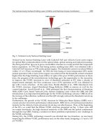
Figure 24.11 show
the amount of the fixation device (construct and tack)
that is available to go through the peritoneum, the pre-
peritoneal fat and into the muscle/fascia. Especially in
the obese patient, who has increased intra-abdominal
pressures and more preperitoneal fat, there is concern
that a no-suture technique might lead to a higher likeli-
hood of recurrence.
To minimize bleeding with suture placement, it
is important to visualize the abdominal wall to iden-
tify and avoid the inferior epigastric vessels and their
branches. Bleeding from accidental injury to an abdom-
inal wall vessel is usually controlled with direct pressure
and/or tying down the suture. Persistent bleeding can
be controlled with suture ligation proximal and distal
to the bleeding site, placing sutures through the same
skin incision.
Suture site pain may be lessened by injecting local
anesthetic prior to skin incision and by tying the knots
gently to avoid entrapping nerves and tissue [23] Tying
the knots gently might also help prevent a rare cause of
recurrence – herniation at the suture site. Placing the
suture about 1 cm inside the edge of the mesh and mak-
ing sure the mesh covers the suture site should also help
to prevent a suture site hernia recurrence. The exact
interval between sutures will vary depending on the
size and type of defect ( Swiss cheese vs. single defect)
and the amount of mesh overlap. In general, the larger
the defect, the closer the suture interval should be. For
example, when repairing a 1-cm recurrent umbilical
hernia using a 10×15 cm mesh, the initial four sutures
(top, bottom and each side) should provide adequate
suture fixation. For a large single defect involving an
entire midline incision, suture intervals of 3–5 cm is
recommended. On the other hand, for a Swiss cheese
defect of the same mid-line incision, an interval of
5–8 cm between sutures should be adequate.
Proper placement of the tacks or other point fixa-
tion devices includes placing the devices within 1 cm
of each other inside the edge of the mesh to prevent in-
ternal herniation between the mesh and the abdominal
wall. It is important to place the point fixation device
as flush with the mesh as possible. Any portion of the
tack that is hanging below the mesh could be a site for
increased adhesion formation, or worse, could cause
injury to abdominal organs. Bowel fistulas, apparently
caused by exposed tacks, have been reported [24, 25].
Other complications from point fixation devices include
pain, bleeding, tack site hernias, and inadvertent injury
to organs outside the abdominal cavity, including the
heart.
Conclusion
In summary, the best approach to prevent recurrence
following the laparoscopic repair of a recurrent ventral/
incisional hernia is to use both permanent full-thick-
ness abdominal wall sutures and point fixation devices.
Initially two to five sutures are placed on the mesh about
1 cm from the edge. After these sutures are brought out
of the abdomen and tied down gently under the skin,
the point fixation device is used to fix the mesh along
the edges at 1 cm or less intervals. Additional sutures are
then placed at the edges of the mesh at smaller intervals
for large single-defect hernias and at larger intervals for
⊡
Fig. 24.11. a The mesh explant shows the abdominal wall side of the mesh and the amount of constructs available for fixation.
b This mesh explant shows the abdominal wall side of the mesh and the amount of the tacks available for fixation
ab
Schumpelick.indd 249Schumpelick.indd 249 05.04.2007 8:52:24 Uhr05.04.2007 8:52:24 Uhr
250 How to Treat the Recurrent Incisional Hernia
24
Swiss-cheese type and smaller hernia defects. Despite
this opinion of a majority of experts in the literature,
various other forms of fixation are being used and have
similar published results. Prospective studies and new
fixation options may lead to improved knowledge and
better techniques for mesh fixation.
References
1. Bageacu S, Brenton C, Blanc P, et al. Laparoscopic repair of
incisional hernia. A retrospective study of 159 patients. Surg
Endosc 2002; 16: 345–348
2. Ben-Haim M, Kuriansky J, Tal R, et al. Pitfalls and complica-
tions with laparoscopic intraperitoneal expanded polytetra-
flurorethylene patch repair of postoperative ventral hernia.
Surg Endosc 2002; 16: 785–788
3. Birgisson g, Mastrangelo MJ, Park A, et al. Obesity and lapa-
roscopic repair of ventral hernias. Surg Endosc 2001; 15:
1419–1422
4. Bower CE, Kirby W, Reade CC, et al. Complications of lapa-
roscopic incisional-ventral hernia repair. Surg Endosc 2004;
18: 672–675
5. Gonzalez R, Duncan T, Mason E, Ramshaw BJ, Wilson R. Lapa-
roscopic versus open umbilical hernia repair. JSLS 2003; 7:
323–328
6. Heniford BT, Park A, Ramshaw BJ, et al. Laparoscopic repair of
ventral hernias, nine years’ experience with 850 consecutive
hernias. Ann Surg 2003; 238(3): 391–400
7. LeBlanc KA, Rhynes VK, Whitaker JM, et al. Laparoscopic in-
cisional and ventral hernioplasty: lessons learned from 200
patients. Hernia 2003; 7: 118–124
8. Park A, Birch DW, Lovrics P. Laparoscopic and open incisional
hernia repair: A comparison study. Surgery 1998; 124(4):
816–822
9.
Ramshaw BJ, Duncan TD, Esartia P, Lucas G, Mason EM, Miller
J, Promes J, Schwab J, Wilson RA. Comparison of laparoscopic
and open ventral herniorrhaphy. AM Surg 1999; 65: 827–832
10. Robbins SB, Gonzalez RP, Pofahl WE. Laparoscopic ventral
hernia repair reduces wound complications. Am Surg 2001;
67(9): 896–900
11. Roth JS, Mastrangelo MJ, Park AE, Witzke D. Laparoscopic in-
cisional/ventral herniorraphy: a five hear experience. Hernia
1999; 4: 209–214
12. Carbajo MA, Blanco JI, de la Cuesta C, Inglada L, Martin F,
Martin JC, Toledano M Vaquero C. Laparoscopic treatment
vs. open surgery in the solution of major incisional and
abdominal wall hernias with mesh. Surg Endosc 1999; 13:
250–252
13. Carbajo MA, Blanco JI, Martin del Olmo JC, et al. Laparo-
scopic approach to incisional hernia. Surg Endosc 2003; 17:
118–122
14. Chari R, Chari V, Chung R, Eisenstat M. A case controlled study
of laparoscopic incisional hernia repair. Surg Endosc 2000;
14: 117–119
15. Eitan A, Bickel A. Laparoscopically assisted approach for
postoperative ventral hernia repair. J Laparoendo Adv Surg
Tech 2003; 12(5): 309–311
16. Frantzides CT, Carlson MA, Zografakis JG, et al. Minimally
invasive incisional herniorrhaphy. Surg Endosc 2004; 18:
1488–1491
17. Gillian GK, Geis WP, Grover G. Laparoscopic incisional and
ventral hernia repair (LIVH): an evolving outpatient tech-
nique. JSLS 2002; 6: 315–322
18. Holzman MD, Eubanks S, Pappas TN, Purut CM, Reintgen
K. Laparoscopic ventral and incisional hernioplasty. Surg
Endosc 1997; 11: 32–35
19. Sanchez LJ, Bencini L, Moretti R. Recurrences after laparo-
scopic ventral hernia repair: results and critical review. Hernia
2004; 8: 138–143
20. Tagaya N, Mikami H, Aoki H, Kubota K. Long-term complica-
tions of laparoscopic ventral and incisional hernia repair.
Surg Laparosc Endosc Percutan Tech 2004; 14(1): 5–8
21. Joels CS, Matthews BD, Kercher KW, Austin C, Norton HJ,
Williams TC, Heniford BT. Evaluation of adhesion forma-
tion, mesh fixation strength, and hydroxyproline content
after intraabdominal placement of polytetrafluoroethylene
mesh secured using titanium spiral tacks, nitinol anchors,
and polypropylene suture or polyglactin 910 suture. Surg
Endosc 2005; 19(6): 780–785
22. van’t Riet M, Steenwijk PJ, Kleinrensink GJ, Steyerberg EW,
Bonjer HJ. Tensile strength of mesh fixation methods in lapa-
roscopic incisional hernia repair. Surg Endosc 2002; 16(12):
1713–1716
23. Carbonell AM, Harold KL, Mahmutovic AJ, Hassan R, Mat-
thews BD, Kercher KW, Sing RF, Heniford BT. Local injection
for the treatment of suture site pain after laparoscopic ven-
tral hernia repair. Am Surg 2003; 69(8): 688–691
24. Ladurner R, Mussack T. Small bowel perforation due to pro-
truding spiral tackers: a rare complication of laparoscopic
incisional hernia repair. Surg Endosc 2004; 18(6): 1001
25. DeMaria EJ, Moss JM, Sugerman HJ. Laparoscopic intraperi-
toneal polytetrafluoroethylene (PTFE) prosthetic patch repair
of ventral hernia. Surg Endosc 2000; 14: 326–329
26. LeBlanc KA. Tack hernia: a new entity. JSLS 2003; 4: 383–
387
Discussion
Kukleta: Don’t you think that the medialization that you
describe in your group is a consequence of shrinkage?
Because you fix it very well and you found it in nearly
90%. Nine out of ten patients had a substantial medial-
ization. That would be the only positive effect of shrink-
age.
Ramshaw:
Actually, on a few re-operations which
we had done with large meshes, we saw a little buck-
ling in the mesh inside. So I don’t think it is shrinkage,
because I think it is a true natural healing contrac-
tor, just as we see with the skin. If you eliminate the
intra-abdominal pressure, it contracts over time. So I
don’t think it is actually contraction of the mesh doing
that.
Schumpelick.indd 250Schumpelick.indd 250 05.04.2007 8:52:25 Uhr05.04.2007 8:52:25 Uhr
251
VII
Laparoscopical Repair
Frantzidis: One issue that hasn’t been raised with these
very large hernias: Do you offer your patients a binder
to reduce seroma formation and may help to incorporate
the mesh into the tissue?
Ramshaw:
With those very large defects I think that dense
spaces are always going to fill with fluid. I offer patients
a binder. I explain to them that it may be helpful in two
ways, to eliminate those dense spaces and possibly with the
security of eliminating movement that can cause especially
early fixation pain postoperatively. So I definitively offer it
and ask them to wear it. I don’t make it mandatory, but if
they wear it, I think they end up with a better result.
Schumpelick.indd 251Schumpelick.indd 251 05.04.2007 8:52:25 Uhr05.04.2007 8:52:25 Uhr
VIII
Primary Inguinal Hernia
25 How to Create a Recurrence 255
26 How to Treat Recurrent Inguinal Hernia 289
Schumpelick.indd 253Schumpelick.indd 253 05.04.2007 8:52:26 Uhr05.04.2007 8:52:26 Uhr
VIII
25 How to Create a Recurrence
Introduction
For many years, repair of inguinal hernias was primarily
based on Bassini-like repairs, aiming to re-enforce or re-
establish a weak or absent posterior wall of the inguinal
canal by using the anatomical structures bordering the
defect, with many of the differences in the various open
surgical techniques described being rather subtle.
Previous studies have shown recurrence rates of non-
mesh repairs in the range of 20–30% with highest recur-
rence rates after Bassini repair [1–4], and in most large
series, the rate of operation for a recurrence approaches
16–18%, confirming the high recurrence rates of past non-
mesh inguinal hernia repairs.
This study presents the results after Bassini repair,
based on data from the Danish Hernia Database.
Material and Methods
The analysis was based on 74,131 elective inguinal her-
niorraphies recorded in the Danish Hernia Database in
the period 1 Jan. 1998 to 30 June 2005 (
⊡
Table 25.1
). The
setup and organization of the Danish Hernia Database
is described elsewhere [5, 6]. In brief, the database re-
cords basic information, including type of repair, on all
(> 98%) inguinal and femoral herniorraphies performed
in Denmark, based on schemes filled out by the operat-
ing surgeon at time of operation. The database uses rate
of operated recurrences as a proxy for recurrence and
patient-specific observation time is calculated by the use
of unique social security numbers. Cumulative re-opera-
25.1 Bassini
M. B-N, H. K
⊡
Table 25.1. Number of herniorraphies, age, operative
findings and rate of operation for recurrence. Danish
Hernia Database 1 Jan. 1998 to 30 June 2005
Bassini Lichtenstein
No. of hernior-
raphies
1383 48,400
Median age 56 years 58 years
Direct/indirect
hernias
60/40% 56/44%
Primary/recurrent
hernias
88/12% 89/11%
Schumpelick.indd 255Schumpelick.indd 255 05.04.2007 8:52:26 Uhr05.04.2007 8:52:26 Uhr
256 VIII Primary Inguinal Hernia
25
tion rates are shown as Kaplan-Meier plots and compared
by use of log rank test. Hazard ratios for risk factors are
calculated using multivariate Cox proportional-hazards
regression. P < 0.05 is considered significant.
Results
Of the 74,131 elective inguinal herniorraphies re-
corded in the Danish Hernia Database, 1383 (1.8%)
were Bassini repairs. The use of Bassini repairs declined,
from 4% in 1998, to < 0.5% in 2005, concomitant to an
increase in the use of Lichtenstein repairs from 34 to
78% (
⊡
Fig. 25.1). Only small differences were found,
comparing age, ratios direct/indirect hernias and pri-
mary/recurrent repairs for Lichtenstein and Bassini
repairs.
Kaplan-Meier estimates of re-operation rates
show a significantly higher re-operation rate af-
ter Bassini repair, compared to Lichtenstein repair
(
⊡
Fig. 25.2) while analysis of re-operation rates after
Bassini repair, shows a re-operation rate after repair
of direct inguinal hernia being twice that of indi-
rect hernias, and recurrent repairs having almost
three times the re-operation rates of primary hernias
(
⊡
Table 25.2).
0
year
20
40
60
80
1998
1999
percentage
2000 2001 2002 2003 2004 2005
Lichtenstein
laparoscopic
other open mesh
other conv. open
Bassini
observation time [months]
2
6
rate of r-eoperations [%]
0
0
4
10
10
20 30 40 50 80
8
60 70 90
p<0.001
Bassini vs. Lichtenstein
hazard ratio 2.5 (95% CI 2.0–3.0)
⊡
Fig. 25.1. Changes in use of operative
techniques, Danish Hernia Database Jan.
1988 to June2005. n = 74,131 elective in-
guinal herniorraphies
⊡
Fig. 25.2. Kaplan-Meier estimates of
re-operation rates, Bassini (n= 1383) and
Lichtenstein (n = 48,000) repair of elective
inguinal hernia, Danish Hernia Database 1
Jan. 1998 to 30 June 2005. p < 0.05 com-
paring Bassini and Lichtenstein repair (log
rank test)
Schumpelick.indd 256Schumpelick.indd 256 05.04.2007 8:52:27 Uhr05.04.2007 8:52:27 Uhr
257
VIII
How to Create a Recurrence
Discussion
These data from the Danish Hernia Database confirm a
high rate of reoperation after Bassini repair (10% after
7 1/2 years).
Although mesh implantation in itself has been sus-
pected to be a factor in chronic postherniorraphy pain,
previous studies do not confirm this relation [7] and
no evidence exists showing an advantage of the Bassini
repair in other outcome parameters.
As a consequence of the unacceptably high risk of
recurrence after Bassini (and other open non-mesh
repairs) and the absence of data supporting the use
of Bassini repair, the use of Bassini repair should be
abandoned.
Conclusion and Consequences
To create a recurrence after a Bassini-type inguinal
herniorraphy is easy: you just do it and leave the rest
to time and gravity. The use of Bassini repair should
be abandoned.
References
1. Tran VK, Putz T, Rohde H (1992) A randomized controlled trial
for inguinal hernia repair to compare the Shouldice and the
Bassini-Kirschner operation. Int Surg 77: 235–237
2. Paul A, Troidl H, Williams JI, Rixen D, Langen R (1994) Ran-
domized trial of modified Bassini versus Shouldice inguinal
hernia repair. The Cologne Hernia Study Group. Br J Surg 81:
1531–1534
3. Hay JM, Boudet MJ, Fingerhut A, Poucher J, Hennet H, Habib
E, Veyrieres M, Flamant Y (1995) Shouldice inguinal hernia
repair in the male adult: the gold standard? A multicenter
controlled trial in 1578 patients. Ann Surg 222: 719–727
4. Strand L (1998) Randomized trial of three types of repair
used in 324 consecutive operations of hernia. A study of the
frequency of recurrence. Ugeskr Laeger 160: 1010–1013
5. Bay-Nielsen M, Kehlet H, Steering committee of the Danish
hernia data base (1999) Establishment of a national Danish
hernia data base: preliminary report. Hernia 3: 81–83
6. Bay-Nielsen M, Kehlet H, Strand L, Malmstrom J, Andersen
FH, Wara P, Juul P, Callesen T (2001) Quality assessment of
26,304 herniorrhaphies in Denmark: a prospective nation-
wide study. Lancet 358: 1124–1128
7. Bay-Nielsen M, Nilsson E, Nordin P, Kehlet H (2004) Chronic
pain after open mesh vs. sutured repair of indirect inguinal
hernia in young males. Br J Surg 91: 1372–1376
Discussion
Campanelli: I don’t agree with your conclusion. If you
follow the original steps of Bassini repair, it’s a perfect
repair. You can do it ambulant under local anesthesia and
you can achieve the same results as with a mesh repair.
So what are your specific steps of Bassini repair?
Bay-Nielsen: It’s not my repair. I just described how sur-
geons do the Bassini in Denmark.
Kingsnorth:
I think the problem is that you don’t have
control over the surgeons. But you have more control over
surgeons doing Lichtenstein because they are able to ap-
ply the principle of the repair better and achieve results
close to Lichtenstein, while general surgeons don’t appear
to be able to apply the basic principles of the Bassini to
get his results.
Bay-Nielsen: It gives us the ability to say: you do a Bassini
repair, these are your results, and you should do some-
thing else.
Read: I was surprised that the incidence of indirect hernia
was less than the incidence of direct hernia in the popu-
lation who are operated upon. This is against the main
experience with this type of hernia.
Bay-Nielsen: I cannot comment on that.
⊡
Table 25.2. Risk factors, comparing elective inguinal
Bassini (n = 1383) and Lichtenstein repairs (n = 48,400)
Risk factor Hazard ratio, comparing
Bassini and Lichtenstein
(95% CI)
Age (> 65, ≤ 65) 1.2 (0.8–1.8)
Direct vs. indirect 2.1 (1.4–3.1)
Recurrence vs. primary 2.7 (1.7–4.2)
Schumpelick.indd 257Schumpelick.indd 257 05.04.2007 8:52:27 Uhr05.04.2007 8:52:27 Uhr
258 VIII Primary Inguinal Hernia
25
Introduction
To the serious and dedicated surgeon, it would be unthink-
able to expect a career without being competent in the
performance of a pure tissue repair for inguinal hernias.
It would be unrealistic if not careless. To accomplish this
competence will not be easy, for it will take valiant and
diligent effort not to be overwhelmed or intimidated by
the manufacturers and salesmen of surgical prosthetics,
instruments and implements. Although it is a necessary
evil of marketing strategies to sell indiscriminately if not
wantonly, the onus is on the surgeon to be steadfast and
show perspicacity, for he is the guardian of his patient’s
most prized possession: his well-being.
This is not a plea for blind conservatism but a call for
an informed liberal choice. A pure tissue repair is always a
proper operation when the pathology consists of an indirect
inguinal hernia. This is nearly always the case for children,
young adults, females as well as many adults who present
with a pure indirect inguinal hernia. That is, unless an indi-
rect sac has a neck wide enough to involve the posterior
inguinal wall. The use of mesh is properly indicated for direct
inguinal hernias, femoral hernias whether or not associated
with an indirect inguinal hernia and recurrent hernias. Per-
haps the most important reason to be adept with a pure
tissue repair is that it imparts knowledge that will enable
you to manage any situation in the groin, particularly during
emergencies when incarceration, strangulation or bacterial
contamination of the operative site may proscribe the use of
prostheses. Important, too, is that you are the one to decide
what is best for your particular need. No one else has made
that decision for you at a sales strategy powwow!
The number of pure tissue repairs derived from Bassi-
ni’s technique is now well over 80 and counting [1]. All
have quietly disappeared but for the Shouldice repair.
The Shouldice repair itself has been recognized by many,
quietly, to be really a Bassini with no difference to justify
a new appellation. The fact, too, is that no particular tech-
nique was ever described by Shouldice himself. The pres-
ent discussion and recommendations will apply therefore
to the Shouldice as well as the Bassini repairs. I always
find it ironic to read that the Shouldice repair yields bet-
ter results than the Bassini repair. The good results of the
Shouldice Hospital are, in no small measure, the result of
an expertise acquired from doing thousands of procedures
to the exclusion of all other surgical operations, by a team
of dedicated surgeons. Who among us does not recall that
the Bassini repair was taught as a “modified Bassini” and
therefore, by not resecting the cremasters and not open-
ing the posterior inguinal wall but imbricating it instead,
one did a corruption of that repair which evidently leads
to poorer results! Shouldice respects the very steps intro-
duced by Bassini, adding a second running suture in the
reconstruction for good measure.
Another digression, about the McVay repair this time,
begs to be made since the dissection is entirely a Bassini-
Shouldice dissection without the resection of the cremas-
ter. It is still performed by a few surgeons, though their
number is dwindling. McVay’s contribution was made at a
time when mesh was not in common use and, when used,
was fraught with and evoked unwarranted fears. The McVay
contribution was one of exquisite understanding of the
anatomy of the groin. As a hernia repair, it was beset by a
moderate incidence of recurrence, suffered from too much
tension and pain and was associated with a constant, if low,
incidence of femoral vein complications. Most notable is
the fact that a recurrence from a true McVay repair is always
the most difficult dissection one can expect while doing
open surgery on a recurrent inguinal hernia.
How does one then, “create” a recurrence while per-
forming a Shouldice repair? The answer must be provided
under five headings:
▬ Magnitude of the problem.
▬ Corruption of the established technique.
▬
Shouldice against odds. Attempting to perform a
Shouldice repair in a class of hernia where a pure tis-
sue repair is known to yield poor results.
▬ Inadequate knowledge of the anatomy and pathology
of the groin.
▬ Specifics.
Magnitude of the Problem
The incidence of recurrence following inguinal hernia
repairs varies between 8 and 33%, and depends on the
operative technique [2]. In the hands of the Shouldice
Hospital surgeons who rely on the Shouldice repair only,
that is to say when mesh is not used, that incidence varies
between 1 and 20% [3]. Looking at pre-mesh days (up to
1983), the results can be assessed from
⊡
Table 25.3.
With reference to recurrence rates following pri-
mary inguinal hernia repairs, the Shouldice Hospital
claims an incidence of less than 1%. However, I have
25.2 Shouldice
R. B
Schumpelick.indd 258Schumpelick.indd 258 05.04.2007 8:52:28 Uhr05.04.2007 8:52:28 Uhr
259
VIII
How to Create a Recurrence
never seen a study emanating from the Shouldice group
analyzing the extent of the follow-up that would be ac-
ceptable to a statistician. My own attempt at follow-
ing 400 patients from their records alone, from 1986 to
1996, yielded a dismal follow-up of 10% only [4]. The
literature reports incidences of recurrences as high as
12.5% at 4 years [5]. On a yearly basis, 13–15% of all
patients presenting at or, referred to the Shouldice Hos-
pital are already recurrences and persisting with the use
of the Shouldice repair may well be a way of “creating”
a hernia! At least by their own admission.
Corruption of Established Techniques
A chronic bane in surgery is the blind improvisation of
a particular step in a well-established operation. This
variation is often perpetrated without the benefit of a
defining study to confirm the premise or pretence of
that variation. This is seen when the Bassini or Shoul-
dice repairs are carried out without the resection of the
cremaster or without the division of the posterior wall
of the inguinal canal or when reconstruction is carried
out by “imbrication” of the transversalis fascia or when
the external oblique aponeurosis is approximated under
the cord, leaving the latter in the subcutaneous position.
These shortcuts usually lead to shortcomings.
Shouldice Against Odds
The Shouldice Hospital, which remains a bastion of
pure tissue repairs, has finally conceded that, in fact,
there are situations when “mesh is indicated”! Like a
man who has long been used to suspenders, wearing a
waist belt only feels somewhat unsafe still! Their own
statistics do reveal at last that mesh must be the order
of the day when dealing with recurrences. Often, unfor-
tunately, recurrences are due to missed and overlooked
hernias during a previous attempt at herniorrhaphy. If
such a missed hernia is an indirect inguinal sac, a Shoul-
dice repair may well be safely attempted. A Shouldice
repair should not be attempted in the presence of a di-
rect inguinal hernia. The reason will be examined under
the next heading. The Shouldice repair should not be
modified to include the ligament of Cooper. This has
been proposed by a surgeon, resulting in a McVay type
of repair in order to correct a co-existent femoral her-
nia [6]. In such a modification, the resulting operation
would be closer to a McVay than to a Shouldice. Though
this modification may perhaps handle a small femoral
hernia, a double-blind study was never carried out and
cases needing mesh were excluded from the series since
they were too large to handle by a suture repair! In other
words patient selection took place, negating the study
and casting much doubt on the results [6, 7].
Inadequate Awareness of Inguinal
Anatomy and Pathology
It is often said that the anatomy of the groin is the most
difficult with which one has to contend. That is probably
the case and this is reflected by the unaltered incidence
of hernia recurrences in the past three decades despite
the addition of prosthetic sheets, prosthetic gadgets and
laparoscopic mastery [8]. Adequate textbooks abound
which discuss the anatomy of the groin; however, many
can be confusing, unless one is to dedicate the necessary
time to study them. The anatomy lab and the operating
room are ideal places to identify, confirm and crystallize
the acquired knowledge.
Hernia pathology used to imply progressive changes
in anatomy secondary to the mechanical strains and
stresses of daily life, work and ageing. Today, there is a
resurgence of interest in the biological and metabolic as-
pect of hernia disease, particularly, more recently, at the
cellular, nuclear, chemical and molecular level. Scientific
activity is centred on the nature and changes taking place
within the collagen tissue as a result of inherited factors
or external deleterious stimuli e.g. smoking [9].
Specifics
How then, specifically, does one “create a recurrence”
during a Shouldice-Bassini repair? The answer is, of
necessity, speculative, since no-one has ever gone about
⊡
Table 25.3. Shouldice Hospital: own re-recurrences
from 1057 operations [3]
1x
recurrent inguinal hernia
18/775 12.3%
2x 15/212 17%
3x 16/49 12%
4x 11/14 17%
5x 11/5 20%
6x 10/2 10%??
Schumpelick.indd 259Schumpelick.indd 259 05.04.2007 8:52:28 Uhr05.04.2007 8:52:28 Uhr
260 VIII Primary Inguinal Hernia
25
to knowingly create such hernias. Logic and a smatter-
ing of detective work will help.
1. The skin incision is often suggested to be, in most
textbooks, 2 to 3 cm above a line joining the anterior
superior iliac spine to the pubic spine. Personal and
practical experience dictates that the incision must
be on and along that very line from the anterior su-
perior iliac spine to the very level of the pubic spine
to provide an optimal exposure of the relevant site
of surgery. This location displays easily the medial
portion of the floor of the canal where recurrences
occur most often. The undersurface of the liga-
ment of Poupart becomes clearly visible at the level
of the femoral triangle, where a femoral hernia can
be routinely searched for and excluded. An incision
so situated minimizes tension by the retractors, for
these are often the very source of marked discomfort
during surgery under local anaesthesia.
2. Resection of the cremasterics permits the accurate
identification, without fail, of an indirect sac at the
medial aspect of the cord at the internal ring. In
the series of 1057 recurrences seen and reported by
Obney and Chan [3], 37% of the recurrences turned
out to be indirect inguinal hernias (missed hernias?)!
45% were direct inguinal hernias, 8% were femoral
hernias (most likely overlooked also) and in 10%,
two or more hernias were discovered.
3. Division of the posterior wall of the inguinal canal
allows the examination of the preperitoneal space,
the identification of femoral pathology and rare her-
nias. But above all, it affords the identification of
good tissue layers which will allow for a solid repair.
The posterior inguinal wall will not be made up of
a weak, thin and translucent transversus abdominis
fascia and its posterior layer, the true transversalis
fascia which is part of the endopelvic fascia.
4. Division of the cribriformis fascia is a small surgical
step, requires little time and pays off handsomely in
terms of discovering a femoral hernia which would
otherwise have become a missed hernia and there-
fore a recurrence.
5. Nowadays, the division of the posterior wall of the
inguinal canal must come under scrutiny. Is it a nec-
essary step in all patients undergoing the Bassini or
Shouldice repairs? Many of the Shouldice surgeons
with whom I shared surgical opinions over many
years varied in their approach. Oftentimes, when
the wall and the tissues were good, the wall was not
divided. Why divide a good structure and run the
risk of a recurrence which, if it takes place, will do so
at the very medial end of this wall just lateral to the
pubic spine? Some surgeons take the middle of the
road by dividing the posterior inguinal wall halfway
only. I very rarely divide the posterior inguinal wall
in women because they seldom have direct hernias
or in patients who have an indirect inguinal her-
nia with a good posterior wall and in children. In
women the occurrence of a direct hernia is low, 1
out of 12 primary inguinal hernias compared to 1
out of 2 in men [10]. If one considers that women
make up 5% of the hernia population, their chance
of having a direct inguinal hernia is 0.4% of all in-
guinal hernias!
6. When direct inguinal hernias are present, they
must be considered to be secondary to metabolic,
genetic and chemical factors which lead to tissue
degeneration and therefore hernia formation. In
these patients, the use of prostheses is justified and
recommended [9]. We have seen above that in the
hands of the Shouldice surgeons, the incidence of
re-recurrence can be between 2.3 and 20% when
they repair recurrences without mesh. The patients
at their hospital present with a recurrence number
12–16% of the total number of patients [11]. Yet,
mesh was used in only 0.86% of recurrent indirect
inguinal hernias and in 5.78% of direct inguinal her-
nias. Somehow, logic is being ignored and a reason-
able conclusion would be that the Shouldice Hospital
is instrumental in “creating hernias” while doing a
Shouldice repair [12]!
7. The relaxing incision is a most trusted manoeuvre
in relieving tension in pure tissue repair. Introduced
by Wolfler in 1892, it was re-introduced by Berger
in 1902 and Halsted in 1903 [13]. It has since been
adapted in 12 variations [13]. Koontz confirmed
experimentally that“ not only does an incision over
fascia over good muscle not weaken the structure,
but the fascial covering is rapidly regenerated [14].
I have used a relaxing incision in over 2200 cases
without a single cause for regret. I have often seen,
while performing a generous relaxing incision as far
as the level of the internal ring that an interstitial or
low Spigelian hernia becomes evident which will
invariably require a mesh repair. In this case, the
hernia was not “created”, it was discovered!
Conclusion
Alexis Carrel, the Nobel laureate in medicine in 1912,
remarked that “the very fame of a specialist renders
him dangerous”. I thought a long time about this. Did
he mean that man becomes welded to his thoughts and
techniques and promotes them to the reckless exclusion
Schumpelick.indd 260Schumpelick.indd 260 05.04.2007 8:52:28 Uhr05.04.2007 8:52:28 Uhr
261
VIII
How to Create a Recurrence
of all logic and deference to worthy and newer chal-
lenges? This may well be. It is a form of slavery from
which man must detach himself. For his sake, for the
sake of science, but above all for the sake of man.
References
1. Bendavid R. New techniques in hernia repair. World J Surg
1989; 13: 522–531
2. Weber A, Garteiz D, Valencia S. Epidemiology of inguinal
hernia: A useful aid for adequate surgical decisions. In: Ben-
david R, Abrahamson J, Arregui ME, Bernhard J, Phillips EH
(eds) Abdominal wall hernias. Springer, Berlin Heidelberg
New York, 2001
3. Obney N, Chan CK. Repair of multiple time recurrent Inguinal
hernias with reference to common causes of recurrence.
Contemp Surg 1984; 25: 25–32
4. Bendavid R. Introduction to pure tissue repairs. In: Bendavid
R, Abrahamson J, Arregui ME, Bernhard J, Phillips EH (eds)
Abdominal wall hernias. Springer, Berlin Heidelberg New
York, 2001, p 353
5. Champault G, Barrat C, Catheline JM, Rizk N. Groin hernias- 4
year result of two randomized prospective studies. Hernia
1998; 2: 19–23
6. Welsh DRJ, Alexander MAJ. The Shouldice repair. Surg Clin
N Am 1993; 73: 451–469
7. Degani CT. Femoral hernia repair. A five year prospective
study. American Hernia Society Meeting; February 9–12/2005
in San Diego, Ca. Abstract #19–1
8. Schumpelick V. Herniosis: Recent understanding. San Diego
conference of American Hernia Society, Feb 9–12, 2005:Ab-
stract Book, # 46-I, page 114
9. Bendavid R. The unified theory of hernia formation. Hernia
2004; 8: 171–176
10. Glassow F. Inguinal hernia in the female. Surgery, gynecology
and obstetrics. 1963; 116: 701–704
11. Bendavid R. The Shouldice repair. In: Nyhus LM, Condon RE
(eds) Hernia IV. Lippincott, Philadelphia, 1995, p 223
12. Shouldice EB. The Shouldice repair for groin hernias. Surg
Clin N Am 2003; 83: 1181
13. Bendavid R. Relaxing incisions. In: Bendavid R, Abraham-
son J, Arregui ME, Bernhard J, Phillips EH (eds) Abdominal
wall hernias. Springer, Berlin Heidelberg New York, 2001,
pp:343–346
14. Koontz AR. Hernia. Appleton-Century-Crofts; New York, 1963,
pp 52–53
Discussion
Kingsnorth: Does the suture material you use contribute
to recurrence? You still use stainless steal wire?
Bendavid: I have switched to polypropylene suture mate-
rial. The danger of stainless steel is that you have sharp
ends and you very often get pricked.
Miserez: I have two questions. Do you advocate doing
anything with the posterior wall in the young adult and
in women, or do you just leave like it is? Do you still use
four layers for a Shouldice repair?
Bendavid:
In women, direct hernias are never a problem,
they normally don’t occur. In indirect hernias we just
narrow the internal ring and we rarely see a recurrence.
Concerning the second question: we still do the four-layer
repair. In about 15 to 20% we add a relaxing incision to
make it as tension-free as possible.
Flament:
Five years ago during a meeting organized by
Prof. Lange, 500 surgeons were asked what to do with a
2 cm lateral hernia and 95% answered that they would
do a Shouldice repair. This is just a statement.
Kingsnorth: This is an important statement.
Kurzer:
When we talk about the Shouldice or the Bassini
repair, we talk about procedures which have a high ef-
ficacy, but only in the hand of experts. But a hernia repair
is only effective if it can be spread easily to the rest of the
surgical community. The advantage of the onlay mesh
repair is that it can be spread to the general surgeon and
still gives good results. The suture repairs have efficacy
but only the onlay mesh repair has effectiveness as an
operation in the surgical community.
Bendavid: I agree with you. The Lichtenstein repair can
be done even by worse surgeons with good results. There
is no doubt that the Shouldice is not an easy operation.
Chan:
There are two things that I should like to say.
First of all, direct hernia recurrence is not only from a
Shouldice repair. If you use big bites and put too much
tension on your suture, you will have a medial recur-
rence with any type of repair. Furthermore, you have to
be aware not to miss a small indirect hernia, which still
accounts for several recurrences. The third is that doing
the Shouldice repair you may open the femoral canal by
putting too much tension on your suture line. If you want
to see the anatomy and the exact technique, come to the
Shouldice Hospital and follow us for a week or two. Mesh
is seldom necessary, we use mesh in only about 1% of
the cases.
Kehlet: I want to come back to the results of the two na-
tionwide registers we have, Sweden and Denmark. Here
we see that Shouldice is a catastrophe. Do you really still
recommend the Shouldice? When you do the Shouldice
it might provide good results because you are experts.
But it has been proven in several studies that it does not
function in the general community.
Bendavid:
The registers just reflect what you put in them.
But you don’t have any control over the surgeons.
Deysine: You cannot learn a Bassini or a Shouldice from
a book or a drawing. The only way for a general surgeon
to learn the Shouldice is to go to Toronto and to watch
you operating. In the region where I live nobody can do
a Shouldice besides Berliner.
Schumpelick.indd 261Schumpelick.indd 261 05.04.2007 8:52:29 Uhr05.04.2007 8:52:29 Uhr
262 VIII Primary Inguinal Hernia
25
Bendavid:
I agree, the Shouldice is not an easy opera-
tion and you have to move and learn it from an ex-
pert.
Schumpelick:
I would like to comment on Dr. Kehlet. I
agree with you. Very often where Shouldice is written
about, it is no Shouldice. A lot of surgeons do the opera-
tion with no knowledge of anatomy. We re-operated more
than 200 of these cases. I think the name Shouldice is not
the operation of Shouldice.
Muschaweck:
Dr. Kehlet, I think the Shouldice is not a
catastrophe. You have to do the Shouldice correctly and
then you will have excellent results.
Bendavid: The advantage of having learned the Shouldice
technique is that afterwards you are able to manage any
problems in the groin and use any other technique of
repair, including all the mesh techniques.
Kehlet:
We are here as hernia experts and we have to give
advice for hernia surgeons around the world. I do not say
that Shouldice is a disaster in expert hands, but when
every surgeon is doing it, it is probably not a Shouldice at
all and they should not do it or even try to do it.
Kingsnorth:
A lot of surgeons want to have a fall-back
operation when they do not want to proceed with a Lich-
tenstein repair. In those countries where surgeons believe
that they should not put a mesh in a group of patients,
they need a tissue repair. There is no doubt that the Shoul-
dice is the best tissue repair if it is applied correctly. In
countries where surgeons believe that the Lichtenstein can
be applied universally, the Shouldice repair is irrelevant.
But in countries where a tissue repair is still supported by
a group of surgeons, it is their duty to apply the correct
Shouldice, which unfortunately is quite difficult.
Introduction
Lichtenstein tension-free hernioplasty began in 1984.
In the late 1980s, analyzing data from our own hernia
registry, published in 1987, we identified the following
flaws [1].
1. The mesh did not extend beyond the pubic tubercle
to overlap the pubic bone.
2. The mesh was too small (only 5×10 cm) to provide
enough mesh tissue contact beyond the inguinal
floor.
3. The mesh was kept flat (
⊡
Fig. 25.3, broken line), and,
therefore, was subject to tension when the patient
stood up from the supine position of the operation.
4. The upper edge of the mesh was fixed using a continu-
ous suture, which potentially left the iliohypogastric
nerve at risk.
5. Passing the genital nerve and external spermatic vessel
through a gap along the suture line of the mesh with
inguinal ligament exposed the nerve to potential risk
of entrapment.
In 1989, to correct the above problems, a set of principles
(outlined below) was established by our group, employed
with satisfactory results, and reported in 1993 [2].
Key Principles of the Lichtenstein Tension-
Free Repair
1. Use a large sheet of mesh that will extend approxi-
mately 2 cm medial to the pubic tubercle, 4–5 cm
above the Hesselbach triangle, and 5–6 cm lateral
to the internal ring (
⊡
Fig. 25.4
). We suggest using
a 7×15 cm sheet of mesh for easy handling, then
trimming 3–4 cm from its lateral side.
25.3 Lichtenstein
P. A
⊡
Fig. 25.3. Cross-section of the tension-free repair demon-
strating an inverted direct hernia sac and the dome-shaped
laxity of the mesh versus a completely flat mesh (dotted
line)
Schumpelick.indd 262Schumpelick.indd 262 05.04.2007 8:52:30 Uhr05.04.2007 8:52:30 Uhr
263
VIII
How to Create a Recurrence
2. Cross the tails of the mesh behind the spermatic cord
to avoid recurrence lateral to the internal ring (see
⊡
Fig. 25.4). Suturing the tails together in a paral-
lel position, without crossing, is a known cause of
recurrence lateral to the internal ring area.
3. Secure the mesh with two interrupted sutures on
the upper edge and one continuous suture with no
more than three to four passes on the lower edge of
the mesh to prevent folding and movement of the
mesh in the mobile area of the groin (see
⊡
Fig. 25.4).
Fixation of the mesh prevents movement, folding,
and wadding of the mesh ( meshoma) (
⊡
Fig. 25.5),
which can cause chronic pain and recurrence of the
hernia [3].
4. Keep the mesh with a slightly relaxed, tented up,
or buckled configuration (see
⊡
Figs. 25.3, 25.4
)
to counteract the forward protrusion of the trans-
versalis fascia when the patient stands up from the
intra-operative supine position, and to compensate
for contraction of the mesh.
5.
Visualize and protect the ilioinguinal, iliohypogas-
tric, and genital nerves throughout the operation
(
⊡
Fig. 25.6
). The iliohypogastric nerve can be iden-
tified easily, while the external oblique aponeurosis
is being separated from the internal oblique layer to
make room for the mesh. Because of a natural ana-
tomic cleavage, separation of these two layers from
each other is easy, fast, and bloodless. The most vul-
nerable part of the iliohypogastric nerve is its intra-
muscular segment (
⊡
Fig. 25.6
, dotted line), which
runs along the lower edge of the internal oblique
muscle (the so-called conjoint tendon). Passing a
suture through the internal oblique muscle to ap-
proximate this layer to the inguinal ligament (dur-
ing tissue approximation repairs) to a plug (during
mesh plug repair) or to the upper edge of the mesh
(during Lichtenstein repair) is liable to injure the
intramuscular portion of the iliohypogastric nerve
with the needle or entrap the nerve with the suture
[4]. The genital nerve is protected by not removing
⊡
Fig. 25.4. Extension of mesh beyond the boundary of the
inguinal floor (dotted line) 1 1.5–2.0 cm medial to the pubic
tubercle, 2 4.0–5.0 cm above the inguinal floor, 3 5.0–6.0 cm
lateral to the internal ring
⊡
Fig. 25.5. CT scan image of a meshoma (above). The explanted
meshoma (below)
⊡
Fig. 25.6. Neuro-anatomy of the inguinal canal
⊡
Fig. 25.7. Paravasal nerves within the lamina propria of vas
deferens
Schumpelick.indd 263Schumpelick.indd 263 05.04.2007 8:52:31 Uhr05.04.2007 8:52:31 Uhr
264 VIII Primary Inguinal Hernia
25
the cremasteric muscle and keeping the easily vis-
ible blue external spermatic vein with the spermatic
cord while it is being lifted from the inguinal floor
[4]. Removing the cremasteric muscles exposes
the genital nerve, paravasal nerves (
⊡
Fig. 25.7),
and the vas deferens to the mesh, which may lead
to chronic inguinodynia, orchalgia, and/or pos-
sible infertility [5] respectively. The ilioinguinal
nerve can easily be located over the spermatic
cord. Manipulating and lifting the nerve from its
natural bed will increase the risk of perineural
fibrosis and chronic postherniorrhaphy inguino-
dynia [4].
Causes of Recurrence after Lichtenstein
Tension-Free Hernia Repair
Causes of recurrence can be grouped in two categories:
(1) material-related causes and (2) technique-related
causes.
Material-Related Causes
▬ Mesh shrinkage: According to our clinical and
laboratory studies reported in 1995, after implan-
tation in vivo, mesh shrinks by approximately 20%.
Shrinkage of mesh can lead to recurrence of hernia.
Recurrence, however, can be prevented by extending
the mesh well beyond the boundary of the inguinal
floor.
▬ Mesh deformity related to the textile engineering
of the mesh: Certain structural designs of meshes
leads to narrowing of the mesh in the perpendicular
direction of stretching the mesh (
⊡
Fig. 25.8). As a
result the narrowed centre of the mesh can pull away
from its attachment to the host tissue and lead to
recurrence.
Technique-Related Causes
These include:
1. Failure to extend the mesh for approximately 1.5–
2.0 cm medial to the pubic tubercle, 4–5 cm above
the inguinal floor, and 5–6 cm lateral to the internal
ring.
2. Failure to keep the mesh slightly relaxed or buckled
up to account for forward protrusion of abdominal
wall in response to increased intra-abdominal pres-
sure when the patient stands up from the surgical
supine position and begins routine daily activi-
ties.
3. Inadequate mesh fixation that can lead to wrinkling
of the mesh and recurrence of hernia.
References
1. Amid PK. Lichtenstein tension-free hernioplasty: Its incep-
tion, evolution, and principles. Hernia 2004; 8: 1–7
2. Amid PK, Shulman AG, Lichtenstein IL. A critical scrutiny of
the open tension-free hernioplasty. Am J Surg 1993; 165:
369–371
⊡
Fig. 25.8. Central narrowing of mesh in
vertical direction with the mesh stretched
in horizontal direction
Schumpelick.indd 264Schumpelick.indd 264 05.04.2007 8:52:32 Uhr05.04.2007 8:52:32 Uhr
265
VIII
How to Create a Recurrence
3. Amid PK. Radiologic images of meshoma. A new phenom-
enon causing chronic pain after prosthetic repair of abdomi-
nal wall hernias. Arch Surg 2004; 139: 1297
4. Amid PK. Causes, prevention, and surgical treatment of
postherniorrhaphy neuropathic inguinodynia: Triple neu-
rectomy with proximal end implantation. Hernia 2004; 8:
343–349
5. Peiper C, Junge K, Klinge U, Strehlau E, Ottinger A,
Schumpelick V. Is there a risk of infertility after inguinal mesh
repair? Experimental studies in the pig and the rabbit. Hernia
2006; 10(1): 7–12
Discussion
Bendavid: In one of your drawings you showed a lateral
recurrence. An indirect hernia should never be a problem
as a recurrence.
Amid: I have seen several recurrences lateral of the cord.
To avoid this, the mesh should be extended lateral to the
internal ring. Except for the one suture, you don’t have
to put sutures lateral to the internal ring because you can
damage the ilioinguinal nerve.
Miserez:
Does chronic testicular pain arise from an in-
ternal ring which is too narrow? How can the ring be
calibrated?
Amid:
It is difficult to prove that the pain arises from a
narrow lateral ring. Much more likely, for example, is a
lesion of the paravascular nerves.
Kingsnorth:
The question is still: how do you calibrate
the ring?
Amid: I use the tip of my finger.
Read:
With the preservation of the cremasteric muscle
I am concerned that the surgeon might miss an indirect
hernia.
Amid: Around the internal ring we do a longitudinal inci-
sion on the cremasteric muscle. Doing this I can identify
any indirect hernia sac and dissect it as high as I want.
Young: Without opening the cremasteric fascia it might
be difficult to dissect the sac correctly in some cases.
Amid:
The vertical incision of the muscle opens the entire
spermatic cord like a book and you can dissect the sac
very easily.
Young: How do you handle the nerves? You showed a
ligation on a nerve which provoked chronic pain. On
the other hand, you have to ligate the nerves to prevent
a neuroma.
Amid: It is a fundamental difference if the nerve is intact
or not. If you put a suture to an intact nerve, you will
provoke pain. If you cut the nerve, you have a dead nerve
and this nerve has to be ligated at the proximal end.
Schumpelick: Y
ou recommended not to resect the cremas-
teric muscle in order to keep the spermatic duct away from
the mesh. But on the back there is no muscle at all.
Amid:
You are right. The muscle is like a half-moon shape.
But on the back you still have the cremasteric fascia,
which will avoid direct contact between duct and mesh.
Introduction
Inguinal hernia repair is one of the most common opera-
tion performed by general surgeons. It is considered a rou-
tine procedure, > 150,000 of these repairs are performed
annually in Italy, and > 730,000 in the United States [1].
Recurrences of hernia represent failure to achieve the
operative goal. It remains a significant clinical problem
despite advances in surgical techniques. Reasonably, we
can say that the most important yardstick for the success
of a hernia repair is still the recurrence rate [2, 3].
Comprehensive audit from national hernia registers
in Sweden [4] and Denmark [5] has shown an incidence
of recurrence of 16–18% following primary repair, but a
recurrence rate of even over 30% has been reported [6].
In a surgical reference centre, with hernia surgery spe-
cialization, this rate is above 0.3%, this means about 1100
recurrences a year, despite the introduction of laparoscopy
and marked increase in the use of prosthethic materials
for the repair of hernia in the wide community [7]. Ap-
plying the meshes has not in fact solved the problem of
recurrence, but called for different strategies for handling
those recurrences [8, 9].
One must examine the factors and influences which
come to bear on the choice of technique and quality in
our performance.
There are factors beyond the control of the surgeon
such as genetics, metabolic disorders, collagen diseases
25.4 Plug and PHS Technique
D. P, M. C, G. C
Schumpelick.indd 265Schumpelick.indd 265 05.04.2007 8:52:33 Uhr05.04.2007 8:52:33 Uhr
266 VIII Primary Inguinal Hernia
25
and smoking, which are now being recognized. Equally,
in the hands of surgeons there are factors like the surgi-
cal technique, the choice of prosthesis and the necessary
knowledge of the inguinal anatomy.
The aim of this work is to clarify some aspects linked
to recurrent inguinal hernia, despite the increasing use of
prostheses like PHS and hernia plug repair.
Materials and Methods
Mesh is a surgically designed, sterile woven material,
made of a synthetic plastic (i.e. polypropylene), specifi-
cally used to repair hernias. Prostheses by definition are
adjunct foreign materials used in the repair of hernias
and traditionally have come usually in flat sheets of
various sizes [7].
The plug is a 3-D filler cap that we insert in internal
inguinal ring to ensure a correct obliteration near the
issue of the spermatic cord (
⊡
Fig. 25.9)
The Prolene Hernia System 3 in 1 is a unique and
innovative design that combines the three most popular
tension-free techniques utilized today in the repair of
inguinal hernias (
⊡
Fig. 25.10).
Its onlay patch covers the entire floor of the canal; it
overlaps the pubic tubercle for added support, and pro-
vides the security of conventional patch techniques.
Its connector provides the simplicity of a plug repair.
Additionally, it secures the underlay patch to prevent
migration. Its profile is a significant improvement over
the bulk of conventional plugs.
The underlay patch, like a laparoscopic repair,
provides posterior support; however, it accomplishes
that support from a much simpler anterior approach.
This underlay patch lies in the preperitoneal space
and opens to cover the entire myopectineal orifice.
This key and exclusive feature of the Prolene Hernia
System ensures that both the femoral and inguinal re-
gions are protected to minimize the possibility of re-
currence.
Discussion
Here again, a thorough understanding of the anatomy
will lead us to applying our prosthesis in the proper
plane. The anatomy of the inguinal hernia region has
never been easily mastered by anyone. This delicate
aspect of anatomy was taken up and discussed with
a clear sense of rediscovery and grateful acknowl-
edgement especially by Cooper, who had enunciated
with uncanny accuracy anatomical features which
have been since overlooked, neglected or simply for-
gotten [10].
One further aspect which illustrates anatomical dif-
ficulty as an obstacle to good surgery is the presence of
a vasculature in the preperitoneal space [11].
The PHS presents an interesting dichotomy; it will
work in exactly the same way as a sheet of polypropyl-
ene will work if properly placed in the preperitoneal
space under direct vision or as practised in the Lich-
tenstein technique with evidently good result.
The most telling series to indict the poor knowledge
of anatomy was the publication of Obney and Chan
[12]; these authors reported a series of 1057 repairs on
recurrent hernias and noted that 37% of the patients
had an indirect inguinal hernia. An accurate knowledge
of the anatomy could not yield this level of recurrence.
It is evident that sacs are not being identified and often
overlooked. A proper dissection would not only dis-
cover hernias but would expose the proper planes and
tissues to be incorporated in a reliable reconstruction
with or without a prosthesis.
Another very difficult point to by-pass is excessive
body weight. Obesity is the bane of all surgery. Obese
people require larger amounts of sedation, local or
general anaesthesia, larger incision and longer operat-
ing time; their tissues show marked fatty infiltration,
lipomas, and therefore proneness to wound infections.
A proper dissection plane to plane is more difficult.
Obesity implies excessive tension along any suture line
and at peripheral sites where suture or staples maintain
a prosthesis in place [3, 8, 13].
⊡
Fig. 25.9. Plug
⊡
Fig. 25.10. The Prolene Hernia System 3 in 1
Schumpelick.indd 266Schumpelick.indd 266 05.04.2007 8:52:34 Uhr05.04.2007 8:52:34 Uhr
267
VIII
How to Create a Recurrence
An inadequate tissue dissection may cause a hae-
matoma, and this could be a cause of mesh lifting and
recurrence [14]. It will be a problem especially in pa-
tients with a difficult dissection of anatomical plane like
obese patients, or with a scar tissue caused by previous
intervention (e.g. appendicectomy, abdominal way for
prostatectomy).
So, we have to evaluate the anatomy of inguinal re-
gion, choosing the correct graft plane, we have to take
more care of our patient tissues, but we have also to
know the surgical technique for positioning properly
our prosthesis.
The technical position of PHS will take more care
and will need a short training. The PHS is inserted as
a plug, into the internal ring (
⊡
Fig. 25.11).
The underlay patch has to be extended in the pre-
peritoneal space.
The knowledge of this space is the most important
because we have already said that in this space ( space
of Bogros) we have a great vascularization [11].
The connector is, like the plug, to obliterate the in-
ternal inguinal ring [15].
The plug repair is one of the most common tech-
niques, and it’s used combined in open anterior ap-
proach ( Trabucco hernia repair) [16].
The plugs secure the larger internal inguinal ring
defect. Most people do not use the plug (Guidelines
European Hernia Society 2005) but choose to close the
internal inguinal ring with one or two stitches using
reabsorbable material and so the mesh on the inguinal
canal floor is the only “device” that protects from prob-
able recurrence.
In overweight patients, the correct plane dissection
and thereby the correct sac isolation just near the inter-
nal inguinal ring is very difficult [3, 8, 13]. The closure
of the internal inguinal ring is also more difficult with
or without the plug.
Conclusion
There are factors beyond the control of the surgeon
(genetics and metabolics), but there are factors in the
hands of the surgeons.
We have a wide spread use of prostheses but this
is not a solution for all recurrences. We have to know
the anatomy, the surgical technique and the proper
position of all prostheses that we use in our repara-
tions.
References
1. Rutkow IM (1998) Epidemiologic, economic and sociologic
aspect of hernia surgery in United States in the 1990’s. Surg
Clin North Am 78: 941–951
2.
Nilsson E, Haapaniemi S, Gruber G (1998) Methods of repair
and risk for reoperation in Sweden. Br J Surg 85: 1686–1691
3.
Bendavid R. Archives of the Shouldice Hospital. Unpubl. data
4. Nilsson E (1999) Outcomes. In: Kurzer M, Kark AE, Wantz GE
(eds) Surgical management of abdomina wall hernias. Martin
Dunitz, London, pp 11–19
5. Bay-Nilsson E, Kehlet H (1999) Steering committee of the
Danish hernia database. Establishment of a national Danish
hernia database: preliminary report. Hernia 3: 81–83
6. Ijzermans JNM, de Wilt H, Hop WCJ (1991) Recurrent ingui-
nal hernia treated by classical hernioplasty. Arch Surg 126:
1097–100
7. Bendavid R (2003) Recurrences: the fault of the surgeon. In:
Schumpelick V, Nyhus LM (eds) Meshes: benefits and risks.
Springer, Berlin Heidelberg New York, pp 51–62
8. Bendavid R (2002) The Shouldice repair. In: Fitzgibbons R
Jr, Greenburg AG (eds) Nyhus and Condon hernia, 5th edn.
Lippincott Williams & Wilkins, Philadelphia, pp 129–138
9. Campanelli G, Pettinari D, Nicolosi FM, Cavalli M, Contessini
Avesani E (2005) Inguinal hernia recurrence: classification
and approach. Hernia 2006; 10(2): 159–161
⊡
Fig. 25.11. Insertion of PHS
Schumpelick.indd 267Schumpelick.indd 267 05.04.2007 8:52:36 Uhr05.04.2007 8:52:36 Uhr
268 VIII Primary Inguinal Hernia
25
Rives procedure. In our experience it is easy to create the
preperitoneal space. The sublay mesh is mandatory for
the repair and the PHS connector is only for to keep the
sublay mesh in place.
Kehlet: I think in inguinal hernia surgery we have too
many procedures. To decide which method can be pre-
ferred we need results. What are the efficacy and the
effectiveness data of the PHS procedure?
Campanelli:
I do not have much experience with the
PHS operation. But I think we really need many differ-
ent procedures to provide a tailored approach in hernia
surgery.
Kingsnorth: You recommended the PHS for indirect but
not for direct hernia. In contrast, Prof. Flament uses his
approach especially for direct hernias.
Campanelli: In direct hernia you often have a large bulge
of the posterior wall and not a defect. In these cases its
better to place a mesh on the bulge than to destroy the
wall which it’s intact.
Schumpelick: You did not talk about the Rutkow plug.
Is there still a place for this procedure?
Campanelli: I don’t use the plug. But the operation tech-
nique and the indication are the same as for the PHS
device.
Köckerling: The plug just creates a meshoma in the pre-
peritoneal space. I will show this in the afternoon.
Kingsnorth:
We don’t have to be too dismissive concerning
the plug. About two million plugs have been implanted
and a lot of surgeons still use this method.
Deysine:
I have to defend the plug. I have put in about 1500
plugs up to now. Normally, I use the plug in indirect hernia.
In direct hernia I prefer the Rives operation. The only six or
seven times I saw a problem with the plug it was because of
incomplete dissection of the preperitoneal space.
Young: We remove more plugs than any other type of
mesh in our practise for recurrence or chronic groin
pain. We have done about 1500 plug repairs during the
past 3 years. Follow-up is difficult because the operation
is rather young, but the method seems to be successful
also in hands of surgeons who are not experts in hernia
surgery.
10. Bendavid R (2001) The transversalis fascia. New observa-
tion. In: Bendavid R (eds) Abdominal wall hernia, chap 10.
Springer, Berlin Heidelberg New York, pp 97–100
11. Bendavid R (1992) The spaces of Bogros and the deep ingui-
nal venous circulations. Surg Gin Obst 174: 355–358
12. Obney N, Chan CK (1984) Repair of multiple time recurrent
inguinal hernia with reference to common causes of recur-
rence. Contemp Surg 25: 25–32
13. Israelsson LA (2002). Wound failure and incisional hernia.
In: Fitzgibbons R Jr, Greenburg AG (eds) Nyhus and Condon
hernia, 5th edn. Lippincott Williams & Wilkins, Philadelphia,
pp 328–340
14. Lowham AS, Filipi CJ, Fitzgibbons R Jr, Stoppa R, Wantz GE,
Felix EL, Crafton WB (1997) Mechanisms of hernia recurrence
after preperitoneal mesh repair: traditional and laparoscopic.
Ann Surg 225 (4): 422–431
15. Nienahuijs SW, Van Oort I, Keemers-Gels ME, Strobbe LJA,
Rosman C. (2005) Randomized clinical trial comparing the
prolene hernia system, mesh plug repair and Lichtenstein
method for open inguinal hernia repair. Brit J Surg 92:
33–38
16. Prieto-Dias-Chavez E, Medina-Chavez JL, Gonzalez-Ojeda A,
Coll-Cardenas R, Uribarren-Berrueta O, Trujllo-Hernandez B,
Vasquez C (2005) Tension-free hernioplasty versus conven-
tional hernioplasty for inguinal hernia repair. Surg Today 35:
1047–1053
Discussion
Bendavid:
Using the PHS in the case of a large direct
hernia you would put two layers of Marlex mesh to the
floor which seems by far too much mesh material.
Campanelli:
You are right, but in cases of large indirect
hernias you really need the connector of the PHS device.
Jeekel: The principle of the operation is good because a
part of the mesh is in the preperitoneal space in a sublay
position. But how can you be sure that you prepared the
right space, that means the preperitoneal space? And the
second question is, what are the results?
Campanelli:
You cannot see the space, therefore you have
to prepare the space very exactly.
Flament:
We have investigated the transinguinal pre-
peritoneal mesh placement for many years. It is just the
Schumpelick.indd 268Schumpelick.indd 268 05.04.2007 8:52:37 Uhr05.04.2007 8:52:37 Uhr
269
VIII
How to Create a Recurrence
Introduction
Laparoscopy has provided surgeons with new and in-
novative ways to treat common surgical problems. Over
the past 10–15 years, this technology has been applied
to the treatment of inguinal hernias, where laparoscopy
has introduced a host of alternative surgical techniques.
Ger et al. first described the application of laparoscopy
to inguinal hernia repair in 1990. In this paper, Ger and
colleagues described repair of indirect inguinal hernias
through laparoscopic stapling of the abdominal open-
ing of the patent processus vaginalis [1]. Other minimally
invasive techniques were later developed including a plug
and patch repair [2] and an intraperitoneal onlay mesh
(IPOM) repair [3]. The plug and patch repair was not widely
adopted due to high recurrence rates coupled with small
bowel obstructions related to adhesions [4]. The IPOM
repair, as described by Fitzgibbons et al., involved plac-
ing a prosthetic mesh over the inguinal hernia defect in-
traabdominally without performing a groin dissection [5].
While the advantage of this technique was its simplicity,
this repair allowed for direct contact of the mesh with
viscera and the potential for mesh erosion into bowel. As
a result, this method of inguinal hernia repair was largely
abandoned.
Today, most laparoscopic inguinal hernia repairs are
performed with placement of a prosthetic mesh into the
preperitoneal space. This can be accomplished in one of
two ways: the totally extraperitoneal (TEP) approach and
the transabdominal preperitoneal (TAPP) approach. The
TAPP approach was the first to be developed and was ini-
tially described by Arregui and colleagues [6]. This repair
starts with entry into the abdominal cavity, followed by
incision into the preperitoneal space and blunt dissection
and reduction of the hernia sac. Once this is done, a piece
of prosthetic mesh is placed over the hernia defects with
subsequent re-approximation of the peritoneum. The TAPP
approach allows for a large working space and a good
view of the inguinal anatomy bilaterally. However, this
technique requires laparoscopic access into the peritoneal
cavity, placing the patient at potential risk of trocar inju-
ries, preperitoneal hernias from the peritoneal incision and
port sites, and intra-abdominal adhesive complications.
TAPP vs. TEP
The TEP approach was eventually developed in re-
sponse to concerns about the need for intra-abdominal
laparoscopic access required in the TAPP repair [7].
This method allows for direct access to the preperi-
toneal space while avoiding the peritoneal incision.
However, this procedure is also felt to be technically
more demanding given the smaller working space
provided compared to the one found in the TAPP
repair.
When Felix et al. compared the two methods to each
other, they found that the TAPP repair had a higher
incidence of intra-abdominal complications than the
TEP repair, including port-site hernias, small bowel
obstruction, and small bowel injury. However, several
TEP repairs needed conversion to a TAPP approach in
the study. Additionally, the study showed no appreciable
differences with regards to postoperative pain and re-
turn to normal activity [8].
In a review of the available literature comparing
TAPP vs. TEP repairs, Wake et al. found no statistical
difference in length of operation, length of stay, time to
return to normal activity, or recurrence rates between
the two techniques. They did find higher rates of intra-
abdominal injuries and port site hernias in TAPP repairs
[9]. In another review, Leibl and colleagues reported
similar findings. Of note, however, they stated that the
TAPP approach, in general, has a shorter learning curve
than the TEP repair [10]. While this conclusion has not
been supported by prospective studies, these authors
suggested that because of the shorter learning curve, the
TAPP repair might be more easily adopted into further
surgical education.
Further randomized controlled trials are needed
to compare the TAPP to the TEP repair to see which
method is superior. Nevertheless, we find the TAPP
repair to be useful in some clinical circumstances, for
example in patients with large indirect inguinal hernias
and in patients with incarcerated inguinal hernias. In
addition, the TAPP approach is easier than the TEP
approach in patients who have had prior operations in
the preperitoneal space.
25.5 Transabdominal Preperitoneal (TAPP) Inguinal Hernia Repair
B. K, Q.Y. D
Schumpelick.indd 269Schumpelick.indd 269 05.04.2007 8:52:37 Uhr05.04.2007 8:52:37 Uhr
270 VIII Primary Inguinal Hernia
25
Operative Steps
The patient is placed supine with both arms tucked
and under general anaesthesia. The operation is per-
formed using three trocars: one 10-mm port subum-
bilically and two 5-mm ports, one in the right lower
quadrant and one in the left lower quadrant. Pneumo-
peritoneum is established, and a 10-mm, 30° angled
laparoscope is inserted. The groin anatomy is identified,
specifically the inferior epigastric vessels and the internal
inguinal ring, through which the spermatic vessels and
the vas deferens run (
⊡
Fig. 25.12). The peritoneum is
incised several centimetres above the hernia defect, from
the edge of the medial umbilical ligament out laterally
towards the anterior superior iliac spine. The peritoneal
incision should be made in close proximity to where the
upper edge of the mesh used for repair will most likely
be positioned. The preperitoneal space is then dissected
bluntly in the avascular plane between the peritoneum
and the transversalis fascia (
⊡
Fig. 25.13
).
Indirect Hernia
The cord structures are dissected free from the surround-
ing tissues, as the indirect hernia sac is isolated out. The
indirect sac is found on the anterolateral side of the cord
and is adherent to it. When separating the sac from the
cord, it is important to handle the vas deferens and the
spermatic vessels with care so as to minimize trauma. If
the sac is small, it can be completely dissected free from
the cord, becoming part of the peritoneum (
⊡
Fig. 25.14
).
A large sac can be divided a few centimetres distal to the
internal ring with the subsequent peritoneal defect closed
with an endoloop suture (
⊡
Fig. 25.15).
Direct Hernia
Direct hernias are typically easier to reduce than in-
direct hernias. Once the preperitoneal space has been
entered, the direct hernia defect is dissected by simply
⊡
Fig. 25.12. Right groin anatomy view from a 30° laparoscope
placed below the umbilicus. Inf inferior epigastric vessels; Int
internal ring; Sper spermatic vessels
⊡
Fig. 25.13. Peritoneal incision exposing the preperitoneal
space in the right groin. Pre preperitoneal space; Cut cut edge
of peritoneum; Per peritoneum
⊡
Fig. 25.14a,b. Right indirect inguinal hernia. a Dissection of indirect hernia sac from cord structures. Inf inferior epigastric vessels;
Int internal ring; Sac indirect hernia sac. b Reduced indirect hernia sac. Per peritoneum; Red reduced hernia sac
ab
Schumpelick.indd 270Schumpelick.indd 270 05.04.2007 8:52:37 Uhr05.04.2007 8:52:37 Uhr
271
VIII
How to Create a Recurrence
separating the peritoneum from the overlying myo-
pectineal orifice. When reducing the direct hernia sac,
a pseudosac is usually encountered (
⊡
Fig. 25.16). The
pseudosac is part of the transversalis fascia that over-
lies and adheres to the peritoneum; it invaginates into
the preperitoneal space when the surgeon pulls on the
true sac during the dissection. The pseudosac must
be separated from the true hernia sac in order for the
peritoneum to be fully released back into the peritoneal
cavity. Once the pseudosac is freed, it will typically re-
tract back into the direct hernia defect.
Placement of Mesh
Prosthetic mesh is required for TAPP repairs. We rou-
tinely use a large piece of polypropylene mesh (16×12 cm)
to cover all the myopectineal orifices, including the direct,
indirect, and femoral hernia spaces. For direct hernias
only, we have also used a preformed, contoured mesh
(Bard 3D Max Mesh) for coverage. For indirect hernias,
we usually use a 16×12 cm mesh that is slit medially. The
lower tail is wrapped around the spermatic vessels and the
vas deferens in a lateral to medial fashion. The upper edge
of the lower tail and the lower end of the upper tail are
then fixed onto Cooper’s ligament (
⊡
Fig. 25.17). The slit
in the mesh allows it to lie flat in the preperitoneal space
while still providing complete coverage of the indirect
hernia defect. It is important that the preperitoneal space
is completely dissected out (Stoppa described this step as
“ parietalization”) so that the mesh does not fold within
this space and compromise the repair. The mesh needs to
be fixed medially at Cooper’s ligament and laterally above
the iliopubic tract to prevent movement of the mesh. The
mesh should also slightly overlap the midline to avoid
recurrence through the direct hernia space.
⊡
Fig. 25.15a,b. Large right indirect inguinal hernia sac divided just distal to the internal ring. a Divided indirect hernia sac with
exposed intra-abdominal cavity. Per cut edge of sac. b Endoloop closure of peritoneal defect. End endoloop suture
⊡
Fig. 25.16. Right direct inguinal hernia. Dissection showing
the “pseudosac”, which is the retracted transversalis fascia. Psd
pseudosac, Inf inferior epigastric vessels
⊡
Fig. 25.17. Placement of slit mesh wrapped around cord struc-
tures in a lateral to medial direction for a right indirect inguinal
hernia. Sper spermatic vessels, Low lower tail of mesh
ab
Schumpelick.indd 271Schumpelick.indd 271 05.04.2007 8:52:40 Uhr05.04.2007 8:52:40 Uhr
272 VIII Primary Inguinal Hernia
25
Issue of Recurrence
In the hands of experienced surgeons, laparoscopic in-
guinal hernia repair should result in a recurrence rate
of 1% or less [11]. Several reviews of recurrence from
TAPP repairs are summarized in
⊡
Table 25.4.
A number of factors have been noted to lead to
higher rates of recurrence. These are:
▬ Surgeon’s inexperience.
▬ Inadequate dissection of the preperitoneal space.
▬ Missed hernia defects.
▬ Insufficient size of the mesh.
▬ Insufficient overlap of the mesh beyond the edges
of the hernia defects.
▬ Improper fixation of the mesh.
Technical reasons appear to dominate these factors [16]
and, as a result, suggest that operative modifications can
lead to better outcomes.
Surgeon inexperience has been well accepted as a
critical factor in determining the risk of recurrence
after a TAPP hernia repair. Bobrzynski et al. specifi-
cally addressed this and found that their recurrence
rates after TAPP repairs dropped from 2.84 to 1.14%
after accounting for their learning curve [17]. Similarly,
Voitk suggested that at least 50 cases were required be-
fore operating times and complication rates stabilized
[18]. Certainly, understanding the preperitoneal groin
anatomy and the subtle layers and variations found in
this area is important, and this understanding is largely
gained through experience. Only by fully understanding
this anatomy can complete dissection of the preperito-
neal space be ensured. This includes full dissection of
the midline and Cooper’s ligament, full dissection and
reduction of direct and indirect hernia sacs, removal
of cord lipomas, and identification of all myopectineal
orifices and cord structures. Incomplete dissection can
result in missed hernias, leading to persistent rather
than recurrent hernias. Complete dissection avoids what
Lowham and colleagues refer to as secondary mecha-
nisms of hernia recurrence, which include insufficient
size and overlap of the mesh, improper fixation of the
mesh, and lipomatous hernia “recurrences” [19].
Factors pertaining to the mesh prosthesis also influ-
ence recurrence rates after TAPP repairs. Several studies
reported decreased recurrence rates as larger pieces of
mesh were used [20–22]. After the preperitoneal space
is dissected and all of the myopectineal orifices are
identified, the mesh used needs to properly cover all
of the existing and potential defects. At least 3 cm of the
mesh should overlap of the edge of the hernia defects for
complete coverage. This is necessary because the hernia
defect tends to enlarge and the mesh tends to shrink
over time. Potential hernia defects need to be covered
to prevent the future development of hernias in those
spaces. We routinely use a 16×12-cm mesh to provide
enough overlap medially and laterally.
Mesh fixation prevents recurrence as well. Mesh can
be fixed in place with tacks, sutures, or staples. Fixing
the mesh avoids early mesh migration and mitigates the
effects of mesh shrinkage. Over time, tissue ingrowth
will keep the mesh in place. Early on, however, the
mesh needs to be anchored. We routinely fix the mesh
to Cooper’s ligament medially and above the iliopubic
tract laterally. This immobilizes the mesh and prevents
it from folding. When fixing the mesh laterally, it is
important to do so above the iliopubic tract to avoid
injury to the lateral femoral cutaneous nerve and sub-
sequent neuralgias.
Using a slit mesh during laparoscopic inguinal her-
nia repairs has not been universally adopted. Some
surgeons believe that if the tails of the slit mesh are
not properly reapproximated, the opening will cause a
recurrence [19]. Felix, however, noted that a slit mesh
is necessary to prevent the cord structures from lifting
the mesh off of the inguinal floor [8]. In our experience,
a slit mesh is preferred to cover indirect hernia defects.
We slit the mesh medially and then fix both tails to Coo-
per’s ligament. For direct hernias, however, slit mesh is
not necessary. We prefer to use a preformed, contoured
mesh (Bard 3D Max Mesh) with no slit made in it.
Finally, a preperitoneal hernia is a unique problem
that may develop after a TAPP repair. In TAPP repairs,
a peritoneal incision is required to gain access to the
preperitoneal space and to dissect and repair all of the
hernia defects. This peritoneal incision needs to be
⊡
Table 25.4. Summary of TAPP repair studies
Author Year
Her-
nias
[n]
Recur-
rence
rate [%]
Bittner et al. [2]
2002 8050 0.70
Kapiris et al. [13]
2001 3530 0.62
Schultz et al.
[14]
2001 2500 1.04
Birth et al. [15]
1996 1000 1.10
Felix et al. [8]
1995 733 0.30
Schumpelick.indd 272Schumpelick.indd 272 05.04.2007 8:52:41 Uhr05.04.2007 8:52:41 Uhr
273
VIII
How to Create a Recurrence
closed to prevent herniation of intra-abdominal con-
tents into the dissected preperitoneal space. Closing
the peritoneal incision also prevents the bowel from
directly contacting the mesh. Because of the need to
incise and close the peritoneum, surgeons who prefer
the TEP technique frequently cite this as a significant
disadvantage of the TAPP repair.
Conclusion
The TAPP approach to inguinal hernia repair has proven
to be a safe and effective alternative to conventional
open hernia repairs. A recurrence rate of 1% or less
should be expected provided the surgeon has gained
the appropriate experience. Several factors contribute
to better outcomes and a reduced recurrence with the
TAPP repair. These include overcoming surgeon inexpe-
rience, adequately dissecting out the preperitoneal space
and identifying all potential hernia defects, using a large
piece of mesh with sufficient overlap beyond the edges
of all myopectineal orifices, and fixing the mesh.
References
1. Ger R, Monroe K, Duvivier R, Mishrick A. Management of
indirect inguinal hernias by laparoscopic closure of the neck
of the sac. Am J Surg 1990; 159(4): 370–373
2. Schultz L, Cartuill J, Graber JN, Hickok DF. Transabdominal
Preperitoneal Procedure. Semin Laparosc Surg. 1994; 1(2):
98–105
3. Kingsley D, Vogt DM, Nelson MT, Curet MJ, Pitcher DE. Lapa-
roscopic intraperitoneal onlay inguinal herniorrhaphy. Am J
Surg 1998; 176(6): 548–553
4. Tetik C, Arregui ME, Dulucq JL, Fitzgibbons RJ, Franklin ME,
McKernan JB, Rosin RD, Schultz LS, Toy FK. Complications
and recurrences associated with laparoscopic repair of groin
hernias. A multi-institutional retrospective analysis. Surg
Endosc 1994; 8(11): 1316–1322; discussion 1322–1323
5. Fitzgibbons RJ Jr, Salerno GM, Filipi CJ, Hunter WJ, Watson P.
A laparoscopic intraperitoneal onlay mesh technique for the
repair of an indirect inguinal hernia. Ann Surg 1994; 219(2):
144–156
6.
Arregui ME, Davis CJ, Yucel O, Nagan RF. Laparoscopic mesh
repair of inguinal hernia using a preperitoneal approach: a
preliminary report. Surg Laparosc Endosc 1992; 2(1): 53–58
7. Soper NJ, Swanstrom LL, Eubanks WS. Mastery of endoscopic
and laparoscopic surgery. Lippincott Williams & Wilkins;
Philadelphia, PA, 2005, p 49
8. Felix EL, Michas CA, Gonzalez MH Jr. Laparoscopic hernio-
plasty. TAPP vs TEP. Surg Endosc 1995; 9(9): 984–989
9. Wake BL, McCormack K, Fraser C, Vale L, Perez J, Grant AM.
Transabdominal pre-peritoneal (TAPP) vs totally extraperi-
toneal (TEP) laparoscopic techniques for inguinal hernia
repair. Cochrane Database Syst Rev. 2005; 25: CD004703
10. Leibl BJ, Jager C, Kraft B, Kraft K, Schwarz J, Ulrich M, Bittner
R. Laparoscopic hernia repair-TAPP or/and TEP? Langenbecks
Arch Surg 2005; 390(2): 77–82
11. Cameron JL et al. Current surgical therapy. Elsevier Mosby;
Philadelphia, PA, 2004, pp 1207–1213
12. Bittner R, Schmedt CG, Schwarz J, Kraft K, Leibl BJ. Laparo-
scopic transperitoneal procedure for routine repair of groin
hernia. Br J Surg 2002; 89(8): 1062–1066
13. Kapiris SA, Brough WA, Royston CM, O‘Boyle C, Sedman PC.
Laparoscopic transabdominal preperitoneal (TAPP) hernia
repair. A 7-year two-center experience in 3017patients. Surg
Endosc 2001; 15(9): 972–975
14. Schultz C, Baca I, Gotzen V. Laparoscopic inguinal hernia
repair. Surg Endosc 2001; 15(6): 582–584
15.
Birth M, Friedman RL, Melullis M, Weiser HF. Laparoscopic
transabdominal preperitoneal hernioplasty: results of 1000
consecutive cases. J Laparoendosc Surg 1996; 6(5): 293–300
16. Phillips EH, Rosenthal R, Fallas M, Carroll B, Arregui M, Corbitt
J, Fitzgibbons R, Seid A, Schultz L, Toy F et al. Reasons for
early recurrence following laparoscopic hernioplasty. Surg
Endosc 1995; 9(2): 140–144; discussion 144–145
17. Bobrzynski A, Budzynski A, Biesiada Z, Kowalczyk M, Lu-
bikowski J, Sienko J. Experience the key factor in success-
ful laparoscopic total extraperitoneal and transabdominal
preperitoneal hernia repair. Hernia 2001; 5(2): 80–83
18. Voitk AJ. The learning curve in laparoscopic inguinal hernia
repair for the community general surgeon. Can J Surg 1998;
41(6): 446–450
19. Lowham AS, Filipi CJ, Fitzgibbons RJ Jr, Stoppa R, Wantz GE,
Felix EL, Crafton WB. Mechanisms of hernia recurrence after
preperitoneal mesh repair. Traditional and laparoscopic. Ann
Surg 1997; 225(4): 422–431
20. Leibl BJ, Schmedt CG, Kraft K, Ulrich M, Bittner R. Recurrence
after endoscopic transperitoneal hernia repair (TAPP): causes,
reparative techniques, and results of the reoperation. J Am
Coll Surg 2000; 190(6): 651–655
21. Felix E, Scott S, Crafton B, Geis P, Duncan T, Sewell R, McKer-
nan B. Causes of recurrence after laparoscopic hernioplasty.
A multicenter study. Surg Endosc 1998; 12(3): 226–231
22. Fitzgibbons RJ Jr, Camps J, Cornet DA et al. Laparoscopic
inguinal herniorrhaphy-results of a multicenter trial. Ann
Surg 1995; 221: 3–13
Discussion
Kukleta: Up to now, I did not understand the bad results
of the Neumayer study. Now I do. Your task was to show
how to create a recurrence and you did.
Duh:
In the Neumayer study 90% of the endoscopic re-
pairs were done with the TEP repair.
Kingsnorth: Dr. Kukleta, you tell us that he is doing a
bad operation. So tell us why?
Kukleta: The strong part of the TAPP is that you visual-
ize everything. You haven’t seen anything on this video.
The 6-cm opening of the peritoneum is not enough to see
down below the important structures. I have not seen the
Coopers ligament, and the mesh was too big as well.
Schumpelick.indd 273Schumpelick.indd 273 05.04.2007 8:52:42 Uhr05.04.2007 8:52:42 Uhr
274 VIII Primary Inguinal Hernia
25
Flament:
I
n France last year we saw three deaths and severe
complications from laparoscopic surgery and none from
open surgery. How would you explain this difference?
Duh: The severe complications after laparoscopic repair
depend a lot on the learning curve problem. Laparoscopic
operation is harder to learn and if you make mistakes,
some of these mistakes can be deadly.
Peiper:
Do you have an age limitation for your laparo-
scopic approach?
Duh: In general, the indication depends on the type of
hernial defect. I examine the patients and then decide
whether a large mesh is necessary or not. Under the age
of 20 I would not do a TAPP.
Halm:
In bilateral hernia I use a large mesh for both
hernias with a special shape and I have achieved excellent
results. Would you comment on this?
Duh:
You are right. The problem is where the weakness
occurs, and in bilateral hernia that is in the middle.
Fitzgibbons:
I want to emphasize that Dr. Duh is an
excellent hernia surgeon and that this video actually does
not show his surgical expertise. Besides, the Neumayer
study was TEP in 90% and not TAPP, and the high re-
currence rate in the endoscopic group was not because
the surgeons were not good. I don’t want to leave this
impression in the audience here.
Kurzer: Dr. Duh, what were your indications? Concerning
the contra-indications you mentioned one contra-indica-
tion is a future prostatectomy, but that’s virtually all the
patients you operated upon.
Duh:
A lot of our hernia patients will be candidates
for prostatectomy and there might be a problem in
the future. We have to think about it. Concerning in-
dications, we know that recurrent and bilateral her-
nias are good but I also do a TAPP in single primary
hernia. I don’t have a recurrence in a TAPP or a TEP
repair.
Kurzer:
So one message from this meeting is that you
should have your TAPP or TEP done by an expert, who
has gone through his learning curve, and a single primary
hernia should be done by an open onlay mesh.
Duh: If I were the patient, I would choose my surgeon
and not my technique.
Ferzli: Concerning future prostatectomy, it is not fair to
discuss this only with TAPP or TEP repair. During all the
years when open preperitoneal mesh repair according to
Rives, Stoppa or Wantz was done, nobody talked about
future prostatectomy.
Duh: I have been working with urologists at our depart-
ment and I know they hate us for putting mesh in the
preperitoneal space.
Introduction
Endoscopic total extraperitoneal repair (TEP) for treatment
of inguinal hernia was first described by Dulucq et al, fol-
lowed by Mc Kernan and Laws in early 1990 and reported
by Schultz [1]. The main advantage of the TEP approach is
that the entire dissection is done in extraperitoneal space
without transgressing into the abdominal cavity. Laparo-
scopic groin hernia repair totally reinforces the myopec-
tineal orifice of Fruchaud.
Recurrence in TEP
Most important end point of any hernia surgery is the
rate of recurrence. Several studies have focused on
causes of recurrence after endoscopic hernia repair.
Some surgeons have cited early displacement, folding
or invagination of mesh during early postoperative
period [2]. Lowan et al. have reported factors leading
to recurrence including surgeons’ inexperience, inad-
equate dissection, insufficient prosthesis, overlap of her-
nial defects, improper fixation, folding and twisting of
prosthesis, missed hernias and mesh lifting secondary
haematoma formation [3].
Recurrence after TEP has been reported to be as
low as 0.4% [4]. Phillips et al. have reported recurrence
in patients with small mesh size (6×10 cm) [5]. Here
we present our experience of more than a decade of
TEP repair and lessons learnt regarding causes of re-
currence.
The Sir Ganga Ram Hospital (SGRH)
The Department of Minimal Access Surgery at the Sir
Ganga Ram Hospital, the first of its kind in the sub-
continent, was founded in 1996 to focus exclusively on
25.6 TEP
P. C
Schumpelick.indd 274Schumpelick.indd 274 05.04.2007 8:52:42 Uhr05.04.2007 8:52:42 Uhr
275
VIII
How to Create a Recurrence
evaluation, development and expansion of procedures
and techniques in minimal access or key hole surgery.
Minimal access surgery (MAS) has introduced a sweep-
ing revolution in surgical practice since its dramatic
entry more than a decade ago. We perform more than
2500 basic and advanced laparoscopic surgeries per
year, including about 500 endoscopic hernia repairs.
Patient-Related Factors
Improper patient selection in early period of experience
may become a leading cause of increased recurrence.
We have proposed a classification system based on
expected level of intraoperative difficulty of endoscopic
hernia repair. This functional classification grades groin
hernias according to the pre-operative predictive level
of difficulty of endoscopic surgery.
SGRH Classification for TEP Repair
Grade I
▬ Small, direct, reducible hernia
Swelling appears on coughing/straining and dis-
appears on lying down
Finger breadth-size defect in the functional direct
floor ( Hesselbach triangle)
Endoscopically – minimal dissection of sac from
fascia transversalis is required
Grade II
▬ Small, indirect, incomplete, reducible hernia
Hernial swelling limited to inguinal canal
Endoscopically – the sac can be reduced com-
pletely and may not require transection or liga-
tion
▬ Moderate-sized direct hernia
Swelling is present in standing and reduces in the
supine position
Thumb-sized defect in the direct floor
Endoscopically, the sac needs to be dissected
from the fascia transversalis
▬ Reducible femoral hernia
Grade III
▬ Moderate-sized indirect reducible inguinal hernia
Hernial swelling (sac) extends beyond superfi-
cial ring, up to the neck of scrotum but does not
descend to the testis
Endoscopically – this type of hernia will require
transection of sac and ligation of its proximal
part of sac
▬ Large reducible direct hernia
Involvement of the entire direct floor
Big bulge on clinical examination over the tri-
angle of Hesselbach
Endoscopically, creation of space in the midline is
difficult. Anatomical distortion – stretching and
lateral displacement of inferior epigastric vessel
▬ Recurrent groin hernia
Endoscopically – difficult dissection in region of
spermatic cord and the space lateral to it.
Grade IV
▬ Large reducible indirect inguino scrotal hernia
Large sac extending up to the testis. The testis
cannot be palpated separately from hernia in
erect position
The sac may contain omentum or small bowel,
which require manual reduction in supine posi-
tion
Endoscopically – the internal ring is enlarged
with a wide mouthed sac. There is difficulty in
dissecting sac from cord structures. Medial dis-
placement and stretching of the inferior epigas-
tric vessels may occur. Inadvertent opening of
peritoneum may lead to pneumoperitoneum and
dissection of sac becomes difficult
There is higher incidence of post-operative se-
roma/haematoma because of traction on sac
The chances of damage to the cord structures are
increased
Grade V
▬
Large, complete, indirect inguinal hernia, which is
only partially reducible or irreducible
▬ Irreducible femoral hernia
The sliding component includes bowel or blad-
der
Endoscopically the sac is bulky. Adhesions between
contents of the sac and sac wall. The sac often
needs to be opened and the contents reduced lapa-
roscopically. Injury to the contents (bowel, bladder
and omentum) while reducing them is likely
Schumpelick.indd 275Schumpelick.indd 275 05.04.2007 8:52:43 Uhr05.04.2007 8:52:43 Uhr