Non-pulmonary Critical Care - part 2 pot
Bạn đang xem bản rút gọn của tài liệu. Xem và tải ngay bản đầy đủ của tài liệu tại đây (190.45 KB, 13 trang )
ventilation, which begets sedatives and/or narcotics to
ensure patient comfort and patient–ventilator synchrony.
Those patients who do not require mechanical ventilation
frequently still evidence frequent disruptions of sleep (i.e.,
from alarming monitors or intravenous pumps, blood
draws, bathing, or changes in positioning to prevent
decubitus ulcer formation); moreover, nonventilated
ICU patients not uncommonly still require sedatives or
narcotics and suffer multiple organ dysfunctions.
For practicality and simpli city, we divide proven
or potential risk factors among three categories: (1) host
characteristics, (2) features of the acute illness, and (3)
environmental or iatrogenic factors (Table 1).
27a
The accumulation of risk factors likely portends
development of delirium. In separate cohorts of non-ICU
patients, Francis et al
28
and Inouye and Charpentier
29
noted at least a 60% risk of developing delirium among
patients with three or more risk factors. Critically ill
patients easily have at least three risk factors. In fact, in a
study of 53 consecutive medical ICU patients, Ely et al
discovered that each patient had a mean ( Æ standard
deviation) of 11 ( Æ 4) delirium risk factors, with a range
of three to 17.
30
The accumulation of risk factors in
critically ill patients undoubtedly contributes to a high
prevalence of delirium.
Perhaps the most universal and potentially mod-
ifiable risk factor among critically ill patients is expo-
sure to psychoactive medications such as sedatives and
analgesics. Ely et al identified exposure to benzodiaze-
pines or narcotics in 98% of patients in an ICU
cohort.
30
In a cohort of 216 medical and surgical ICU
patients, Dubois et al found that morphine was the
strongest predictor of delirium in a multivariable model,
with an increase in odds of at least sixfold over
5 months.
16
Additionally, in a cohort of 198 mechanically
ventilated medical and cardiac ICU patients, Pandhar-
ipande et al found that lorazepam had an independent
and dose-related temporal association with delirium.
31
The risk for daily transition to delirium increased by 20%
per milligram of lorazepam [odds ratio (OR) ¼ 1.2 per
mg; 95% confidence interval (CI), 1.1 to 1.4; p ¼ .003]
after adjusting for 11 relevant covariates. Despite a trend
toward significance, midazolam and fentanyl did not
significantly portend the same risk, likely because the
study was underpowered to detect a difference with these
medications. It is possible that delirium represents an
idiosyncratic reaction to psychoactive (or other) med-
ications, accounting for some of the difference in the
limited data to date. Further evaluation of the role of
psychoactive medications commonly used in the ICU
is warranted to elucidate mechanisms and the role of
these medications in the development of delirium.
Efforts to minimize the use of sedatives and analgesics
have been strongly encouraged for ventilated patients.
32
Additionally, alternative medications should be sought,
such as dexmedetomidine in place of benzodiazepines
for sedation.
Another important and common risk factor,
though unalterable, among hospitalized patients is older
age. Increasing age has been shown to independently
predict delirium among non-ICU patients.
33,34
How-
ever, this relationship has been unproven among ICU
patients > 65 years old, who account for nearly 60% of all
ICU patient-days.
35
Some have suggested that this
disconnect may be due to a generally faulty assumption
that the risk factors outside the ICU are the same as
those within. For example, in a cohort of 216 critically ill
medical and surgical patients in whom the overall
prevalence of delirium was low (19%), the authors
were unable to show that common risk factors for
delirium (e.g., age) outside the ICU were significant
among the critically ill.
16
Likewise in a cohort of 818
surgical ICU patients, age was not associated with
delirium using logistic regression. However, this pop-
ulation was young compared with typical ICU popula-
tions.
13
Moreover, both of these studies suffered from
insufficient power or methodological problems, poten-
tially limiting their ability to detect differences among
patients > 65 years old. Finally, in 2006 Pandharipande
et al demonstrated that age significantly increases risk
for development of ICU delirium.
31
The authors noted
a statistically significant, 2% increase in probability of
transition to delirium for each year beyond age 65, even
after controlling for relevant covariates.
HOW DO WE MEASURE DELIRIUM
IN THE ICU?
At best, and despite its prevalence, medical doctors and
intensivists have markedly underdiagnosed delirium
Table 1 Risk Factors for Delirium
Baseline Characteristics Disease Factors Iatrogenic/Environmental Factors
Cognitive impairment Sepsis Sedative medications
Comorbidities Hypoxemia Analgesic medications
Age Metabolic derangements Use of bladder catheter
Severity of illness score Anticholinergic medications
Sleep quality/quantity
Adapted from Pandharipande et al.
27a
See American Psychiatric Association, Practice guidelines for the
treatment of delirium, for more details.
63
212 SEMINARS IN RESPIRATORY AND CRITICAL CARE MEDICINE/VOLUME 27, NUMBER 3 2006
since its nosological inception in 1980.
36–38
After the
1987 revision of the DSM, Inouye et al
6
developed and
validated a tool (the CAM) to assess hospitalized pa-
tients for delirium. Other tools for use outside the ICU
were subsequently developed and tested with varying
degrees of success.
39,40
However, they were not practical
for use in critically ill patients because they required
verbal responses, took at least 5 to 10 minutes to
complete, were not found valid and reliable among
ICU patients, or were not demonstrated to be associated
with clinical outcomes. Hence, delirium continued to be
underrecognized in the ICU population.
Efforts to create an instrument to objectively
evaluate for delirium among ICU patients began in
1996 and led to the development and validation of the
Cognitive Test for Delirium
41,42
and the Intensive
Care Delirium Screening Checklist.
17
The abbreviated
version of the Cognitive Test for Delirium was pre-
sented in 1997 by Hart et al
42
afterevaluationin19
deliriouspatients(15criticallyill).Althoughittookbut
a few minutes to complete and was shown to discrim-
inate well among delirium, dementia, depression, and
schizophrenia (p < .0001), the abbreviated Cognitive
Test for Delirium has not been prospectively validated
in a cohort of ICU patients. In contrast, when the
Intensive Care Delirium Screening Checklist was first
reported in 2001, it was heralded as valid in a general
population of ICU patients. Using a c onsultant ps y-
chiatrist as the reference standard rater and relying
largely upon chart review, the checklist of eight differ-
ent delirium features derived from DSM criteria pre-
dicted delirium with reported 99% sensitivity but only
64% specificity, using a cutoff of four or more features
as indicating delirium. Because of the low specificity,
the authors concluded that th e checklist was most
appropriate for use in screening for (rather than diag-
nosing) delirium.
Developed in 2001, the CAM-ICU is the only
delirium assessment tool for use in critically ill patients
that has been validated against a reference standard rater
in multiple cohorts.
14,15,21
Building upon the work of
Inouye et al,
6
Hart et al,
41,42
and others
43
as well as the
DSM-IV,
44
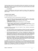
Ely et al
14
created the CAM-ICU (Fig. 1)
for use by nonpsychiatrists in mechanically ventilated
patients. When features 1 and 2 and either feature 3 or
feature 4 are present, a patient is said to be delirious, or
‘‘CAM-ICU positive.’’ In the largest validation cohort of
111 medical ICU patients using the CAM-ICU and as
compared with reference standard raters, two study
nurses demonstrated high sensitivity (93 to 100%),
high specificity (98 to 100%), and high interrater reli-
ability (k ¼ 0.96; 95% CI, 0.92 to 0.99).
14
Importantly,
the CAM-ICU also distinguishes delirium from
coma—defined as the state of unarousability, unaware-
ness of the environment, and absence of spontaneous
interaction or awareness of the interviewer—and de-
mentia. Requiring on average less than 2 minutes to
complete, the CAM-ICU has been validated, and its
reliability has been confirmed in other languages and at
least one other region of the world.
21
Its ease of use
among nonpsychiatrists also makes the CAM-ICU
practical.
45
Subsequent revisions of the CAM-ICU,
including use of the validated Richmond Agitation
Sedation Scale to monitor level of consciousness
46–48
and a greater reliance upon a form of the Vigilance A
random letter test
43
rather than the picture recognition
tool for the attention screening examination, are being
incorporated in ongoing clinical trials. The develop ment
of a simple, objective, brief, valid, reliable, and widely
accepted, bedside assessment tool for critically ill, often
Figure 1 Diagnosis of delirium with the Confusion Assessment Method for the intensive care unit (CAM-ICU). Adapted from Inouye
et al
6
and copyright # 2002, E. Wesley Ely, M.D., M.P.H. and Vanderbilt University, all rights reserved. See
for more information.
DE LIRIU M AND COGNITIVE D YSFUNCTION I N THE I CU / MILLER, ELY 213
mechanically ventilated, patients has revolutionized
the detection of delirium but also revealed its serious
sequelae.
OUTCOMES AND PROGNOSTIC
SIGNIFICANCE OF DELIRIUM IN THE
INTENSIVE CARE UNIT
With an effective tool to diagnose delirium, the next step
was to determine the independent assoc iation of delir-
ium with multiple, clinically significant end points. In
the last 5 years, much has bee n learned about the impact
of delirium both during hospitalization and for up to
2 years later. On a daily basis, delirium and/or altered
levels of consciousness contribute to an increased risk of
complications related to the comp lex bedside monitor-
ing, drug delivery,
16
and life-supporting therapies that
are commonplace in ICUs. Given that most patients
who develop deli rium survive to hospital discharge, these
long-term sequelae will likely prove to be more burden-
some than currently imagined.
Mortality
Increased risk of death among delirious, noncritically ill
patients for up to 2 years has been previously identi-
fied.
49,50
In the last 5 years, additional data on critically
ill patients have shown a similar impact of delirium,
though not always during the immediate hospitalization
period.
12,16,24
Lin et al demonstrated a 13-fold increased
risk for in-hospital death among 111 mechanically
ventilated patients.
21
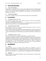
Furthermore, Ely et al prospec-
tively followed 275 mechanically ventilated, medical
ICU patients for 6 months from the time of hospital
discharge and noted a threefold increase in the risk of
death by 6 months, even after adjustment for age,
severity of illness, comorbid conditions, coma, and the
use of sedatives and analgesics (Fig. 2, adjusted hazard
ratio ¼ 3.2; 95% CI, 1.4 to 7.7; p ¼ 0.008).
20
Cost
Costs of care in the ICU are significantly higher among
those who develop delirium than in those who do not.
Milbrandt et al reported in 2004 that the costs to care for
a delirious, critically ill patient were significantly higher
than for patients who never became delirious, even after
controlling for several covariates.
51
The costs of caring
for delirious, critically ill patients were a median $9014
and $14,730 higher for ICU and hospital stays, respec-
tively. At least part o f this increase would likely be
accounted for by longer lengths of stay.
Length of Stay
That ICU delirium independently prolongs hospital
length of stay has been demonstrated in two cohorts.
The first of these, a prospective cohort of 275 mechan-
ically ventilated ICU patients, demonstrated an ad-
justed hazard ratio of 2.0 ( p < .001) for longer
hospital stay among delirious patients, even after
controlling for covariates.
20
Similarly, in a cohort of
261 consecutive, non-intubated medical ICU patients,
patients who had experienced delirium in the ICU had
a 41% greater risk of remaining in the hospital relative
to non-delirious patients (p ¼ .023).
25
Whether intu-
bated or not, it appears delirium may prolong length of
hospital stay.
Figure 2 Kaplan-Meier analysis of delirium in the intensive care unit and 6-month survival. Adapted from Ely et al.
20
214 SEMINARS IN RESPIRATORY AND CRITICAL CARE MEDICINE/VOLUME 27, NUMBER 3 2006
Failure of Extubation
As is the case with longer ICU and hospital stays among
delirious patients, the potential for delirium to contrib-
ute to extubation failure necessarily subjects patients to
risk of complications. For example, failure of extubation
poses risks of prolonged ventilation or of reintubation,
including such complications as nosocomial pneumo-
nia
52
and death.
53
That abnormal mental status signifi-
cantly predicts failure of extubation has been previously
studied.
54,55
Nearly 10% of delirious, intubated patients
in one study versus 2.3% of nondelirious, intubated
patients self-extubated.
16
Recently we have also demon-
strated that the presen ce of delirium, specifically as
determined by the CAM-ICU, is associated with a
threefold increased risk for reintubation within 48 hours
of predominantly planned extubations.
56
Long-Term Cognitive Impairment
In addition to the predisposition of cognitive impair-
ment among critically ill patients for the development
of delirium, this condition may also independently
cause or result in greater occurrence and severity of
cognitive impairment for up to 3 years after ICU stay.
In a review of nine prospective studies evaluating a
total of 1885 hospitalized medical and surgical (non-
ICU) patients, Jackson et al found that intrahospital
delirium increased the likelihood of dementia as much
as threefold up to 3 years from the time of hospital
discharge.
57
A smaller study that stringently examined
34 previously mechanically ventilated patients demon-
strated that, although ‘‘normal’’ at baseline almost one
third were neuropsychologically impaired by standar-
dized neuropsychological batteries at 6 months follow-
ing hospital discharge.
58
More recently, Hopkins et al
have confirmed the high prevalence of cognitive im-
pairment among previously critically ill patients 2 years
after discharge from the hospital.
59
An area ripe for
future investigation, the likely role of delirium in
contributing to long-term cognitive impairment sug-
gests the need for larger, prospective cohort studies to
better identify risk factors for persistence of neuro-
cognitive dysfunction. As with mortality, cost, length
ofstay,andfailureofextubation,thepresenceoflong-
term cognitive impairment among critically ill patients
represents a significant, lasting b urden for patients and
their families.
STRATEGIES FOR OPTIMAL
MANAGEMENT
With the development of effective diagnostic tools such
as the CAM-ICU and the Richmond Agitation Seda-
tion Scale, the Society of Critical Care Medicine’s
recommendations to monitor for delirium and sedation
level in all critically ill patients are easier to follow—and
to do so frequently. However, for them to take their
place alongside such stalwart, multidisciplinary inter-
ventions in the ICU as deep venous thrombosis preven-
tion or antibiotics, evidence that altering the occurrence
or course of delirium decreases the frequency of unto-
ward outcomes is essential. Likely, this will require
targeted prevention strategies and specialized, multi-
disciplinary interventions, both nonpharmacological
and pharmacological.
Nonpharmacological Approach
Although several clinical trials of multicomponent, non-
pharmacological interventions have been per-
formed,
11,60,61
none targeted the ICU and all have met
with modest, postdischarge success. The largest such
trials in the literature are those conducted by Inou ye
et al
11
and Lundstro
¨
metal.
61
The former, a non-
randomized trial of patients > 70 years old admitted
either to a specialized ward or to a regular unit, featured
an intervention protocol. The protocol targeted such
features as cognitive stimulation, reorientation prompts,
a sleep protocol, visua l and hearing aids, reminders to
prevent volume depletion, and walking/exercise. The
incidence of delirium among the intervention as com-
pared with those who received usual care was signifi-
cantly lower, 9.9% versus 15.0%, respectively (p ¼ .02).
Unfortunately, neither the severity of delirium nor the
recurrence rates differed between the two groups, and
subsequent follow-up of the patients 6 months after
hospital discharge did not demonstrate sustained bene-
fits overall.
62
However, the highest-risk deli rious pa-
tients (arguably those m ost similar to critically ill
patients) did report higher health and functional status
scores at 6 months than did high-risk, nondelirious
patients. The more recent trial by Lundstro
¨
metal
incorporating a nurse-driven, multifactorial intervention
program among 400 hundred patients > 70 years old
resulted in significant reductions in duration of delirium
(30% absolute risk reduction, p ¼ .001) and hospital
length of stay (3 day reduction, p < .001).
61
We hope
long-term follow-up will yield promising results and
anticipate that interventions to prevent or diminish the
consequences of delirium among both noncritically ill
and ICU patients will remain fundamental to manage-
ment of delirium in the future.
Drug Therapy
That nonpharmacological interventions to prevent a
disease are prudent is no surprise. However, with a
widening array of medications at our disposal, physi-
cians are tempted to act when faced with medical
abnormalities. Accordingly, the temptation to seek
pharmacological interventions for diseases such as de-
lirium in the ICU frequently outpaces the scientific
DE LIRIU M AND COGNITIVE D YSFUNCTION I N THE I CU / MILLER, ELY 215
evidence to support their use. One need look no further
than a 2004 survey of 912 intensivists, of whom 92%
considered delirium a substantial problem in the ICU
despite only 40% routinely screening for it. Yet, 79%
reported that delirium requires active intervention and
66% felt haloperidol should be the treatment of choice,
followed by lorazepam (12%) and atypical antipsy-
chotics (4%). Despite this impulse to intervene phar-
macologically with haloperidol and the support of this
position by both the Society of Critical Care Medicine
1
and the American Psychiatric Association,
63
there are
no randomized, placebo-controlled trials to confirm
the efficacy of haloperidol in either the prevention or
the treatment of delirium. Not surprisingly, no drugs
have Food and Drug Administration approval for the
prevention or the treatment of delirium. Through 2005,
anecdotal evidence, uncontrolled trials, and a few
randomized trials comparing haloperidol to neurolep-
tics or benzodiazepines constitute the basis for profes-
sional societies’ recommendation of haloperidol.
Medications such as haloperidol and the so-called
atypical antipsychotics (e.g., olanzapine, risperidone,
ziprasidone, aripiprazole, quetiapine) are thought to
exert their antidelirium effect in at least two ways. First,
the drugs are thought to ‘‘normalize’’ cerebral fun ction by
disinhibition of acetylcholine, blockade of dopamine
receptors, and activation of serotonin receptors. Second,
some data suggest that haloperidol may exhibit antiin-
flammatory effects upon the production of proinflam-
matory cyto kines.
64,65
The atypical antipsychotics do not have intra-
venous formulations. As such, those who have or are
investigating haloperidol as compared with an atypical
antipsychotic must rely upon either enteral or intra-
muscular administration. Nonetheless, intravenous hal-
operidol is not only common but recommended,
1,66,67
with a protocol of escalating dose (e.g., doubling every
30 minutes until desired effect) noted in professional
guidelines.
1
Pharmacokinetics is important with antipsy-
chotics, as with all drugs, in portending risk of adverse
events. The half-life of haloperidol is $21 hours, with
peak plasma concentrations noted within 2 to 6 hours
of dosing (enteral) or 20 minutes (intramuscular).
Notable adverse effects can include hypotension that
antagonizes adrenaline (especially in parenteral
form), and dose-dependent QTc prolongation leading
to cardiac tachyarrhythmias such as torsades de
pointes
68–70
—particularly among patients with preex-
isting cardiac disease, those receiving other medica-
tions that prolong the QTc, those receiving > 35 mg
cumulative dose, and those with extrapyramidal symp-
toms,
71
neuroleptic malignant syndrome,
72
dyspho-
ria,
73
or laryngospasm.
74
Meanwhile, the atypical
antipsychotics typically have half-lives of 20 þ hours,
with the exception of ziprasidone ($7hours).Peak
plasma concentrations are typically reached within 5 to 8
hours following enteral ingestion, though risperidone
reaches peak concentration within 1 hour, or within
1 hour for drugs administered intramuscularly. In con-
trast to haloperidol, the atypical antipsychotics cause few
side effects usually, though weight gain and hypotension
are not uncommon, and there may be an increased risk of
hyperglycemia or diabetes.
75
The risk of extrapyramidal
symptoms or neuroleptic malignant syndrome is lower
than with haloperidol.
Data supporting the use of haloperidol either
alone or versus other medications is limited. Evidence
for the potential benefit of haloperidol, however, was
recognized by Milbrandt et al. In a cohort of 989
mechanically ventilated ICU patients, those who re-
ceived haloperidol within the first 48 hours were 16%
less likely to die during the hospitalization.
76
One
explanation for this finding is the purported antiinflam-
matory effect of haloperidol.
The only prospective trials to our knowledge to
evaluate the efficacy of treatment of delirium in the ICU
with antipsychotics are fraught with limitations but
provide reassurance as to the safety of the atypical
antipsychotics. One unblinded study evaluated the effi-
cacy and safety of enteral olanzapine as compared with
enteral haloperidol in the treatment of 73 ICU patients
who screened positive for delirium according to DSM-
IV criteria.
22
Although there was no difference in the
development of delirium between the treatment groups,
the study confirmed the safety of olanzapine because
none of the patients in the olanzapine arm but six
patients in the haloperidol arm experienced extrapyra-
midal symptoms. In addition to a simplistic random-
ization scheme, a predominance of surgical ICU
patients, and a low severity of illness score (mean Acute
Physiology and Chronic Health II score of 12.7), the low
overall prevalence of delirium (21%) relative to other
ICU cohorts calls into question the generalizability of
this study’s results. Although the trial did not intend to
answer the question of whether olanzapine is equal or
superior to haloperidol, it did suggest that the former is
no more, and perhaps less, harmful. Moreover, similar
results were found in a separate trial comp aring haloper-
idol to risperidone.
77
Again, delirium scores decreased
during drug administration in both groups (p < .05), but
there was no difference in the scores between the two
treatment groups (p ¼ .51). A placebo-controlled study
comparing placebo to haloperidol and to an atypical
antipsychotic would most appropriately address this
question.
As suggested by the first admonition of the
Hippocratic oath, it may be as important what medi-
cations we do not use as those we do use to alter the
occurrence and course of delirium. Almost all ICU
patients require and receive sedatives and analgesics,
particularly during the early part of their ICU stay, and
216 SEMINARS IN RESPIRATORY AND CRITICAL CARE MEDICINE/VOLUME 27, NUMBER 3 2006
avoidance of harmful medications during that time
seems prudent. Little evidence exists to date to guide
this type of decision, but in a study by Breitbart et al,
78
the authors randomized 30 hospitalized and subse-
quently delirious acquired immunodeficiency syn-
drome patients to receive either lorazepam (n ¼ 6),
chlorpromazine (n ¼ 13), or haloperidol (n ¼ 11).
78
They found that either haloperidol or chlorpromazine
significantly reduced the delirium score as compared
with lorazepam (p < .001), leading the authors to
suggest that lorazepam alone is ineffective in decreas-
ing delirium symptoms. That it is a known risk factor
further validates th e assertion that outside well-known
drug withdrawal syndromes, benzodiazepines are not
recommended for routine treatment of delirium.
Moreover, when used, they should be used in the
context of sedation protocols that employ intermittent
bolus sedation, if at all possible.
32,79,80
Although minimal data beyond consensus opinion
exist currently to guide prevention or treatment decisions
in ICU delirium, the CAM-ICU provides an effective
way to evaluate the safety and efficacy of nonpharmaco-
logical and pharmacological interventions alike. Several,
ongoing, randomized, and/or placebo-controlled trials
should begin to clarify the indications for use of the
typical and atypical antipsychotics and the indications for
use of agents other than benzodiazepines for sedation.
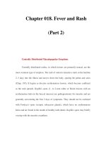
SPECTRUM OF ACUTE BRAIN
DYSFUNCTION
In addition to delirium, other forms of brain dysfunction
such as coma, stupor, and the more recently described
subsyndromal delirium may represent a continuum of
acute brain dysfunction (Fig. 3). Subsyndromal delirium
is perhaps best defined as the presence of some, b ut not
all, of the criteria for delirium.
81–84
In the case of the
CAM-ICU, that could mean disorganized thinking
despite normal levels of consciousness and attention or
a fluctuating level of consciousness despite preserved
attention and thinking. Distinction of these subtypes
of brain dysfunction may be pertinent in light of the
prevailing notion that increasing severity of delirium is
associated with worse outcomes outside the ICU.
81–83
For example, Marcantonio et al
83
compared 504 patients
who remained CAM negative (not delirious) or who
became CAM positive (delirious) or CAM intermediate
(subsyndromal delirium, those with one or more CAM
features but not diagnostic of delirium) during their stay
at postacute skilled nursing facilities. They found that
patients with subsyndromal delirium had intermediate
6-month mortality rates (25.0, 18.3, and 5.7% for
delirious, subsyndromal delirium, and ‘‘normal’’ patients,
respectively), intermediate rehospitalization rates, and
intermediate rates of complications, even after adjusting
for age, preexisting dementia, and medical comorbidity.
Identification of similar phenomenonology of acute
brain dysfunction in the ICU might help better target
therapeutic interventions.
SUMMARY
Since 2 001 our knowledge of delirium in the ICU has
changed dramatically. There has been greater under-
standing, if persistent underappreciation, of the occur-
rence of delirium among the critically ill. Also efforts to
identify risk factors for delirium in the ICU have
increased and are suggesting targets for refinements of
ICU practice that will hopefully diminish the sequelae of
delirium. With the additional development of the
CAM-ICU, the independent association of delirium
with untoward outcomes from mortality to long-term
cognitive impairment has been possible to describe.
Now, further efforts to determine appropriate interven-
tions and management of delirium must come to the
fore. Placebo-controlled tri als for treatment of delirium
are on the horizon. In avoiding harmful medications or
pursuing beneficial ones, the prevention and treatment
of delirium or any of the various subtypes along the
continuum of acute brain dysfunction in the ICU will
require a multidisciplinary approach. Diligent investiga-
tion, including large cohort studies, will help identify
potential new targets for intervention in the years ahead.
GRANT SUPPORT
Dr. Ely is a recipient of a K23 from the National
Institute of Health (#AG01023–01A1).
REFERENCES
1. Jacobi J, Fraser GL, Coursin DB, et al. Clinical practice
guidelines for the sustained use of sedatives and analgesics in
the critically ill adult. Crit Care Med 2002;30:119–141
2. Ely EW, Siegel MD, Inouye SK. Delirium in the intensive
care unit: an under-recognized syndrome of organ dysfunc-
tion. Semin Respir Crit Care Med 2001;22:115–126
Figure 3 Proposed continuum of acute brain dysfunction in the ICU.
DE LIRIU M AND COGNITIVE D YSFUNCTION IN THE ICU / MILLER, ELY 217
3. Meagher DJ, Hanlon DO, Mahony EO, Casey PR,
Trzepacz PT. Relati onship between symptoms and motoric
subtype of delirium. J Neuropsychiatry Clin Neurosci 2000;
12:51–56
4. Lipowski ZJ. Delirium: Acute Confusional States. Rev. ed.
New York: Oxford University Press; 1990
5. Peterson JF, Pun BT, Dittus RS, et al. Delirium and its
motoric subtypes: a study of 614 critically ill patients. J Am
Geriatr Soc 2006;54:479–484
6. Inouye SK, van Dyck CH, Alessi CA, Balkin S, Siegal AP,
Horwitz RI. Clarifying confusion: the confusion assessment
method: a new method for detection of delirium. Ann Intern
Med 1990;113:941–948
7. American Psychiatric Association. Diagnostic and Statistical
Manual of Mental Disorders. 3rd rev. ed. Washington, DC:
American Psychiatric Association; 1987
8. O’Keeffe S, Lavan J. The prognostic significance of delirium
in older hospital patients. J Am Geriatr Soc 1997;45:174–
178
9. Levkoff SE, Evans DA, Liptzin B, et al. Delirium: the
occurrence and persistence of symptoms among elderly
hospitalized patients. Arch Intern Med 1992;152:334–340
10. Williams-Russo P, Urquhart BL, Sharrock NE, Charlson ME.
Postoperative delirium: predictors and prognosis in elderly
orthopedic patients. J Am Geriatr Soc 1992;40:759–767
11. Inouye SK, Bogardus ST Jr, Charpentier PA, et al. A
multicomponent intervention to prevent delirium in hospi-
talized older patients. N Engl J Med 1999;340:669–676
12. Kishi Y, Iwasaki Y, Takezawa K, Kurosawa H, Endo S.
Delirium in critical care unit patients admitted through an
emergency room. Gen Hosp Psychiatry 1995;17:371–379
13. Aldemir M, O
¨
zen S, Kara IH, Sir A, Bac¸ B. Predisposing
factors for delirium in the surgical intensive care unit. Crit
Care 2001;5:265–270
14. Ely EW, Inouye SK, Bernard GR, et al. Delirium in
mechanically ventilated patients: validity and reliability of
the confusion assessment method for the intensive care unit
(CAM-ICU). JAMA 2001;286:2703–2710
15. Ely EW, Margolin R, Francis J, et al. Evaluation of delirium
in critically ill patients: validation of the Confusion Assess-
ment Method for the Intensive Care Unit (CAM-ICU). Crit
Care Med 2001;29:1370–1379
16. Dubois MJ, Bergeron N, Dumont M, Dial S, Skrobik Y.
Delirium in an intensive care unit: a study of risk factors.
Intensive Care Med 2001;27:1297–1304
17. BergeronN,DuboisMJ,DumontM,DialS,SkrobikY.
Intensive Care Delirium Screening Checklist: evaluation of
a new screening tool. Intensive Care Med 2001;27:859–
864
18. Rincon HG, Granados M, Unutzer J, et al. Prevalence,
detection and treatment of anxiety, depression, and delirium
in the adult critical care unit. Psychosomatics 2001;42:391–
396
19. McNicoll L, Pisani MA, Zhang Y, Ely EW, Siegel MD,
Inouye SK. Delirium in the intensive care unit: occurrence
and clinical course in older patients. J Am Geriatr Soc 2003;
51:591–598
20. Ely EW, Shintani A, Truman B, et al. Delirium as a predictor
of mortality in mechanically ventilated patients in the
intensive care unit. JAMA 2004;291:1753–1762
21. Lin SM, Liu CY, Wang CH, et al. The impact of delirium on
the survival of mechanically ventilated patients. Crit Care
Med 2004;32:2254–2259
22. Skrobik YK, Bergeron N, Dumont M, Gottfried SB.
Olanzapine vs haloperidol: treating delirium in a critical care
setting. Intensive Care Med 2004;30:444–449
23. McNicoll L, Pisani MA, Ely EW, Gifford D, Inouye SK.
Detection of delirium in the intensive care unit: comparison of
confusion assessment method for the intensive care unit with
confusion assessment method ratings. J Am Geriatr Soc 2005;
53:495–500
24. Micek ST, Anand NJ, Laible BR, Shannon WD, Kollef MH.
Delirium as detected by the CAM-ICU predicts restraint use
among mechanically ventilated medical patients. Crit Care
Med 2005;33:1260–1265
25. Thomason JW, Shintani A, Peterson JF, Pun BT, Jackson JC,
Ely EW. Intensive care unit delirium is an independent
predictor of longer hospital stay: a prospective analysis of 261
non-ventilated patients. Crit Care 2005;9:R375–R381
26. Cole MG, Dendukuri N, McCusker J, Han L. An empirical
study of different diagnostic criteria for delirium among
elderly medical inpatients. J Neuropsychiatry Clin Neurosci
2003;15:200–207
27. Laurila JV, Pitkala KH, Strandberg TE, Tilvis RS. Delirium
among patients with and without dementia: does the
diagnosis according to the DSM-IV differ from the previous
classifications? Int J Geriatr Psychiatry 2004;19:271–277
27a. Pandharipande P, Jackson J, Ely EW. Delirium: acute
cognitive dysfunction in the critically ill. Curr Opin Crit
Care 2005;11:360–368
28. Francis J, Martin D, Kapoor WN. A prospective study of
delirium in hospitalized elderly. JAMA 1990;263:1097–
1101
29. Inouye SK, Charpentier PA. Precipitating factors for delirium
in hospitalized elderly persons: predictive model and inter-
relationship with baseline vulnerability. JAMA 1996;275:
852–857
30. Ely EW, Gautam S, Margolin R, et al. The impact of
delirium in the intensive care unit on hospital length of stay.
Intensive Care Med 2001;27:1892–1900
31. Pandharipande PP, Shintani A, Peterson J, et al. Lorazepam
is an independent risk factor for transitioning to delirium
in intensive care unit patients. Anesthesiology 2006;104:21–
26
32. Kress JP, Pohlman AS, O’Connor MF, Hall JB. Daily
interruption of sedative infusions in critically ill patients
undergoing mechanical ventilation. N Engl J Med 2000;342:
1471–1477
33. Schor JD, Levkoff SE, Lipsitz LA, et al. Risk factors for
delirium in hospitalized elderly. JAMA 1992;267:827–831
34. Marcantonio ER, Goldman L, Mangione CM, et al. A
clinical prediction rule for delirium after elective noncardiac
surgery. JAMA 1994;271:134–139
35. Angus DC, Kelley MA, Schmitz RJ, White A, Popovich J Jr.
Caring for the critically ill patient: current and projected
workforce requirements for care of the critically ill and
patients with pulmonary disease: can we meet the require-
ments of an aging population? JAMA 2000;284:2762–
2770
36. Kakuma R, du Fort GG, Arsenault L, et al. Delirium in older
emergency department patients discharged home: effect on
survival. J Am Geriatr Soc 2003;51:443–450
37. Inouye SK, Foreman MD, Mion LC, Katz KH, Cooney LM
Jr. Nurses’ recognition of delirium and its symptoms: com-
parison of nurse and researcher ratings. Arch Intern Med 2001;
161:2467–2473
218 SEMINARS IN RESPIRATORY AND CRITICAL CARE MEDICINE/VOLUME 27, NUMBER 3 2006
38. Ely EW, Stephens RK, Jackson JC, et al. Current opinions
regarding the importance, diagnosis, and management of
delirium in the intensive care unit: a survey of 912 healthcare
professionals. Crit Care Med 2004;32:106–112
39. Rapp CG, Wakefield B, Kundrat M, et al. Acute confusion
assessment instruments: clinical versus research usability.
Appl Nurs Res 2000;13:37–45
40. Breitbart W, Rosenfeld B, Roth A, Smith MJ, Cohen K,
Passik S. The Memorial Delirium Assessment Scale. J Pain
Symptom Manage 1997;13:128–137
41. Hart RP, Levenson JL, Sessler CN, Best AM, Schwar tz
SM, Rutherford LE. Validation of a cognitive test for
delirium i n medical ICU patients. Psychosomatics 1996;37:
533–546
42. Hart RP, Best AM, Sessler CN, Levenson JL. Abbreviated
cognitive test for delirium. J Psychosom Res 1997;43:417–
423
43. Strub RL, Black FW. The Mental Status Examination in
Neurology. 4th ed. Philadelphia: FA Davis; 2000
44. American Psychiatric Association. Delirium, dementia and
amnestic and other cognitive disorders. In: Diagnostic and
Statistical Manual of Mental Disorders. 4th ed. Washington,
DC: American Psychiatric Association. 1994
45. Pun BT, Gordon SM, Peterson JF, et al. Large-scale
implementation of sedation and delirium monitoring in the
intensive care unit: a report from two medical centers. Crit
Care Med 2005;33:1199–1205
46. Ely EW, Gautam S, May L, et al. A comparison of different
sedation scales in the ICU and validation of the Richmond
Agitation Sedation Scale (RASS) [abstract]. Am J Respir Crit
Care Med 2001;163:A954
47. Ely EW, Truman B, Shintani A, et al. Monitoring sedation
status over time in ICU patients: reliability and validity of the
Richmond Agitation-Sedation Scale (RASS). JAMA 2003;
289:2983–2991
48. Sessler CN, Gosnell MS, Grap MJ, et al. The Richmond
Agitation-Sedation Scale: validity and reliability in adult
intensive care unit patients. Am J Respir Crit Care Med
2002;166:1338–1344
49. Francis J, Kapoor WN. Prognosis after hospital discharge of
older medical patients with delirium. J Am Geriatr Soc 1992;
40:601–606
50. McCusker J, Cole M, Abrahamowicz M, Primeau F, Belzile
E. Delirium predicts 12-month mortality. Arch Intern Med
2002;162:457–463
51. Milbrandt EB, Deppen S, Harrison PL, et al. Costs associated
with delirium in mechanically ventilated patients. Crit Care
Med 2004;32:955–962
52. Torres A, Gatell JM, Aznar E. Reintubation increases the
risk of nosocomial pne umonia i n patients nee ding mec han-
ical ventilation. Am J Respir Crit Care Med 1995;152:
137–141
53. Esteban A, Alia I, Gordo F, et al. Extubation outcome
after spontaneous breathing trials with T-tube or pressure
support ventilation. The Spanish Lung Failure Collabora-
tive Group. Am J Respir Crit Care Med 19 97;156(2 Pt 1):
459–465
54. Namen AM, Ely EW, Tatter SB, et al. Predictors of
successful extubation in neurosurgical patients. Am J Respir
Crit Care Med 2001;163(3 Pt 1):658–664
55. Salam A, Tilluckdharry L, Amoateng-Adjepong Y, Man-
thous CA. Neurologic status, cough, secretions and extuba-
tion outcomes. Intensive Care Med 2004;30:1334–1339
56. Miller RR, Shintani AK, Girard TD, et al. Delirium predicts
extubation failure [abstract]. Proceedings of the American
Thoracic Society 2006;3:A42
57. Jackson JC, Gordon SM, Hart RP, Hopkins RO, Ely EW.
The association between delirium and cognitive decline: a
review of the empirical literature. Neuropsychol Rev 2004;14:
87–98
58. Jackson JC, Hart RP, Gordon SM, et al. Six-month
neuropsychological outcome of medical intensive care unit
patients. Crit Care Med 2003;31:1226–1234
59. Hopkins RO, Brett S. Chronic neurocognitive effects of
critical illness. Curr Opin Crit Care 2005;11:369–375
60. Marcantonio ER, Flacker JM, Wright RJ, Resnick NM.
Reducing delirium after hip fracture: a randomized trial. J Am
Geriatr Soc 2001;49:516–522
61. Lundstro
¨
m M, Edlund A, Karlsson S, Brannstrom B, Bucht
G, Gustafson Y. A multifactorial intervention program
reduces the duration of delirium, length of hospitalization,
and mortality in delirious patients. J Am Geriatr Soc 2005;
53:622–628
62. Bogardus ST Jr, Desai MM, Williams CS, Leo-Summers L,
Acampora D, Inouye SK. The effects of a targeted multi-
component delirium intervention on postdischarge out-
comes for hospitalized older adults. Am J Med 2003;114:
383–390
63. American Psychiatric Association. Practice guideline for the
treatment of patients with delirium. Am J Psychiatry 1999;
156(Suppl 5):1–20
64. Moots RJ, Al Saffar Z, Hutchinson D, et al. Old drug, new
tricks: haloperidol inhibits secretion of proinflammatory
cytokines. Ann Rheum Dis 1999;58:585–587
65. Song C, Lin A, Kenis G, Bosmans E, Maes M. Immuno-
suppressive effects of clozapine and haloperidol: enhanced
production of the interleukin-1 receptor antagonist. Schiz-
ophr Res 2000;42:157–164
66. Tesar GE, Stern TA. Rapid tranquilization of the agitated
intensive care unit patient. J Intensive Care Med 1988;3:195–
201
67. Tesar GE, Murray GB, Cassem NH. Use of high-dose
intravenous haloperidol in the treatment of agitated cardiac
patients. J Clin Psychopharmacol 1985;5:344–347
68. Lawrence KR, Nasraway SA. Conduction disturbances
associated with administration of butyrophenone antipsy-
chotics in the critically ill: a review of the literature.
Pharmacotherapy 1997;17:531–537
69. Perrault LP, Denault AY, Carrier M, Cartier R, Belisle S.
Torsades de pointes secondary to intravenous haloperidol after
coronary bypass grafting surgery. Can J Anaesth 2000;47:251–
254
70. Sharma ND, Rosman HS, Padhi ID, Tisdale JE. Torsades de
pointes associated with intravenous haloperidol in critically ill
patients. Am J Cardiol 1998;81:238–240
71. Bashford G, Bradd P. Drug-induced Parkinsonism associated
with dysphagia and aspiration: a brief report. J Geriatr Psy-
chiatry Neurol 1996;9:133–135
72. Adnet P, Lestavel P, Krivosic-Horber R. Neuroleptic
malignant syndrome. Br J Anaesth 2000;85:129–135
73. King DJ, Burke M, Lucas RA. Antipsychotic drug-induced
dysphoria. Br J Psychiatry 1995;167:480–482
74. Ilchef R. Neuroleptic-induced laryngeal dystonia can mimic
anaphylaxis. Aust N Z J Psychiatry 1997;31:877–879
75. Lindenmayer JP, Czobor P, Volavka J, et al. Changes in
glucose and cholesterol levels in patients with schizophrenia
DE LIRIU M AND COGNITIVE D YSFUNCTION IN THE ICU / MILLER, ELY 219
treated with typical or atypical antipsychotics. Am J Psychiatry
2003;160:290–296
76. Milbrandt EB, Kersten A, Kong L, et al. Haloperidol use is
associated with lower hospital mortality in mechanically
ventilated patients. Crit Care Med 2005;33:226–229
77. Han CS, Kim YK. A double-blind trial of risperidone and
haloperidol for the treatment of delirium. Psychosomatics
2004;45:297–301
78. Breitbart W, Marotta R, Platt MM, et al. A double-blind
trial of haloperidol, chlorpromazine, and lorazepam in the
treatment of delirium in hospitalized AIDS patients. Am J
Psychiatry 1996;153:231–237
79. Brook AD, Ahrens TS, Schaiff R, et al. Effect of a nursing-
implemented sedation protocol on the duration of mechanical
ventilation. Crit Care Med 1999;27:2609–2615
80. Kollef MH, Levy NT, Ahrens TS, Schaiff R, Prentice D,
Sherman G. The use of continuous I.V. sedation is associated
with prolongation of mechanical ventilation. Chest 1998;114:
541–548
81. Levkoff SE, Y ang FM, Liptzin B. Delirium: the impor-
tance of subsyndromal states. Primary Psychiatry 2004;11:
40–44
82. Cole M, McCusker J, Dendukuri N, Han L. The prognostic
significance of subsyndromal delirium in elderly medical
inpatients. J Am Geriatr Soc 2003;51:754–760
83. Marcantonio ER, Kiely DK, Simon SE, et al. Outcomes of
older people admitted to postacute facilities with delirium.
J Am Geriatr Soc 2005;53:963–969
84. Liptzin B. What criteria should be used for the diagnosis of
delirium? Dement Geriatr Cogn Disord 1999;10:364–367
220 SEMINARS IN RESPIRATORY AND CRITICAL CARE MEDICINE/VOLUME 27, NUMBER 3 2006
Cardiac Arrhythmias in the Intensive Care Unit
Daniel J. Tarditi, D.O.
1
and Steven M. Hollenberg, M.D.
1
ABSTRACT
Cardiac arrhythmias are a common problem encountered in the intensive care unit
(ICU) and represent a major source of morbidity. Arrhythmias are most likely to occur in
patients with structural heart disease. The inci ting factor for an arrhythmia in a given
patient may be an insult such as hypoxia, infection, cardiac ischemia, catecholamine excess
(endogenous or exogenous), or an electrolyte abnormality. Management includes correc-
tion of these imbalances as well as medical therapy directed at the arrhythmia itself. The
physiological impact of arrhythmias depends on ventricular response rate and duration, and
the impact of a given arrhythmia in a given situation depends on the patient’s cardiac
physiology and function. Similarly, urgency and type of treatment are determined by the
physiological impact of the arrhythmia as well as by underlying cardiac status. The purpose
of this review is to provide an update regarding current concepts of diagnosis and acute
management of arrhythmias in the ICU. A systematic approach to diagnosis and evaluation
will be presented, followed by consideration of specific arrhythmias.
KEYWORDS: Arrhythmia, ICU, ventricular tachycardia, AV nodal reentrant tachycardia,
atrial fibrillation, atrial flutter, sinus tachycardia, Wolff-Parkinson-White syndrome,
electrical storm, bradycardia
Arrhythmias are a common dilemma confront-
ing the intensivist. They represent a major source of
morbidity, and they lengthen hospital stay. Arrhythmias
are most likely to occur in patients with structural heart
disease. The inciting factor for an arrhythmia in a given
patient may be an insult such as hypoxia, infection,
cardiac ischemia, catecholamine excess (endogenous or
exogenous), or an electroly te abnormality. Management
includes correction of these imbalances as well as medical
therapy directed at the arrhythmia itself.
The physiologi cal impact of arrhythmias depends
on ventricular response rate and duration as well as on
the underlying cardiac function. Bradyarrhythmias may
decrease cardiac output due to heart rate alone in
patients with relatively fixed stroke volumes, and loss
of an atrial kick may cause a dramatic increase in
pulmonary pressures in patients with diastolic dysfunc-
tion. Similarly, tachyarrhythmi as can decrease diastolic
filling and reduce cardiac output, resulting in hypoten-
sion, in addition to producing myocardial ischemia.
Clearly, the impact of a given arrhythmia in a given
situation depends on the patient’s cardiac physiology and
function. Similarly, urgency and type of treatment are
determined by the physiological impact of the arrhyth-
mia as well as by underlying cardiac status.
This review pro vides an update regarding current
concepts of diagnosis and acute management of arrhyth-
mias in the intensive care unit (ICU). A systematic
approach to diagnosis and evaluation will be presented,
followed by consideration of specific arrhythmias.
1
Divisions of Cardiovascular Disease and Critical Care Medicine,
Cooper University Hospital, Camden, New Jersey.
Address for correspondence and reprint requests: Steven M.
Hollenberg, M.D., Divisions of Cardiovascular Disease and Critical
Care Medicine, Cooper University Hospital, One Cooper Plaza,
366 Dorrance, Camden, NJ 08103. E-mail: Hollenberg-Steven@
cooperhealth.edu.
Non-pulmonary Critical Care: Managing Multisystem Critical Ill-
ness;GuestEditor,CurtisN.Sessler,M.D.
Semin Respir Crit Care Med 2006;27:221–229. Copyright # 2006
by Thieme Medical Publishers, Inc., 333 Seventh Avenue, New York,
NY 10001, USA. Tel: +1(212) 584-4662.
DOI 10.1055/s-2006-945525. ISSN 1069-3424.
221
EVALUATION OF TACHYARRHYTHMIAS
The first step in the evaluation of the critically ill patient
with an arrhythmia is to assess hemodynamic stability. If
hemodynamics are compromised due to the arrhythmia,
cardioversion should be performed unless pharmacolog-
ical treatment is immediately successful. However, be-
fore proceeding with car dioversion, one should consider
whether the arrhythmia is in fact the basis for the
deterioration in hemodynamics.
The next step in evaluation is to determine
whether the arrhythmia is supraventricular or ventricular
in origin. First, one examines QRS width. A narrow
QRS complex (< 0.12 sec) indicates a supraventricular
tachycardia (SVT). Narrow complex tachycardias in-
clude atrial fibrillation (AF), sinus tachycardia, atrioven-
tricular nodal reentrant tachycardia (AVNRT), AV
reentry from the accessory pathway [Wolff-Parkinson-
White syndrome (WPW)], atrial flutter, and atrial
tachycardia. Wide QRS tachycardias include ventricular
tachycardia (VT), SVT with preexisting bundle branch
block, aberrant ventricular conduction, or SVT from
AV reentry using an antegrade accessory pathway
(WPW).
One should try not to rely solely on a rhythm strip
from one monitor lead for diagnosis; there can be
variability in Q RS width depending on which lead is
examined. A 12-lead electrocardiogram (ECG) is more
useful. Also, scrutiny of a previous ECG is often useful;
for example, to identify preexisting bundle branch block
or QTc interval prolongation. Marke d left axis deviation
(À60 to À120 degrees) may indicate a ventricular origin
of the arrhythmia. It is noteworthy that ST segment
depression during SVT lacks specificity in predicting
ischemia. In one series of 100 patients with SVT,
associated ST segment deviation was only 51% specific
(with a positive predictive value of only 6%) for sig nifi-
cant angiographic coronary artery disease or scinti-
graphic evidence of ischemia.
1
Carotid sinus massage and other maneuvers that
increase vagal tone slow AV conduction time and in-
crease refractoriness, and this can aid in the diagnosis
through demonstration of p waves or interruption
of AVNRT or AV reentrant tachycardia (AVRT).
Adenosine can also be used for this purpose. Responses
to vagal maneuvers or adenosine are listed in Table 1.
Adenosine is given as a rapid intravenous (IV) bolus
of 6 mg, and a second dose of 12 mg can be given 1 to
2 minutes later. The effects are more pronounced when
given through a central venous line, in which case
the dosage is then usually halved. The half-life of
adenosine is only 6 to 10 seconds. Severe broncho-
spasm or wheezing can result from its use. Adenosine
can be proarrhythmic, most commonly the induction of
AF (2.7%),
2
and there have been reports of asystole,
VT, and ventricular fibrillation (VF) following its
administration.
3
NARROW COMPLEX TACHYCARDIA
Regular narrow complex SVTs include sinus tachycar-
dia, AVNRT, AVRT, ectopic atrial tachycardia, and
atrial flutter. Irregular narrow complex SVTs include
AF, multifocal atrial tachycardia, atrial flutter with
variable block, and sinus tachycardia with frequent
premature atrial complexes.
The p wave morphology can suggest the origin of
the atrial impulse. The p wave should be upright in lead
II with a normal sinus mechanism. If inverted, this is
suggestive of AVRT, AVNRT, or ectopic atrial tachy-
cardia. P waves may be absent or difficult to discern in
the setting of tachycardia. The RP interval should be
assessed on the 12-lead ECG, with a short RP interval
(RP shorter than PR, and less than 70 msec) suggesting
AVNRT, and a long RP interval most likely indicating
AVRT via a slowly conducting accessory pathway. A
heart rate of 150 beats per minute (bpm) should raise the
suspicion of atrial flutter with 2:1 conduction.
Regular Rhythms
SINUS TACHYCARDIA
Sinus tachycardia often occurs as a response to a sympa-
thetic stimulus (hypoxia, vasopressors, inotropes, pain,
dehydration, hyperthyroidism, etc.). The first step is to
review patient medications, including infusions, to ex-
clude an iatrogenic etiology of the tachyarrhythmia.
Treatment focuses on identifying and trying to correct
the underlying cause. If ischemia is the cause and treat-
ment is warranted, b-blockers are the first treatment
Table 1 Responses to Vagal Maneuvers or Adenosine
Arrhythmia Response to Vagal Maneuvers/Adenosine
Sinus tachycardia Gradual slowing with resumption of the tachycardia
Atrioventricular nodal reentrant
tachycardia
Abrupt termination or only very transient slowing
Atrial fibrillation/flutter Increased atrioventricular block briefly with slowed ventricular response rate
Multifocal atrial tachycardia Increased atrioventricular block briefly with slowed ventricular response rate
Ventricular tachycardia Usually no response
222 SEMINARS IN RESPIRATORY AND CRITICAL CARE MEDICINE/VOLUME 27, NUMBER 3 2006
option. However, it is worth considering that the sinus
tachycardia may be an appropriate hemodynamic response
to hypotension, hypovolemia, or low cardiac output; if this
is the case, overzealous use of b-blockers can reduce
cardiac output, with potentially disastrous consequences.
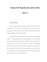
ATRIOVENTRICULAR NODAL REENTRANT TACHYCARDIA
AVNRT typically occurs at a heart rate of 140 to
180 bpm. It is more prevalent in females and is not
usually associated with structural heart disease. AVNRT
involves dual AV nodal pathways, usually with slow
conduction antegrade and retrograde conduction via a
transiently refractory second pathway (Fig. 1). Therefore,
the key to treatment is to block AV conduction. Acute
treatment includes vagal maneuvers and IV adenosine.
Long-term preventative therapy includes medications
that suppress the initiating premature atrial contractions
(b-blockers) or slow AV conduction (nondihydropyridine
calcium-channel blockers, b-blockers, and digoxin),
4
or
catheter ablation of one of the pathways.
ATRIAL FLUTTER
Atrial flutter is a macroreentrant arrhythmia identified
by flutter waves, often best seen in the inferior leads, at
250 to 350 bpm. Patients often present with 2:1 AV
conduction with a ventricular rate of 150 bpm,
although the AV conduction ratio can change abrupt ly.
Acute treatment consists of AV-nodal-blocking drugs
for rate control. If the patient becomes clinically un-
stable, direct current–synchronized (DC-synchronized)
cardioversion with 50 J is usually sufficient, with success
rate of 95 to 100%.
5
IV ibutilide has an efficacy rate of
$76% for conversion to sinus rhythm in clinical trials
but prolongs the QT interval and can provoke sustained
polymorphic VT in 1 to 2% of cases.
6,7
Ibutilide should
not be used in patients with a prolonged QTc interval
(greater than 420 msec), or in those with underlying
sinus node disease. Other antiarrhythmics such as
sotalol, procainamide, and flecainide have demon-
strated less efficacy for acute conversion.
8–10
If a tem-
porary or permanent pacemaker is in place, atrial
overdrive (burst) pacing can sometimes restore sinus
rhythm via overdrive suppression.
Long-term treatment of the ventricular rate in
atrial flutter usually consists of diltiazem, verapamil, b-
blockers, or digoxin. Class IC drugs (flecainide) are very
effective in preventing atrial flutter, but by slowing the
atrial rate, they have the potential to cause 1:1 AV
Figure 1 (A) Atrioventricular (AV) node demonstrating dual pathways: slow (a) pathway with short refractory period and fast (b)
pathway with long refractory period. (B) Premature impulse conducts down slow pathway while fast pathway is still refractory to
conduction. (C) As impulse conducts down slow pathway, the fast pathway recovers. (D) Impulse goes up fast pathway as it conducts to
the ventricle. (E) Impulse reenters cycle in AV node completing reentrant circuit.
CARDIAC ARRHYTHM IAS IN TH E ICU /TARDITI, HOLLENBERG 223
conduction, and should always be combined with AV-
nodal-blocking agents.
Irregular Rhythms
Irregular narrow complex SVT includes AF, multifocal
atrial tachycardia, atrial flutter with variable block, and
sinus tachycardia with frequent premature atrial com-
plexes.
ATRIAL FIBRILLATION
AF is the most com mon narrow comp lex tachyarrhyth-
mia in the ICU (second to VT overall).
11
The preva-
lence of AF in the general pop ulat ion increases
exponentially with age, from 0.9% at age 40 to 5.9%
in those over age 65.
12
The most important risk factors
for develo pment of AF in the general population are
structural heart disease (70% in Framingham study
over 22-year follo w-up), hypertension (50%),
13
valvular
heart disease (34%),
14
and left ventricular hypertrophy.
AF should be approached in the following manner:
find the cause, fix the cause , control the rate, consider
rhythm control, and consider anticoagulation. Pharma-
cological agents for acute rate control include b-block-
ers, nondihydropyridine calcium channel bloc kers, and
digoxin.
Beta-blockers provide more effective rate control
than calcium channel blockers at rest and during exer-
cise.
15
Both oral and IV formulations are available. The
most often used IV medication is metoprolol given at 2.5
to 5.0 mg IV over 1 to 2 minutes every 5 to 10 minutes
for a total of 15 mg as blood pressure tolerates. Esmolol,
0.5 mg/kg bolus, then 0.05 mg/kg/min infusion, is an
alternative with a more rapid onset and offset, which can
be useful in unstable patients.
Nondihydropyridine calcium channel blockers
(diltiazem and verapamil) are also effective AV nodal
blockers. Verapamil may have more negative inotropic
properties than diltiazem and thus may induce hypo-
tension in patients with left ventricular dysfunction and
borderline blood pressure.
16
Diltiazem is available in IV
form and is commonly used as a continuous infusion at a
rate of 5 to 15 mg per hour. Up to 93% of patients will
maintain a ventricular response rate < 100 bpm during a
24-hour infusion.
17
Digoxin controls ventricular response through a
centrally mediated vagal mechanism and by direct action
on the AV node. It controls resting heart rates in patients
who do not have increased catecholamine levels but is
less effective in the ICU. IV digoxin begins to slow the
heart rate in 30 minutes.
18
Cardioversion of a patient with AF carries a
stroke risk from 1.1% if anticoagulated for 3 weeks to
7% if not anticoagulated, even if AF duration is less than
1 week.
19
Due to delay between resumption of organized
atrial electrical activity and of organized mechanical
contraction, there can be delay between cardioversion
and embolic events ranging from 6 hours to 7 days.
20
Postcardiac surgery AF occurs in 25 to 40% of
patients, with peak inci dence on day 2.
21,22
Use of b-
blockers, amiodarone, sotalol and biatrial overdrive pac-
ing to prevent postoperative AF has been studied in
clinical trials.
23
Preoperative administration of sotalol
and amiodarone is equally effective, but side effects of
sotalol limit its use in comparison to amiodarone or b-
blockers. Standard treatment for postoperative AF is to
establish rate control, initially with IV (Table 2) and
then with oral AV nodal blocking medications. There
are numerous risk factors for postoperative AF, with
advanced age being the most important. AF often runs a
self-correcting course in this setting, with resumption of
sinus rhythm in more than 90% of patients by 6 to 8
weeks after surgery, and so cardioversion is not always
necessary.
24
Immediate cardioversion should be per-
formed in patients with recent onset AF accompanied
by symptoms or signs of hemodynamic instability result-
ing in angina, myocardial ischemia, shock, or pulmonary
edema without waiting for prior anticoagulation.
Anticoagulation with IV heparin should be con-
sidered if AF persists for greater than 48 hours. The
stroke risk in unanticoagulat ed patients taken as a whole
is $ 2% per year (0.05% per day), but individual factors
modulate that risk. The risk factors for stroke are heart
failure, hypertension, age > 75 years, diabetes, prior
history of transient ischemic attack (TIA) or stroke,
and female gender.
25
MULTIFOCAL ATRIAL TACHYCARDIA
MAT is an irregular atrial tachycardia diagnosed by
identification of three or more p wave morphologies
Table 2 Intravenous Medications for Heart Rate Control in Atrial Fibrillation
Drug Loading Dose Onset Maintenance Dose
Diltiazem 0.25 mg/kg over 2 min 2–7 min 5–15 mg/h infusion
Esmolol 0.5 mg/kg over 1 min 5 min 0.05–0.2 mg/kg/min
Metoprolol 2.5–5.0 mg over 2 min up to three doses 5 min NA
Propanolol 0.15 mg/kg 5 min NA
Verapamil 0.075–0.15 mg/kg over 2 min 3–5 min NA
Digoxin 0.25 mg each 2 h up to 1.5 mg 2 h 0.125–0.25 mg daily
NA, not applicable.
224 SEMINARS IN RESPIRATORY AND CRITICAL CARE MEDICINE/VOLUME 27, NUMBER 3 2006