Báo cáo y học: "Combined left hepatectomy with fenestration and using a harmonic scalpel, fibrin glue and closed suction drainage to prevent bile leakage and ascites in the management of symptomatic polycystic liver disease: a case report" pps
Bạn đang xem bản rút gọn của tài liệu. Xem và tải ngay bản đầy đủ của tài liệu tại đây (1.78 MB, 5 trang )
Case report
Open Access
Combined left hepatectomy with fenestration and using
a harmonic scalpel, fibrin glue and closed suction drainage
to prevent bile leakage and ascites in the management of
symptomatic polycystic liver disease: a case report
Christopher Kosmidis
1
*, Christopher Efthi miadis
1
, George Anthimidis
1
,
Sofia Levva
1
, John Prousalidis
2
, Konstantinos Papapolychroniadis
2
and
Epaminondas Fachantidis
2
Addresses:
1
Department of Surgery, Interbalkan European Medical Center, Thessaloniki, Greece
2
1
st
Propedeutic Surgical Clinic, Aristotle University of Thessaloniki, AHEPA Hospital, Greece
Email: CK* - ; CE - ; GA - ; SL - ;
JP - ; KP - ; EF -
* Corresponding author
Received: 25 September 2008 Accepted: 3 February 2009 Published: 27 August 2009
Journal of Medical Case Reports 2009, 3:7442 doi: 10.4076/1752-1947-3-7442
This article is available from: />© 2009 Kosmidis et al.; licensee Cases Network Ltd.
This is an Open Access article distributed under the terms of the Creative Commons Attribution License (
/>which permits unrestricted use, distribution, and reproduction in any medium, provided the original work is properly cited.
Abstract
Introduction: Surgical treatment is the usual therapy for patients with polycystic liver disease and
with severe symptoms, yet the results of surgery are often disappointing and the optimal surgical
approach is uncertain.
Case presentation: We present the case of a 41-year-old Greek woman who underwent
combined left hepatectomy with fenestration for symptomatic polycystic liver disease using
ultrasound scalpel, fibrin glue and closed suction drain to prevent bile leakage, haemorrhage and
ascites. Liver resection using the ultrasound scissors allowed quick parenchyma dissection under
haemostatic conditions with safe coagulation of small vessels and bile ducts. Moreover, the ultrasound
scalpel was applied to the cyst cavities exposed on the peritoneum to ablate the fluid-producing
epithelial cyst lining. We also covered the cut cystic cavities exposed to the peritoneum surface of the
liver with fibrin glue. Instead of allowing the opened cysts to drain into the abdominal cavity, we used
two wide bore closed suction fluted drains. We did not observe excessive fluid loss through the
drainage after the second postoperative day. The drain tubes were removed on the third
postoperative day.
Conclusion: In our patient, effective treatment of ascites and prevention of bile leakage and bleeding
indicate that this new approach is promising and may become a useful surgical technique for
polycystic liver disease.
Page 1 of 5
(page number not for citation purposes)
Introduction
Polycystic liver disease (PLD) is a common manifestation
of polycystic kidney disease and is associated with an
autosomal dominant inheritance. Patients are usually
asymptomatic [1]. Symptomatic PLD has been treated by
percutaneous aspiration with or without sclerotherapy,
drainage of the superficial cysts into the abdominal cavity
and fenestration of deeper cysts into the superficial cyst
cavities via laparotomy or laparoscopy, hepatic resection
or orthotopic liver transplant. The results of surgery are
often disappointing, with quick return of symptoms, bile
leakage and symptomatic ascites in many patients [1-3].
We present the case of a patient who underwent combined
left hepatectomy with fenestration for symptomatic PLD
using the harmonic scalpel, fibrin glue and closed suction
drain to prevent bile leakage and ascites.
Case presentation
A 41-year-old Greek woman presented with chronic and
unrelenting right upper quadrant pain, epigastric fullness,
early satiety, nausea, vomiting and dysphagia. On physical
examination, hepatomegaly and tenderness in the right
upper quadrant were found. Abdominal ultrasound (US),
computed tomography (CT), magnetic resonance imaging
(MRI) and magnetic resonance cholangiopancreatography
(MRCP) revealed multiple liver cysts, particularly in the
left hepatic lobe, with characteristics similar to simple
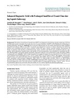
hepatic cysts. Multiple cysts were also found in the kidneys
and the anterior surface of the pancreas (Figure 1). The left
hepatic lobe was enlarged, compressing the stomach to the
spleen. Serum biochemical analysis demonstrated a mild
impairment of liver function: serum glutamic oxaloacetic
transaminase (SGOT) 126 U/L (10-31 U/L), Serum glutamic
pyruvic transaminase (SGPT) 85 U/L (10-31 U/L),
while urea and creatinine were within the normal range.
The patient’s family history was positive for the presence of
PLD. Her 63-year-old mother had multiple non-parasitic
asymptomatic cysts in the liver and kidneys. Additionally,
her 17-year-old daughter and 13-year-old son had multi-
ple cysts in the kidneys, while the liver, the pancreas and
the spleen were normal. Given the family history and the
presence of multiple cysts in the liver, kidneys and the
anterior surface of the pancreas, the diagnosis of PLD
associated with polycystic kidney disease was made.
A double Kocher incision was made to provide excellent
access to the upper abdomen. The left hepatic lobe was
enlarged and full of multiple cysts, the maximum diameter
of which was 9 cm. The stomach was compressed between
the left lobe of the liver and the spleen, explaining the cyst-
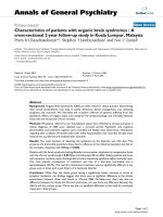
related complaints of the patient. Furthermore, multiple
small cysts and three large dominant cysts (diameter: 7-
13 cm) were located at the right hepatic lobe (Figure 2).
There were huge cysts throughout both kidneys and small
cysts at the anterior surface of the pancreas. The
hepatoduodenal ligament was exposed to provide access
for vascular clamping and identification of major vascular
Figure 1. Pre-operative computed tomography
demonstrating multiple cysts in the liver, anterior surface
of the pancreas and kidneys.
Figure 2. Intra-operative view of multiple small cysts all
over the left hepatic lobe and one of the three large
dominant cysts located in the right hepatic lobe.
Page 2 of 5
(page number not for citation purposes)
Journal of Medical Case Reports 2009, 3:7442 />and biliary structures. The liver was mobilized by the
division of hepatic peritoneal attachments. A left hepatic
lobectomy, that is of segments II, III, IV, was made using
ultrasound scissors (Harmonic Scalpel, UltraCision, Ethi-
con Endosurgery, Cincinnati, Ohio, USA). The diameter of
the removed lobe was 17 cm (Figure 3). Cysts located on
the surface of the right hepatic lobe, including the three
dominant cysts, were surgically unroofed and windows
were created by fenestration between superficial cysts and
adjacent deep cysts. The fluid from the opened cysts was
rapidly aspirated under continuous suction.
After completion of the resection, the tourniquet was
opened and the remaining cut surface carefully inspected
for residual bleeding or nonoccluded bile ducts. The cut
surface was plain and brownish; biliary leaks or persistent
bleeding were e asily detected and sutured with 4-0
polypropylene. The ultrasound scalpel was applied to
the cyst cavities exposed on the peritoneum in order to
attempt ablation of the fluid-producing epithelial cyst
lining. To avoid bile leakage and haemorrhage, fibrin glue
was spread over the raw surface of the liver. Cysts in the
pancreas and kidneys were not treated. Two wide-bore
closed suction fluted drains (30 F) were brought out
through a separate stab wound; one p laced in the
subhepatic space and one in the right subdiaphragmatic
space. Postoperatively, the patient remained well and
without complications. The drain tubes were removed on
the third postoperat ive day, when the drainage had
decreased to less than 30 mL in 8 hours. Symptomatic
relief and reduction in abdominal girth were achieved.
Histologic examination showed von Meyenburg’s com-
plexes. The patient was followed up at clinic - special data
included hepatic and renal function, symptomatic relief,
the patient’s working capacity and CT scans. The follow-up
showed post-resection hypertrophy of the spared liver and
lack of clinically significant cyst progression. Four years
after the procedure, the patient had an improved quality of
life and functional status without deterioration in her
hepatic or renal function.
Discussion
With the widespread use of sensitive imaging techniques,
the frequency of non-parasitic hepatic cysts is reported
more often. Adult polycystic disease is the most common
cystic disease. Liver cysts in patients with polycystic kidney
disease are generally asymptomatic, but in a few patients,
hepatomegal y from numerous large cysts may cause
symptoms [1,4].
Treatment is usually only carried out in patients with
severe symptoms related to large cysts or complications.
Associated medical problems, especially intracranial
aneurysms and valvular heart disease need to be evaluated
in patients with PLD [1,2]. Surgical management differs
from that for patients with simple cysts or cystadenomas
because multiple cysts continue to grow and appear de
novo after treatment [5]. Therefore, the therapeutic aim is
to significantly reduce the size of the polycystic liver
without compromising liver function, and to provide
long-term relief of symptoms. The optimal treatment for
symptomatic PLD is uncertain. There is no clear consensus
regarding the optimum timing of intervention and the
surgical approach is based in part on the number, size and
location of the cysts. The surgical therapy should be
tailored to the extent of disease in each patient.
In our case, the patient was classified as Type II, based on
Gigot’s classification, that is, diffuse involvement of liver
parenchyma by medium-sized cysts with remaining large
areas of non-cystic parenchyma [3]. Therefore, the
combination of hepatic resection with fenestration
appeared to be a valuable option, allowing for the removal
of multiple segments grossly affected (II, III, IV) and
reduction in liver mass with maximal preservation of liver
parenchyma. Fenestration alone would probably be
unsuccessful because the liver parenchyma might be
more rigid due to the fibrosis around the cysts, and the
cysts would not collapse as expected after fenestration.
Likewise, the large superficial and deep-seated cysts within
the right hepatic lobe with more normal parenchyma
should undergo fenestration.
The most commonly reported morbidities with combined
fenestration and resection are pleural effusions, wound
infection, ascites, transient biliary leaks and bleeding
[6-10]. The surgical technique is an important factor in
preventing intra-operative and postoperative complica-
tions. Various techniques have been developed for safe
and careful dissection of the liver parenchyma [9,10]. The
high number of techniques used worldwide shows the lack
of a generally accepted gold standard. Technical
Figure 3. The specimen from the left hepatic lobe.
Page 3 of 5
(page number not for citation purposes)
Journal of Medical Case Reports 2009, 3:7442 />improvement seems to be possible and desirable. The aim
of our study was to prove the suitability of the ultrasound
scissors, closed suction drain and fibrin glue in surgery for
PLD. In our clinic, this cutting device is mainly used in
laparoscopic surgery for dissection of tissue, but we
consider it an appropriate instrument for liver dissection.
Because of its simultaneous haemostatic and coagulating
effect, it might theoretically offer a considerable advantage
in surgery for PLD [10].
Handling of the instrument, cutting and coagulation
quality were satisfactory and safe. To achieve a better
and more effective coagulating effect, the portal structures
were occluded by a tourniquet which did not last longer
than 30 minutes, together with lowering of the central
venous pressure during resection. The liver resection using
the ultrasound scissors allowed quick parenchyma dissec-
tion under haemostatic conditions with safe coagulation
of small vessels and bile ducts of up to 2 to 3 mm in
diameter. Larger vessels and biliary ducts were divided
with clamps and sutured with 4-0 polypropylene. The
major advantage of the ultrasound scalpel was the modest
trauma that it produced and the controlled dissection of
the tissue. Especially in the periphery, the UltraCision was
an ideal dissection instrument: with the absence of large
vessels and bile ducts, nearly all of the parenchyma was
easily divided without causing bleeding, bile leakage or
trauma. Moreover, use of ultrasound scissors on the cyst
cavities exposed on the peritoneum was attempted to
facilitate ablation of the secretory epithelium and reduc-
tion of postoperative continual peritoneal fluid losses.
However, we also covered the cut cystic cavities exposed to
the peritoneum surface of the liver with fibrin glue [11,12].
Fibrin glue causes less intra-abdominal adhesions while
allowing shorter haemostasis time than primary suture
[13]. Moreover, instead of allowing the opened cysts to
drain into the abdominal cavity, two wide-bore closed
suction fluted drains were used.
Ascites is the commonest complication specific to surgery
for PLD, occurring in all patients undergoing resection
[1,2]. We at no time encountered excessive fluid loss
through the drainage after the second postoperative day.
Effective treatment of ascites in our patient may be related
to some extent to the use of the UltraCision instrument as
well as the particular type of drain, which prevents
accumulation of ascitic fluid and ensures complete
evacuation of the collection and collapse of the opened
cystic cavities. However, other factors may play a major
role, such as the use of fibrin glue to seal the cut liver
surface or the type of surgery.
Conclusions
Surgical management of patients with PLD remains a
challenging issue for physicians. The aim of the present
study was to investigate the ability of the UltraCision
instrument, fibrin glue and closed suction drainage in
hepatic resection combined with fenestration for PLD.
This method appears to be an advantageous new
technique. This case report is not sufficient to draw any
final conclusions. Therefore, the benefits of this surgical
approach should be further evaluated. However, our
initial experience is promising, and we believe that it
may become a valuable means in surgery for PLD.
Abbreviations
CT, computed tomography; MRCP, magnetic resonance
cholangiopancreatography; MRI, magnetic resonance ima-
ging; PLD, polycystic liver disease; SGOT, serum glutamic
oxaloacetic transaminase; SGPT, serum glutamic pyruvic
transaminase; US, ultrasound.
Consent
Written informed consent was obtained from the patient
for publication of this case report and any accompanying
images. A copy of the written consent is available for
review by the Editor-in-Chief of this journal.
Competing interests
The authors declare that they have no competing interests.
Authors’ contributions
CK, CE and EF performed the operation and together with
GA and SL contributed to the conception and design of the
manuscript. JP and KP analyzed and interpreted the
patient regarding the polycystic disease. GA and SL were
major contributors in writing the manuscript. All authors
read and approved the final manuscript.
References
1. Everson GT, Taylor MRG, Doctor RB: Management of polycystic
liver disease. Curr Gastroenterol Rep 2005, 7:19-25.
2. Qian Q, Li A, King BF, Kamath PS, Lager DJ, Huston J III, Shub C,
Davila S, Somlo S, Torres VE: Clinical profile of autosomal
dominant polycystic liver disease. Hepatology 2003, 37:164-171.
3. Gigot JF, Jadoul P, Que F, Van Beers BE, Etienne J, Horsmans Y,
Collard A, Geubel A, Pringot J, Kestens PJ: Adult polycystic liver
disease: is fenestration the most adequate operation for long-
term management? Ann Surg 1997, 225:286-294.
4. Martin IJ, McKinley AJ, Currie EJ, Holmes P, Garden OJ: Tailoring
the management of nonparasitic liver cysts. Ann Surg 1998,
228:167-172.
5. Forrest ME, Cho KJ, Shields JJ, Wicks JD, Silver TM, McCormick TL:
Biliary cystadenomas: sonographic-angiographic-pathologic
correlations. AJR 1980, 135:723-727.
6. Soravia C, Mentha G, Giostra E, Morel P, Rohner A: Surgery for
adult polycystic liver disease. Surgery 1995, 117:272-275.
7. Vons C, Chauveau D, Martinod E, Smadja C, Capron F, Grunfeld JP,
Franco D: Liver resection in patients with polycystic liver
disease. Gastroenterol Clin Biol 1998, 22:50-54.
8. Hansman MF, Ryan JA Jr, Holmes JH 4th, Hogan S, Lee FT, Kramer D,
Biehl T: Management and long-term follow-up of hepatic
cysts. Am J Surg 2001, 181:404-410.
9. Yang GS, Li QG, Lu JH, Yang N, Zhang HB, Zhou XP: Combined
hepatic resection with fenestration for highly symptomatic
polycystic liver disease: A report on seven patients. World J
Gastroenterol 2004, 10:2598-2601.
Page 4 of 5
(page number not for citation purposes)
Journal of Medical Case Reports 2009, 3:7442 />10. Schmidbauer S, Hallfeldt KK, Sitzmann G, Kantelhardt T, Trupka A:
Experience with ultrasound scissors and blades (UltraCision)
in open and laparoscopic liver resection. Ann Surg 2002, 235:
27-30.
11. Saxton ML: Hemostasis in minimally invasive liver. Surgery 2007,
42:46-49.
12. Beckingham IJ, Krige JE: ABC of diseases of liver, pancreas, and
biliary system. BMJ 2001, 322:477-480.
13. Demirel AH, Basar OT, Ongoren AU, Bayram E, Kisakurek M: Effects
of primary suture and fibrin sealant on hemostasis and liver
regeneration in an experimental liver injury. World J Gastro-
enterol 2008, 14:81-84.
Do you have a case to share?
Submit your case report today
• Rapid peer review
• Fast publication
• PubMed indexing
• Inclusion in Cases Database
Any patient, any case, can teach us
something
www.casesnetwork.com
Page 5 of 5
(page number not for citation purposes)
Journal of Medical Case Reports 2009, 3:7442 />