Báo cáo y học: " Whole body bone scintigraphy in tenofovir-related osteomalacia: a case report" pps
Bạn đang xem bản rút gọn của tài liệu. Xem và tải ngay bản đầy đủ của tài liệu tại đây (1.36 MB, 4 trang )
Case report
Open Access
Whole body bone scintigraphy in tenofovir-related osteomalacia:
a case report
Antonio Di Biagio
1
*, Raffaella Rosso
1
, Patrizia Monteforte
2
, Rodolfo Russo
3
,
Guido Rovetta
2
and Claudio Viscoli
1
Addresses:
1
Department of Infectious Diseases, San Martino Hospital, University of Genoa, Genoa 16132, Italy
2
Department of Endocrinology and Metabolism Sciences, University of Genoa, Genoa 16132, Italy
3
Nephrology Division, Department of Internal Medicine, San Martino Hospital, University of Genoa, Genoa 16132, Italy
Email: ADB* - ; RR - ; PM - ; RR - ;
GR - ; CV -
* Corresponding author
Received: 27 January 2008 Accepted: 18 February 2009 Published: 22 July 2009
Journal of Medical Case Reports 2009, 3:8136 doi: 10.4076/1752-1947-3-8136
This article is available from: />© 2009 Di Biagio et al.; licensee Cases Network Ltd.
This is an Open Access article distributed under the terms of the Creative Commons Attribution License (
/>which permits unrestricted use, distribution, and reproduction in any medium, provided the original work is properly cited.
Abstract
Introduction: Tenofovir disoproxil fumarate (Viread
®
) is the only nucleotide reverse transcriptase
inhibitor currently approved for the treatment of HIV. It is frequently prescribed not only for its
efficacy but also for its decreased side effect profile compared with other nucleotide analogs. In
addition, it is now increasingly recognized as a cause of acquired Fanconi’s syndrome in individuals
with HIV.
Case presentation: We describe a 48-year-old woman infected with HIV, with chronic renal
insufficiency, who developed Fanconi’s syndrome after inclusion of tenofovir disoproxil fumarate in
her antiretroviral therapy. A whole body bone scintigraphy was performed, revealing an abnormal
distribution of radiotracer uptake, with characteristic changes compatible with osteomalacia. All
symptoms disappeared after tenofovir discontinuation and mineral supplementation. No other
explanation for the sudden and complete resolution of the bone disease was found.
Conclusion: The case highlights the role of whole body bone scintigraphy in the diagnosis of
tenofovir-related osteomalacia.
Introduction
Tenofovir disoproxil fumarate (TDF) is an oral prodrug of
tenofovir, a nucleotide reverse transcriptase inhibitor
(NRTI). Because of its favorable resistance profile and its
activity against HIV-1 strains, TDF is widely used as part of
highly active antiretroviral therapy (HAART). TDF is rapidly
hydrolyzed and is mainly eliminated unchanged by the
kidney, by a combination of glomerular filtration and active
tubular secretion [1]. Nephrotoxicities owing to TDF have
been reported over the past few years. In particular, TDF is
capable of causing a Fanconi-like syndrome with renal
phosphate wasting and concomitant osteomalacia [2-5].
Page 1 of 4
(page number not for citation purposes)
Since th e advent of HAART, multiple epidemiologic
studies have shown that osteopenia and osteoporosis are
common among patients with HIV infection [6]. Osteo-
porosis is well-documented by means of bone densito-
metry (DEXA), while bone scintigraphy is useful for
identifying fractures. In patients with HIV, osteomalacia
has been documented in only a few cases by DEXA, it
appears as bone demineralization and its reversibility has
been described [7,8].
A whole body bone scintigraphy, in order to evaluate
patients who are HIV-negative with bone disorders, is well-
defined [9]. We report a severe case of osteomalacia in a
woman infected with HIV, diagnosed by bone scan and
resolved after TDF discontinuation.
Case presentation
A 48-year-old Caucasian woman was diagnosed with HIV-
1 infection in 1992. Past medical history included primary
amenorrhea (1970), psoriasis (1974), detection of anti-
hepatitis C virus (HCV) antibodies and psoriasis-related
mild chronic renal insufficiency (1995). In May 2002, she
was diagnosed with osteoporosis, based on DEXA, and
started therapy with bisphosphonates (disodium clodro-
nate 100 mg intramuscular/die) associated with inhibitor
COX-2 (celecoxib 200 mg/die). At the same time, renal
ecotomography showed reduced renal size (right kidney
89 × 40 × 43 mm; left kidney 86 × 37 × 49 mm) and
parenchymal thickness, but regular morphology. In
October 2002, she switched from disodium clodronate
to raloxifene (60 mg/die). She was on HAART since 1997:
zidovudine 300 mg twice a day (bid) plus lamivudine
150 mg bid plus indinavir 800 mg three times a day from
July 1997 to July 2001, changed for simplification;
zidovudine 300 mg bid plus lamivudine 150 mg bid
plus efavirenz 600 mg once daily (qd) from July 2001 to
December 2001, stopped for zidovudine-related anemia,
stavudine 30 mg bid plus lamivudine 150 mg bid plus
efavirenz 600 mg qd from December 2001 to May 2002,
stopped for paresthesia; finally, because of good immu-
nological restoration, she had a lymphocyte T CD4+-
guided structured antiretroviral treatment interruption
from May 2002 to February 2004.
During the off-therapy period, her serum creatinine level
had a peak of 1.8 mg/dl (normal range: 0.5-1.3), and it
was 1.7 mg/dl (estimated clearance creatinine 59.4 ml/
min) just before starting the new tenofovir-containing
regimen. On November 2002, her HCV-RNA concentra-
tion was high (3.05 × 10
5
copies/ml, Amplicore HCV
monitor assays: Roche Diagnostic System, USA), while
serum transaminase levels were stable (AST 57 U/l; ALT
70 U/l) and within twice the normal range (0-40 UI/l).
The patient refused to undergo a liver biopsy.
On February 2004, a new qd regimen including tenofovir
300 mg qd, plus lamivudine 300 mg qd, plus efavirenz
600 mg qd was started; her CD4+ cell count and viral
load were 267 cells/mL (21%) and 30000 copies/mL,
respectively.
Baseline renal function was mildly impaired: serum
alkaline phosphate level was 378 U/l (normal range:
98-280) and urinalysis showed urine protein 1 g/dl, urine
glucose 5 g/dl, urine pH6 and traces of hemoglobin.
After four weeks, the patient was admitted to our out-
patient clinic for her follow-up visit, and she referred mild
bone pain in the proximal area of her tibias and in
her ankles; the serum creatinine level at this time was
1.7 mg/dl.
Two weeks later, the patient’s bone pain had worsened
substantially, especially in her lower-limbs and chest, with
myalgia and difficulties in staying upright. She also
complained of increasing fatigue and polyuria.
Her laboratory test results revealed serum creatinine
level 1.8 mg/dl , alkaline phosphatase 1247 U/I, phos-
phate 1.6 mg/dl (normal values: 2.5-4.5), potassium
3.1 mEq/litre (nor mal value: 3.5-5), bicarbonate
11.30 mmol/litre (normal values 22.00-26.00). Urina-
lysis demonstrated urine glucose >10.0 g/litre, urine
pH 6, protein 1.0 g/litre, and traces of ketones. The
24-hour urine collection showed a level of urinary
potassium of 1.20 mEq/litre.
Fanconi’s syndrome was suspected in this patient given the
presence of hypokalemia, hypophoshatemia, glucosuria
and proteinuria. Despite a very good response to HAART
(CD4+ cells count 373 cell/mm
3
(32%) and viral load
<50 copies/mL), tenofovir was switched to abacavir
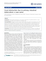
300 mg bid. At that time, radiological evaluation included
a skeletal bone scan: it showed costal and vertebral
multiple hypercaptations, as well as pelvic hypercapta-
tions of bone tracer, indicating areas of increased bone
turnover. The pelvis bone activity was present in locations
typical for osteomalacia (Figure 1). Calcitriol regimen, at
the recommended doses (50 mcg/die), was started and
continued for a year.
Gradually, bone pains and arthralgia improved, and
serum alkaline phosphatase normalized. Serum creati-
nine, urine protein and urine glucose diminished to
1.7 mg/dl, 1.0 g/litre and 5.0 g/litre, respectively; while
serum phosphate and potassium increased to 2.4 mg/dL
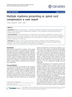
and 3.3 mEq/litre, respectively. After one year, the patient
again underwent a bone scintigraphy, which showed the
complete absence of hypercaptation (Figure 2).
Page 2 of 4
(page number not for citation purposes)
Journal of Medical Case Reports 2009, 3:8136 />Discussion
HAART can readily achieve long-term remission of HIV
disease, but it can also have both short- and long-term
adverse events [10]. The patient described developed
acute renal failure, during TDF-based therapy. However,
renal involvement could be explained by HCV infection
alone, although we cannot rule out tenofovir as the
primary cause. Indeed, the development of nephrotoxicity
has a temporal relationship with TDF administration.
Moreover, disability was progressively reversed by drug
withdrawal and pharmacological interventions. In the
differential diagnosis of Fanconi’s syndrome, we have
also considered other pathologies, such as myeloma,
amyloidosis, Sjögren’s syndrome, vitamin D deficiency
and antibiotic use, but its appearance seemed more
related to the combination of chronic renal disease, renal
tubular acidosis and nephrotoxicity, possibly secondary
to TDF medications.
Osteomalacia is a part of Fanconi’s syndrome, and we
made a diagnosis of hypophosphataemic osteomalacia
because of the patient’s lowered serum phosphate level
and a rising alkaline phosphatase level, associated with
bone pain and typical areas of scintigraphic pelvic activity.
Fanconi’s syndrome occurs as a loss of proximal tubular
function resulting in its fail ure to reabsorb various
substances, among which glucose, bicarbonate, phos-
phates, uric acid, potassium, sodium and amino acids,
with subsequent loss of these in the urine. The syndrome
is defined by a hypokalemic, metabolic acidosis with
hypophosphatemia and glucosuria; however, the presence
of any combination of these features can occur when the
proximal tubule is affected.
Unfortunately, we did not evaluate vitamin D levels,
which would have been useful to confirm the diagnosis; in
addition, the patient refused a bone biopsy.
Skeletal scintigraphy can be of great value in the diagnosis
and evaluation of therapy of many benign bone disorders.
Figure 1. Whole body bone scintigraphy shows multiple foci
of increased radiotracer uptake in the rib cage, the lumbar
spine, the sacroiliac region, the bilateral knee and the right
tibiotarsus.
Figure 2. Repeat scan one year after tenofovir
sparing-regimen shows largely disappeared focal lesions.
Page 3 of 4
(page number not for citation purposes)
Journal of Medical Case Reports 2009, 3:8136 />In our patient we used this technique in order to diagnose
and monitor her severe osteomalacia. However, skeletal
scintigraphy has a number of applications, and many
metabolic bone disorders characterized by altered blood
flow or osteoblastic reaction may be readily identified by
the procedure. In contrast to metastatic disease where focal
abnormalities are characteristically seen, the bone scan
diagnosis of metabolic bone disease generally depends
upon recognition of a generalized increase. This bone scan
appearance in osteomalacia strongly suggests the presence
of a metabolic bone disorder because of a generalized
increase in the tracer uptake in the skeleton producing
“super scan” with nonvisualisation of kidneys. Since in
severe osteomalacia, pseudofractures are common,
increased focal bone uptake of radiopharmaceutical is
also found [9].
Conclusions
We have described a patient with HIV/HCV-infection and
psoriasis-related chronic renal failure. Bone scintigraphy
has proven to be useful for the diagnosis of osteomalacia,
a possible consequence of Fanconi’s syndrome that can
occur with TDF therapy.
It is acknowledged that the subject reported in this case
report was antiretroviral treatment-experienced and con-
sequently received TDF as part of a salvage regimen; these
type of patients, often also HCV co-infected, may have
been subje cted to different antiretrovirals pr escribing
patterns which may add to the clinical presentation
described.
A whole body bone scintigraphy can be indicated in
patients on TDF with bone and joint pain. The bone scan
pattern in typical osteomalacia can be focal, similar to
osseous metastases, or diffuse, as in our patient.
Mineral supplementation and cessation of TDF appear to
be the treatment of choice in patients who show features
of the Fanconi’s syndrome. Patients with underlying renal
abnormality, or who show features of the Fanconi’s
syndrome during follow-up, should discontinue TDF;
equally in patients who develop bone pain or myopathy
on TDF treatment, hypophosphatemia should be looked
for and treated and the drug stopped.
Adverse effects have been reported with virtually all
antiretroviral drugs and are the most common reasons
for switching or discontinuation of therapy and for
medication nonadherence: a better understanding is of
interest not only for HIV specialists as they try to optimize
therapy, but also for other physicians who provide care for
patients infected with HIV.
Abbreviations
HIV, Human Immunodeficiency Virus; HAART, Highly
Active Anti-Retroviral Therapy; COX, cyclooxygenase.
Consent
Written informed consent was obtained from the patient
for publication of this case report and any accompanying
images. A copy of the written consent is available for
review by the Editor-in-Chief of this journal.
Competing interests
The authors declare that they have no competing interests.
Authors’ contributions
AD, RaR and CV revised the article for intellectual content
and helped to draft the manuscript. RoR interpreted the
renal results. PM and GD supervised the acquisition
process and interpreted the scintigraphic images. All
authors read and approved the final manuscript.
References
1. Schooley RT, Ruane P, Myers RA, Beall G, Lampiris H, Berger D,
Chen SS, Miller MD, Isaacson E, Cheng AK Study 902 Team:
Tenofovir DF in antiretroviral-experienced patients: results
from a 48 week, randomised, double blind study. AIDS 2002,
16:1257-1263.
2. Verhelst D, Monge M, Meynard JL, Fouqueray B, Mougenot B,
Girard PM, Ronco P, Rossert J: Fanconi Syndrome and renal
failure induced by tenofovir: a first case report. Am J Kidney Dis
2002, 40:1331-1333.
3. Karras A, Lafaurie M, Furco A, Bourgarit A, Droz D, Sereni D,
Legendre C, Martinez F, Molina JM: Tenofovir-related nephro-
toxicity in human immunodeficiency virus-infected patients:
three cases of renal failure. Fanconi Syndrome and nephro-
genic diabetes insipidus. Clin Infect Dis 2003, 36:1070-1072.
4. Schaaf B, Aires SP, Kramme E, Steinhoff J, Dalhoff K: Acute renal
failure associated with tenofovir treatment in a patient with
acquired immunodeficiency syndrome. Clin Infect Dis 2003, 37:
e41-e43.
5. Earle KE, Senevirartre T, Shaker J, Shoback D: Fanconi’s syndrome
in HIV+ adults: report of three cases and literature review.
J Bone Miner Res 2004, 19:714-721.
6. Amorosa V, Tebas P: Bone disease in HIV infection. Clin Infect Dis
2006, 42:108-114.
7. Parsonage MJ, Wilkins EG, Snowden N, Issa BG, Savage MW: The
development of hypophosphataemic osteomalacia with myo-
pathy in two patients with HIV infection receiving tenofovir
therapy. HIV Med 2005, 6:341-346.
8. Torres Isidro MV, García Benayas T, del Val Gómez Martínez M,
Gonzáles Gallardo F, Gambí Pisonero N, Castilla Miguel S, Gonzáles-
Lahoz J, Gallego Sanz D: Role of bone gammagraphy in the
diagnosis of secondary osteomalacia in a patient treated with
tenofovir. Rev Esp Med Nucl 2006, 25:103-106.
9. Fogelman I, McKillop JA, Bessent RG, Boyle IT, Turner JG, Greig WR:
The role of bone scanning in osteomalacia. J Nucl Med 1978,
19:245-248.
10. Montessori V, Press N, Harris M, Akagi L, Montaner JS: Adverse
effects of antiretroviral therapy for HIV infection. CMJA 2004,
170:229-238.
Page 4 of 4
(page number not for citation purposes)
Journal of Medical Case Reports 2009, 3:8136 />