Báo cáo y học: "Atrial myxoma presenting with orthostatic hypotension in an 84-year-old Hispanic man: a case report" pps
Bạn đang xem bản rút gọn của tài liệu. Xem và tải ngay bản đầy đủ của tài liệu tại đây (483.01 KB, 3 trang )
BioMed Central
Page 1 of 3
(page number not for citation purposes)
Journal of Medical Case Reports
Open Access
Case report
Atrial myxoma presenting with orthostatic hypotension in an
84-year-old Hispanic man: a case report
Ralph M Vicari*
1
, Enrique Polanco
1
, Norberto Schechtmann
1
,
José O Santiago
1
, Kautilya Shaurya
2
, Michael Halstead
3
, Danielle Marszal
4
,
Tamsin Grosskreutz
5
and Shalini Thareja
6
Address:
1
Mima Century Research, E. Sheridan Rd, Melbourne, FL 32901, USA,
2
Miller School of Medicine, NW 14th St, Miami, FL 33136, USA,
3
Tulane University, St Charles Avenue, New Orleans, LA 70118, USA,
4
University of Central Florida, Central Florida Blvd, Orlando, FL 32816, USA,
5
Florida Atlantic University, Glades Rd, Boca Raton, FL 33431, USA and
6
Columbia University, Haven Ave, New York, NY 10032, USA
Email: Ralph M Vicari* - ; Enrique Polanco - ;
Norberto Schechtmann - ; José O Santiago - ;
Kautilya Shaurya - ; Michael Halstead - ; Danielle Marszal - ;
Tamsin Grosskreutz - ; Shalini Thareja -
* Corresponding author
Abstract
Introduction: Left atrial myxomas remain the most common benign primary cardiac tumors, and
these cardiac growths can masquerade as mitral stenosis, infective endocarditis and collagen
vascular disease. Atrial myxomas are found in approximately 14-20% of the population and can lead
to embolization, intercardiac obstructions, conduction disturbances and lethal valve obstructions.
Case presentation: An 84-year-old Hispanic man presented with complaints of dizziness upon
standing, and with no prior history of heart murmurs, syncope, shortness of breath, or chest pain.
Physical examination revealed evidence of orthostatic hypotension and a soft grade 1/6 systolic
murmur at the left sternal border. A transthoracic echocardiogram revealed a large atrial myxoma
occupying the majority of the left atrium, with the posterior border of the large atrial mass defined
by eccentric mitral regurgitation identified during cardiac catheterization. Left atrial myxoma
excision was performed, revealing a 7 × 6.5 × 4.5 cm atrial tumor attached to a 4 × 3 × 2 cm stalk
of atrial septal tissue.
Conclusion: This patient didn't present with the common symptoms associated with an atrial
myxoma, which may include chest pain, dyspnea, orthopnea, peripheral embolism or syncope.
Two-dimensional echocardiography provides substantial advantages in detecting intracardiac
tumors. We recommend a two-dimensional echocardiogram in the workup of orthostatic
hypotension of unknown etiology after the common causes such as autonomic disorders,
dehydration, and vasodilative dysfunctions have been ruled out. By illustrating this correlation
between orthostasis and an atrial myxoma, we hope to facilitate earlier identification of these
intracardiac growths.
Published: 14 December 2009
Journal of Medical Case Reports 2009, 3:9328 doi:10.1186/1752-1947-3-9328
Received: 30 September 2008
Accepted: 14 December 2009
This article is available from: />© 2009 Vicari et al; licensee BioMed Central Ltd.
This is an Open Access article distributed under the terms of the Creative Commons Attribution License ( />),
which permits unrestricted use, distribution, and reproduction in any medium, provided the original work is properly cited.
Journal of Medical Case Reports 2009, 3:9328 />Page 2 of 3
(page number not for citation purposes)
Introduction
Although quite rare, left atrial myxomas account for 80%
of all cardiac tumors. Diagnosis is often difficult due to
the wide array of presenting symptoms. Atrial myxomas
are associated with systemic embolization in 30 to 40% of
cases [1]. These intracardiac growths may masquerade as
mitral stenosis, infective endocarditis, and collagen vascu-
lar disease, which can further impede accurate diagnosis.
The discriminatory marker for an atrial myxoma is often a
tumor 'plop' heard upon auscultation at the apex of the
heart.
We present the case of an 84-year-old man with a large
atrial myxoma, who presented with complaints of posi-
tional dizziness and who was found to have a grade 1/6
systolic murmur, and significant orthostatic hypotension.
Case presentation
An 84-year-old Hispanic man presented with complaints
of dizziness upon standing, which was relieved by lying
down. Physical examination revealed a drop in the
patient's blood pressure from 124/80 mmHg supine to
99/70 mmHg one minute after standing. Pulse rate during
the examination remained static. The patient had no prior
history of heart murmurs, syncope, shortness of breath, or
chest pain. Further physical examination revealed a soft
grade 1/6 systolic murmur at the left sternal border, with
no diastolic murmur present. There was no evidence of a
tumor 'plop'.
A transthoracic echocardiogram was performed that
revealed a large atrial myxoma occupying the majority of
the left atrium. Cardiac catheterization showed eccentric
mitral regurgitation, defining the posterior border of the
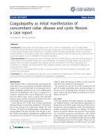
large atrial mass. Transesophageal echocardiography, car-
ried out at the time of surgery, revealed a large myxoma
prolapsing through the mitral valve leaflets into the left
ventricle (Figure 1).
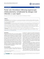
A left atrial myxoma excision was performed, resulting in
successful removal of the tumor. Pathological analysis of
the atrial mass revealed it to be 7 × 6.5 × 4.5 cm attached
to a 4 × 3 × 2 cm stalk of atrial septal tissue (Figure 2). Four
weeks postoperatively, the patient stated that the original
complaint of 'dizziness upon standing' had disappeared,
with no evidence of orthostatic hypotension during a fol-
low-up physical examination. A follow-up echocardio-
gram showed no evidence of atrial myxoma recurrence,
and the mitral valve leaflets separated normally without
regurgitation.
Discussion
Our patient failed to present with the common symptoms
associated with atrial myxoma including chest pain, dysp-
nea, orthopnea, peripheral embolism or syncope. Though
cardiac myxomas are known to present with various non-
specific clinical symptoms [2], orthostatic hypotension is
not listed as a presenting symptom of atrial tumors in
most textbooks of internal medicine or cardiology [3,4].
An extensive literature search revealed one case that
reported orthostasis as a presenting symptom of a left
atrial myxoma [5]. The patient in that case report had a
principal complaint of dizziness upon standing, and
orthostasis was observed with a blood pressure change
from 90/50 mmHg supine to 64/40 mmHg standing.
Upon echocardiographic investigation, a large atrial
Atrial myxoma: prolapsing through mitral valveFigure 1
Atrial myxoma: prolapsing through mitral valve.
Eleven days pre-operatively, the left atrium and left ventricle
are visualized in this transesophageal echocardiogram.
Postoperative atrial myxomaFigure 2
Postoperative atrial myxoma. The atrial tumor seen
directly postoperatively was a mass of 7 × 6.5 × 4.5 cm
attached to a 4 × 3 × 2 cm piece of atrial septal tissue.
Publish with Bio Med Central and every
scientist can read your work free of charge
"BioMed Central will be the most significant development for
disseminating the results of biomedical research in our lifetime."
Sir Paul Nurse, Cancer Research UK
Your research papers will be:
available free of charge to the entire biomedical community
peer reviewed and published immediately upon acceptance
cited in PubMed and archived on PubMed Central
yours — you keep the copyright
Submit your manuscript here:
/>BioMedcentral
Journal of Medical Case Reports 2009, 3:9328 />Page 3 of 3
(page number not for citation purposes)
myxoma was found impeding inflow into the ventricular
cavity upon standing. The myxoma was 3.5 cm in diame-
ter and attached to the postero-inferior portion of the left
atrial wall. This atrial tumor, smaller than the one we
describe, brought on symptoms of orthostasis similar in
severity to those that we observed in the patient in this
case report. Upon removal of the myxoma in the Take-
mura case and in the case we describe, all clinical symp-
toms of orthostasis were relieved [5].
Orthostasis is relatively common in the elderly, being
found in nearly 5-30% of the population with common
causes including neurogenic dysfunction, autonomic fail-
ure, antihypertensive medications and intravascular vol-
ume depletion [6]. Since orthostatic hypotension is
frequent in the elderly and there are numerous known
causes for its occurrence, atrial tumors may be overlooked
as the culprit for the manifestation. Since both cases dis-
cussed presented positional dizziness as the sole present-
ing symptom, we believe it is important to include atrial
myxomas in the differential diagnosis of orthostasis.
Conclusion
Two-dimensional echocardiography provides substantial
advantages in detecting intracardiac tumors. A two-
dimensional echocardiogram is recommended by the
authors of this report in the workup of orthostatic hypo-
tension of unknown etiology. Although atrial myxomas
are usually benign or asymptomatic, there is the possibil-
ity of diastolic embolization [7], conduction alterations
and disturbances, and lethal valve obstructions occurring
[4]. Since surgical excision has been reported to alleviate
symptoms associated with cardiac myxomas, early identi-
fication and removal is preferable. By illustrating this cor-
relation between orthostasis and atrial myxomas, we hope
to facilitate earlier identification of these intracardiac
growths.
Consent
Written informed consent was obtained from the patient
for publication of this case report and any accompanying
images. A copy of the written consent is available for
review by the Editor-in-Chief of this journal.
Competing interests
The authors declare that they have no competing interests.
Authors' contributions
RV came up with original conception and design. RV, EP,
NS, and JS scientifically reviewed and edited the study. KS,
MH, DM, TG, and ST reviewed the medical literature, and
were major contributors in writing the manuscript. KS,
DM, and TG formatted the media. All authors read and
approved the final manuscript.
References
1. Burke AP, Virmani R: Cardiac myxoma: a clinicopathologic
study. Am J Clin Pathol 1993, 100:671-680.
2. Jelic J, Milici D, Alfirevi I, Ani D, Baudoin Z, Bulat C, Coric V, Dadic
D, Husar J, Ivancan V, Korda Z, Letica D, Predrijevac M, Ugljen R,
Vucemilo I: Cardiac myxoma: diagnostic approach, surgical
treatment and follow-up. J Cardiovasc Surg 1996, 37(6):113-117.
3. Engstrom JW, Martin J: Chapter 366. In Disorders of the Autonomic
Nervous System. Harrison's Principles of Internal Medicine Volume 2. 15th
edition. Edited by: Braunwald E, Fauci A, Kasper D, Hauser S, Longo
D, Jameson JL. Blacklick, OH, USA: McGraw-Hill Professional Book
Group; 2001:2417-2421.
4. Colucci WS, Schoen FJ: Chapter 49. In Primary Tumors of the Heart.
Heart Disease: A Textbook of Cardiovascular Medicine Volume 2. 6th edi-
tion. Edited by: Braunwald E, Zipes D, Libby P. Philadelphia, Pennsyl-
vania, USA: W.B. Saunders Company; 2001:1809-1819.
5. Takemura G, Kotoura H, Nishioka A, Kobayashi T, Uegaito T, Miura
A, Inagaki M, Wada T, Watanabe R: A case of cardiac tumor
found during examination in orthostatic hypotension. Koykyu
To Junkan 1990, 38(3):257-259.
6. Low P: Prevalence of orthostatic hypotension. Clin Auton Res
2008, 18:8-13.
7. Braun S, Schrotter H, Reynen K, Schwencke C, Strasser RH: Myo-
cardial infarction as complication of left atrial myxoma. Int J
Cardiol 2005, 101(1):115-121.