Báo cáo y học: " Ileocaecal recurrence of Merkel cell carcinoma of the skin: a case report" pot
Bạn đang xem bản rút gọn của tài liệu. Xem và tải ngay bản đầy đủ của tài liệu tại đây (1.46 MB, 5 trang )
CAS E REP O R T Open Access
Ileocaecal recurrence of Merkel cell carcinoma of
the skin: a case report
Michelle Cheung
*
, Henry Lee, Sanjay Purkayastha, Robert Goldin, Paul Ziprin
Abstract
Introduction: Merkel cell carcinoma is an uncommon skin malignancy that has a high propensity for metastatic
spread. A systematic literature search identified 17 cases describing metastasis to the gastrointestinal tract, with 7
cases involving the small or large bowel. To the best of our knowledge, this is the only case described of Merkel
cell carcinoma metastasising to the ileocaecal valve.
Case presentation: We present a 74-year-old Filipino woman diagnosed with Merkel cell carcinoma of the skin
with regional node involvement. Following excision and radiotherapy, the tumour recurred with metastasis to the
ileocaecal valve. The patient died 28 months after the initial diagnosis.
Conclusion: The prognosis of metastatic Merkel cell carcinoma is poor. Currently the optimal management for
metastatic disease is unclear and lacks a firm evidence base due to the small number of cases reporte d.
Introduction
Merkel cell carcinoma (MCC) is an uncommon and
highly aggressive skin malignancy. It arises from Merkel
cells at the dermo-epidermal junction, which are of neu-
roendocrine origin. Since its first description by Toker
in 1972 [1], more than 2000 cases have been reported in
the literature [2]. Its aetiolog y is not entirely kno wn, but
thereisconvincingevidencefortheroleofultraviolet
radiation. MCC has a predilection for sun-exposed areas
of the body and is associated with other sun-related skin
cancers such as basal cell carcinoma and squamous cell
carcinoma. The occurrence of MCC in areas that are
not exposed to the sun suggests additional causes.
Reports of MCC in organ transplant, human immuno-
deficiency virus (HIV) infection and lymphohemopoietic
malignancies, such as chronic lymphocytic leukemia,
implicate a role for immunosuppression [2,3].
The incidence of MCC is 0.23 per 100,000 in Cauca-
sians [2], which is about 20 times the incidence com-
pared to the Afro-Caribbean population. MCC is also
more common in older men with a mean age of diagno-
sis at 69 years old [2]. In a r eview of 1024 patients, the
primary tumour was found in the head and neck in
40%, in the extremities in 33%, and in the trunk in 23%
of patients [4]. At presentation the regional lymph
nodes are involved in around 25% of cases, and distant
metastases are found in 4% [4]. Metastasis usually
occurs in the skin (28%), lymph nodes (27%), liver
(13%), lung (10%), bone (10%), and the brain (6%) [2,4].
Metastasis can also involve the gastrointestinal (GI)
tract in very rare cases.
A systematic search of the literature (see Appendix)
found 17 ca ses involving GI metastases most commonly
involving the stomach. Seven of these cases described
bowel metastasis ( Table 1). Shalhub et al. described the
case of a 6 2-year- old man with axillary lymphade nopa-
thy and metastasis to the stomach and the descending
colon. The patient had a skin lesio n excision which was
initially diagnosed as bas al cell carcinoma [5]. There are
two case reports of stomach and small bowel MCC pre-
senting with upper GI bleed from Krasagakis et al.[6]
and Canales et al.[7].An85-year-oldJapanesewoman
was also diagnosed with widespread upper GI tract
MCC metastasis on autopsy following intestinal obstruc-
tion [8]. Naunton Morgan and Henderson reported a
man with an enlarging nodule on his shin, who pre-
sented a month later with melaena and where a meta-
static MCC lesion in the proximal jejunum was found
on surgical exploration [9]. Meanwhile, Foste r et al.also
described a case of Merkel cell metastasizing to the
small b owel after a protracted time course [10]. In
* Correspondence:
Academic Surgical Unit, St Mary’s Hospital, Imperial College Healthcare,
Praed Street, London W2 1NY, UK
Cheung et al. Journal of Medical Case Reports 2010, 4:43
/>JOURNAL OF MEDICAL
CASE REPORTS
© 2010 Cheung et al; licensee BioMed Central Ltd. This is an Open Access article distributed under the terms of the Creative Commons
Attribution License ( which permits unrestricted use, distribution, and reproduction in
any medium, provided the original work is properly cited.
addition, there are cases reported of metastasis to the
rectum, the anal canal, and the pancreas [11-21].
Case presentation
A 74-year-old Filipino woman presented with a skin
lesion in her right antecubital fossa. It was a 2 cm soft,
mobile, well-circumscribed m ass which appeared over
three weeks. Suspecting liposarcoma, her general practi-
tioner referred her to the hospital’ s surgical team.
Urgent excision biopsy revealed metastatic carcinoma
expressing neuroendocrine markers. Immunohis tochem-
ical staining showed that the patient had a strong posi-
tivity for CK20, which was a sensitive and specific
marker for Merkel cell carcinoma [22]. She was also
highly positive for chromogr anin, synaptophysin and
CD117, thus confirming the diagnosis of MCC. She
showed no staining for TTF-1, which excluded lung pri-
mary small cell carcinoma.
Subsequent computed tomography (CT) examinations
of her chest, abdomen and pelvis identified significant
right-sided axillary lymphadenopathy, with the largest
node measuring 6 cm in diameter. The CT was con-
ducted two months after our patient’s initial clinical pre-
sentation, and the axillary adenopathy was now clinically
apparent. There was no hilar or mediastinal l ymphade-
nopathy observed. Her lungs, bones and intra-abdominal
organs were all clear of metastasis. A positron emission
tomography (PET) body scan showed high uptake in her
right axilla, with moderate heterogeneous uptake in
some bowel loops. This was thought to represent
inflammatory change in the presence of diverticular
disease. No definite primary source, however, was
identified.
At axillary lymph node dissection, eight of 26 lymph
nodes, as well as the axillary vein, were found to contain
tumour. Adjuvant radiotherapy was administered to our
patient’s axillary and subclavicular regions (42 Gy, 21
fractions, 31 days), but it was postponed until six
months postoperatively due to the presence of seroma
and lymphoedema. Surgical and oncological follow-up at
intervals of two, three and six months found our patient
asymptomatic.
At 18 months from the initial diagnosis, our patient
was referred by her general practitioner for urgent
review with symptoms of obst ruction, specifically early
satiety, bloating, colicky pains and occasional vomiting.
On examination, a firm mass in her right upper quad-
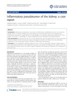
rant was c linically detectable. A whole body CT s can
found a tumour in the ileocaecal valve that extended
into the caecal lumen (Figure 1 and Figure 2). The
tumour was associated with mesenteric lymphadenopa-
thy and local infiltration into the pericolic fat and
vessels.
At colonoscopy a tumour was seen at 70 cm from our
patient’s anus. The morphology and immunop rofile of
the biopsy confirmed it as a rec urrence of MCC. Mar-
kers for colorectal cancer (CA 19.9, CEA) were negative.
Our patient underwent a laparoscopic right hemico-
lectomy. At surgery the tumour was seen to be fungat-
ing and involved the full thickness of her bowel wall.
Histological examination showed clear resection margins
but vascular invasion and multiple lymph n ode involve-
ment were also noted. Our patient’ spostoperative
Table 1 Reported cases of gastrointestinal metastases of
Merkel call cancer.
Author(s) Site of Metastasis
Li M and Liu C [11] Stomach
Cubiella J, et al. [12] Stomach
Idowu M, et al. [13] Stomach
Wolov K, et al. [14] Stomach
Krasagakis K, et al. [6] Stomach, small bowel
Canales L, et al. [7] Stomach, small bowel
Shalhub S, et al. [5] Stomach, descending colon
Hizawa K, et al. [8] Stomach, distal duodenum,
pancreas
Olivero G, et al. [15] Intestinal
Naunton M and Henderson RG [9] Jejunum
Foster R, et al. [10] Small bowel
Huang W S, et al. [16] Rectum
Paterson C, et al. [17] Anal canal
Adsay NV, et al. [18] Pancreas
Bachmann J, et al. [19] Pancreas
Dim DC, et al. [20] Pancreas
Ouellett JR, et al. [21] Pancreas
Figure 1 Computed tomography showing a recur rence of
Merkel cell carcinoma in the caecum at 18 months after the
initial diagnosis. There is a 6 × 4.3 cm soft-tissue mass seen in the
region of the ileocecal valve (A) which extends into the lumen of
the caecum. There are multiple abnormal lymph nodes measuring
up to 2 cm within the ileocolic mesentery. There is nodularity and
irregularity seen around the tumour extending into the pericolic fat
suggestive of a local tumour infiltration.
Cheung et al. Journal of Medical Case Reports 2010, 4:43
/>Page 2 of 5
recovery was uneventful. Follow-up CT scan 3 months
after the resection revealed peritoneal deposits with no
recurrence in the right axilla (Figure 3). She was then
scheduled for palliative chemotherapy.
Prior to chemotherapy our patient became increas-
ingly frail and deteriorated rapidly. She died of bronch-
opneumonia 28 months after the initial diagnosis of
metastatic Merkel cell carcinoma.
Discussion
Merkel cell carcinoma has been described as one the
most aggressive of the skin malignance s [4,22]. In Med-
ina-Franco’s review of 1024 cases, this metastatic disease
affects 31% of patients either at presentation or at later
stage [2]. Excluding the patients with metastasis at pre-
sentation, 31% develop local recurrence, and the average
disease-free interval is only 7.4 months [2]. Of the fac-
tors related to prognosis in MCC, lymph node status
has been shown to be most consistent with clinical
outcome [22-25]. This is reflected in the staging of the
disease, where stage I describes lesions confined to the
skin, stage II describes the involvement of regional
nodes, and stage III describes the presence of metastasis.
Five-year survival rates are 64%, 47% and 11%, respec-
tively [2,23,25,26]. This compares with a 7% to 19% sur-
vival rate for malignant melanoma with metastasis [27].
A retrospective review of 109 patients by Allen et al.
revealed that in patients with stage I disease, the tumour
size at presentation was also an independent predictor
of survival. In view of this, a 4-tiered system is also
widely used to reflect a more accurate prognosis. It clas-
sifies patients with lymph node-negative cutaneous dis-
ease of <2 cm as stage I, and cutaneous lesions of >2
cm as stage II. Lymph node-positive disease is classified
as st age III, while the presence of distant metastases is
stage IV [25].
The treatment of choice for MCC is surgical excision
with wide margins. There is no consensus on the width
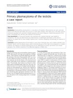
Figure 2 Microscopic appearance of the ileocaecal valve tumour. The mucosa shows infiltration by malignant cells with glassy nuclei,
prominent nucleoli and scant cytoplasm. There is a high proliferation fraction. Immunostaining was positive for CK20, chromgranin A,
synaptophysin, CD56, and MNF116. It was negative for CD3, CD30, CD10, CD20, CD23, CD5. The morphology and immunoprofile confirms this to
be a metastatic Merkel cell carcinoma.
Cheung et al. Journal of Medical Case Reports 2010, 4:43
/>Page 3 of 5
of excision required, b ut many surgical series have sug-
gested margins of 2.5 cm to 3 cm [4]. There is evidence
to show, however, that low local recurrence rates (8%)
can be achieved after margin-negative excisions with
margins that averaged 1.1 cm. This has lead to the sug-
gestion that larger excision margins of at least 2 cm
should be reserved for larger lesions of >2 cm in dia-
meter [23].
Given that MCC occurs so frequently in the head and
the neck, where wide local excision or even Moh’ssur-
gery may be difficult, the role of adjuvant therapy has
been much considered. One large case series review
involving over 400 patients found that radiotherapy
improves local control from a recurrence rate of 52% to
10.5% [4] . Some studies, however, showed cont rary
results where radiotherapy did not improve recur rence
rates in patients with node- negative diseases. They
instead recommend that adjuvant therapy may be bene-
ficial in patients with clinically positive nodes or those
who have undergone elective or sentinel node biopsy
with positive nodes [23].
Due to the importance of lymph node status in treat-
ment outcome, sentinel lymph node biopsy (SLNB) as a
staging procedure is considered standard practice in
some centers, and its use has been recommended by
many others. A number of studies have reported SLNB
positivity rate of 20% to 30% involving patients who are
clinically lymph node negative [23,28]. Where microme-
tastasis is found on SLNB, however, complete lymph
node dissection is considered as first-line management,
while radiotherapy is second-line [26,28].
As a neuroendocri ne tumour, MCC demonstrates che-
mosensitivity to agents used in treating small-cell lung
cancer, such as carboplatin, vincristine and etoposide. The
role of chemotherapy, however, is much less established
than that of radiotherapy. There is some support for its
use either as a radiation sensitizer or as adjuvant therapy
in high-risk cases with the disease at stage II or above [2].
At stage IV of the disease chemotherapy has shown high
but relatively short response rates of 8 months on the
average, and d oes not appear to add significantly where
the median survival in metastasis is 10 months [28].
Overall, the optimal management for patients with
MCC remains unclear. Treatment is often guided by
opinion r ather than evidence-based practice [ 2]. Bichak-
jian et al. proposed an algorithm reflecti ng the evidence
as discussed above [28]. Again it suggests wide local
excision margins of 1 cm for lesions measuring <2 cm,
and 1 cm to 2 cm for lesions measuring >2 cm. Radio-
therapy is best used when clear margins cannot be
obtained. Furthermore, sentinel node biopsy should be
performed in all instances.
Meanwhile, negative SLNB should be observed at inter-
vals between 2 to 6 months for 2 years, with decreasing
intervals thereafter . The use of imaging should be guided
by clinical findings rather than by routine due to high
false-positive rates, and the lack of survival benefit in
detecting asymptomatic distant metastases. Positive sen-
tinel nodes should be treated with complete node dissec-
tion and radiation depending on the level of the nodal
disease. Once there is involvement in distant sites, ther-
apy is primarily for palliation. The literature on the man-
agement of recurrent and metastatic diseases is
particularly limited. The only consensus seems to be that
the outcome of high stage MCC is gravely unfavorable.
Conclusion
The management of Merkel cell car cinoma poses a chal-
lenge to clinicians for several reasons. For one, it is aggres-
sive and has a propensity for spread and recurrence.
Optimal therapy in high stage disease or recurrence is less
clearly established. Its low incidence warrants the colla-
boration of different institutions to produce large multi-
cent er controlled trials in order to compare responses to
different treatment modalities. At any stage, its manage-
ment is likely to benefit from a multidisciplinary approach,
which is highlighted in this case report.
Consent
Written informed consent was obtained from the
patient’s next-of-kin for publication of this case report
and any accompanying images. A copy of the written
consent is available for review by the Editor-in-Chief of
this journal.
Figure 3 Posto perative computed tomography scan following
laparoscopic right hemicolectomy. There is an end-to-side
anastamosis between the distal ileum and the colon.
Cheung et al. Journal of Medical Case Reports 2010, 4:43
/>Page 4 of 5
Appendix
Systematic search algorithm employed.
Literatur e search was conducted u sing MeSH terms
“oesophagus” [All Fields] OR “ esophagus” [All Fields]
OR “ stomach” [All Fields] OR “duodenum” [All Fields]
OR “jejunum” [All Fields] OR “ ileum” [All Fields] OR
“ bowel” [All F ields] OR “ intestine ” [All Fields] OR
“ intestinal” [All Fields] OR “caecum” [All Fields] OR
“rectum” [All Fields] OR “rectal” [All Fields] OR “colon”
[All Fields] AND “merkel cell” [All Fields].
This produced 61 results on PubMed of which 17 are
reports of metastatic Merkel cell carcinoma in the gas-
trointestinal tract (excluding the liver).
Authors’ contributions
MC reviewed the case notes and the literature. She also drafted the
manuscript. HL and SP revised the references and finalized the manuscript.
RG performed the histopathology examinations and provided the specime n
image. PZ performed the patient’s operation and conceived of the article. All
authors read and approved the final manuscript.
Competing interests
The authors declare that they have no competing interests.
Received: 4 November 2009
Accepted: 8 February 2010 Published: 8 February 2010
References
1. Toker C: Trabecular carcinoma of the skin. Arch Dermatol 1972,
105(1):7-10.
2. Poulsen M: Merkel-cell carcinoma of the skin. Lancet Oncol 2004,
5(10):593-599.
3. Ziprin P, Smith S, Salerno G, Rosin RD: Two cases of Merkel cell tumour
arising in patients with chronic lymphocytic leukemia. Br J Dermatol
2000, 142(3):525-528.
4. Medina-Franco H, Urist MM, Fiveash J, Heslin MJ, Bland KI, Beenken SW:
Multimodality treatment of Merkel cell carcinoma: case series and
literature review of 1024 cases. Ann Surg Oncol 2001, 8(3):204-208.
5. Shalhub S, Clarke L, Morgan MB: Metastatic Merkel cell carcinoma
masquerading as colon cancer. Gastrointest Endosc 2004, 60(5):856-858.
6. Krasagakis K, Almond-Roesler B, Zouboulis CC, Tebbe B, Wartenberg E,
Wolff KD, Orfanos CE: Merkel cell carcinoma: report of 10 cases with
emphasis on clinical course, treatment, and in vitro drug sensitivity. J
Am Acad Dermatol 1997, 36(5 Pt 1):727-732.
7. Canales LI, Parker A, Kadakia S: Upper gastrointestinal bleeding from
Merkel cell carcinoma. Am J Gastroenterol 1992, 87(10):1464-1466.
8. Hizawa K, Kurihara S, Nakamori M, Nakahara T, Matsumoto T, Iida M: An
autopsy case of Merkel cell carcinoma presenting aggressive intra-
abdominal metastasis and duodenal obstruction. Nippon Shokakibyo
Gakkai Zasshi 2007, 104(9):1383-1386.
9. Naunton Morgan TC, Henderson RG: Small bowel metastases from a
Merkel cell tumor. Br J Radiol 1985, 58(696):1212-1213.
10. Foster R, Stevens G, Egan M: An unusual pattern of metastases from
Merkel cell carcinoma. Australas Radiol 1994, 38(3):231-232.
11. Li M, Liu C: Cytokeratin 20 confirms Merkel cell metastasis to stomach.
Appl Immunohistochem Mol Morpho 2004, 12(4):346-349.
12. Cubiella J, Salgado M, Riu M, Garcia-Mata J, Sanchez E, Diez MS,
Rodriguez R, Vega M: Gastric metastatis due to Merkel cell carcinoma: a
rare cause of gastric bleeding. Rev Esp Enferm Dig 2004, 96(2):150-151.
13. Idowu MO, Contos M, Gill S, Powers C: Merkel cell carcinoma: a report of
gastrointestinal metastasis and review of the literature. Arch Pathol Lab
Med 2003, 127(3):367-369.
14. Wolov K, Tully O, Mercogliano G: Gastric metastasis of Merkel cell
carcinoma. Clin Gastroenterol Hepatol 2009, 7(3)
:A26.
15. Olivero G, Franchello A, Pacchioni D, Enrichens F, Mao P, Benedetto G: A
rare case of Merkel’s tumor with intestinal metastases. Ann Ital Chir 1990,
61(3):277-280.
16. Huang WS, Lin PY, Lee IL, Chin CC, Wang JY, Yang WG: Metastatic Merkel
cell carcinoma in the rectum: report of a case. Dis Colon Rectum 2007,
50(11):1992-1995.
17. Paterson C, Musselman L, Chorneyko K, Reid S, Rawlinson J: Merkel cell
(neuroendocrine) carcinoma of the anal canal: report of a case. Dis Colon
Rectum 2003, 46(5):676-678.
18. Adsay NV, Andea A, Basturk O, Kilinc N, Nassar H, Cheng JD: Secondary
tumors of the pancreas: an analysis of a surgical and autopsy database
and review of the literature. Virchows Arch 2004, 444(6):527-535.
19. Bachmann J, Kleeff J, Bergmann F, Shrikhande SV, Hartschuh W,
Buchler MW, Friess H: Pancreatic metastasis of Merkel cell carcinoma and
concomitant insulinoma: case report and literature review. World J Surg
Oncol 2005, 3:58.
20. Dim DC, N20. Idowu MO, Contos M, Gill S, Powers C: Merkel cell
carcinoma: a report of gastrointestinal metastasis and review of the
literature. Arch Pathol Lab Med 2003, 127(3):367-369.
21. Ouellette JR, Woodyard L, Toth L, Termuhlen PM: Merkel cell carcinoma
metastatic to the head of the pancreas. JOP 2004, 5(2):92-96.
22. Eng TY, Boersma MG, Fuller CD, Goytia V, Jones WE, Joyner M, Nguyen DD:
A comprehensive review of the treatment of Merkel cell carcinoma. Am
J Clin Oncol 2007, 30(6):624-636.
23. Allen PJ, Bowne WB, Jaques DP, Brennan MF, Busam K, Coit DG: Merkel cell
carcinoma: prognosis and treatment of patients from a single institution.
J Clin Oncol 2005, 23(10):2300-2309.
24. Pectasides D, Pectasides M, Psyrri A, Koumarianou A, Xiros N, Pectasides E,
Gaglia A, Lianos E, Papaxoinis G, Lampadiari V, Economopoulos T: Cisplatin-
based chemotherapy for Merkel cell carcinoma of the skin. Cancer Invest
2006, 24(8):780-785.
25. Allen PJ, Zhang ZF, Coit DG: Surgical management of Merkel cell
carcinoma. Ann Surg 1999, 229(1):97-105.
26. Pectasides D, Pectasides M, Economopoulos T: Merkel cell cancer of the
skin. Ann Oncol 2006, 17(10):1489-1495.
27. Mosca PJ, Teicher E, Nair SP, Pockaj BA: Can surgeons improve survival in
stage IV melanoma?. J Surg Oncol 2008, 97(5):462-468.
28. Bichakjian CK, Lowe L, Lao CD, Sandler HM, Bradford CR, Johnson TM,
Wong SL:
Merkel cell carcinoma: critical review with guidelines for
multidisciplinary management. Cancer 2007, 110(1):1-12.
doi:10.1186/1752-1947-4-43
Cite this article as: Cheung et al.: Ileocaecal recurrence of Merkel cell
carcinoma of the skin: a case report. Journal of Medical Case Reports 2010
4:43.
Submit your next manuscript to BioMed Central
and take full advantage of:
• Convenient online submission
• Thorough peer review
• No space constraints or color figure charges
• Immediate publication on acceptance
• Inclusion in PubMed, CAS, Scopus and Google Scholar
• Research which is freely available for redistribution
Submit your manuscript at
www.biomedcentral.com/submit
Cheung et al. Journal of Medical Case Reports 2010, 4:43
/>Page 5 of 5