Báo cáo y học: "Review Cells of the synovium in rheumatoid arthritis" pot
Bạn đang xem bản rút gọn của tài liệu. Xem và tải ngay bản đầy đủ của tài liệu tại đây (250.97 KB, 13 trang )
Page 1 of 13
(page number not for citation purposes)
Available online />Abstract
Rheumatoid arthritis (RA) is one of the inflammatory joint diseases
in a heterogeneous group of disorders that share features of
destruction of the extracellular matrices of articular cartilage and
bone. The underlying disturbance in immune regulation that is
responsible for the localized joint pathology results in the release of
inflammatory mediators in the synovial fluid and synovium that
directly and indirectly influence cartilage homeostasis. Analysis of
the breakdown products of the matrix components of joint cartilage
in body fluids and quantitative imaging techniques have been used
to assess the effects of the inflammatory joint disease on the local
remodeling of joint structures. The role of the chondrocyte itself in
cartilage destruction in the human rheumatoid joint has been
difficult to address but has been inferred from studies in vitro and
in animal models. This review covers current knowledge about the
specific cellular and biochemical mechanisms that account for the
disruption of the integrity of the cartilage matrix in RA.
Rheumatoid arthritis
Rheumatoid arthritis (RA) is an inflammatory joint disease that
most frequently affects the anatomical components of
articular and juxta-articular tissues of diarthrodial joints. The
diarthrodial joints join two opposing bone surfaces that are
covered by a specialized hyaline cartilage providing a low-
friction, articulating interface. The synovium lines the joint
cavity and is the site of production of synovial fluid, which
provides the nutrition for the articular cartilage and lubricates
the cartilage surfaces. In RA, the synovial lining of diarthrodial
joints is the site of the initial inflammatory process [1,2]. This
lesion is characterized by proliferation of the synovial lining
cells, increased vascularization, and infiltration of the tissue
by inflammatory cells, including lymphocytes, plasma cells,
and activated macrophages [3-5]. With the growth and
expansion of the synovial lining, there is eventual extension of
the inflammatory tissue mass to the adjacent articular
cartilage with progressive overgrowth of the articular surface
and formation of the so-called pannus, which is derived from
the Latin word meaning ‘cloth’ and the Greek word meaning
‘web’. At the interface between the RA synovium and articular
cartilage, tongues of proliferating cells can be seen
penetrating the extracellular matrix of the cartilage. Similarly,
at the interface between the inflamed synovium and adjacent
subchondral bone, there is evidence of local activation of
bone resorption with destruction of the mineralized bone
matrix, accompanied by cells expressing phenotypic features
of osteoclasts, including calcitonin receptor mRNA, cathepsin
K, and tartrate-resistant acid phosphatase (TRAP) [6,7]. RA
synovium produces a broad spectrum of factors possessing
the capacity to stimulate cartilage matrix destruction and
bone erosion [3,4]. Although there is an association between
inflammation and the development of joint damage, the
destruction may progress in spite of attenuated inflammatory
activity, and cartilage and bone erosions may develop in the
absence of overt clinical signs of inflammation [8-11]. Recent
evidence from human and animal studies indicates that
although the specific cellular mechanisms of cartilage and
bone destruction are different, TNF-α, IL-1, and additional
proinflammatory cytokines and mediators can drive elements
of both processes [10,12]. The recent development of assays
for specific biological markers that reflect quantitative and
dynamic changes in the synthetic and degradation products
Review
Cells of the synovium in rheumatoid arthritis
Chondrocytes
Miguel Otero and Mary B Goldring
Research Division of the Hospital for Special Surgery, Weill College of Medicine of Cornell University, Caspary Research Building, 535 E. 70th Street,
New York, NY 10021, USA
Corresponding author: Mary B Goldring,
Published: 26 October 2007 Arthritis Research & Therapy 2007, 9:220 (doi:10.1186/ar2292)
This article is online at />© 2007 BioMed Central Ltd
AIA = antigen-induced arthritis; ADAM = a disintegrin and metalloproteinase; ADAMTS = ADAM with thrombospondin-1 domains; CD-RAP = carti-
lage-derived retinoic-acid-sensitive protein; CH3L1 = chitinase 3-like protein 1; CIA = collagen-induced arthritis; COMP = cartilage oligomeric
matrix protein; COX = cyclooxygenase; GLUT = glucose transporter protein; HIF = hypoxia-inducible factor; IGF = insulin-like growth factor; IL =
interleukin; IL-1Ra = IL-1 receptor antagonist; iNOS = inducible nitric oxide synthetase; MCP = monocyte chemoattractant protein; MIP =
macrophage inflammatory protein; MMP = matrix metalloproteinase; mPGES-1 = microsomal PGE synthase-1; NF = nuclear factor; OSM = onco-
statin M; PGE = prostaglandin E; PPAR = peroxisome proliferator-activated receptor; RA = rheumatoid arthritis; RANKL = receptor activator of
NF-κB ligand; TGF = transforming growth factor; Th = T helper; TIMP = tissue inhibitor of metalloproteinases; TLR = Toll-like receptor; TNF =
tumor necrosis factor; TRAP = tartrate-resistant acid phosphatase; VCAM = vascular cell adhesion molecule; VEGF = vascular endothelial growth
factor.
Page 2 of 13
(page number not for citation purposes)
Arthritis Research & Therapy Vol 9 No 5 Otero and Goldring
of cartilage and bone matrix components has offered the
possibility of identifying patients at risk for rapid joint damage
and also the possibility of early monitoring of the efficacy of
disease-modifying anti-rheumatic therapies [13-15]. This
review will focus on the unique ways in which the
chondrocyte responds to the inflammatory milieu and
contributes to the disease process in the cartilage.
The chondrocyte in adult articular cartilage
Adult human articular cartilage, which covers the articulating
surfaces of long bones, is populated exclusively by
chondrocytes that are somewhat unique to this tissue. The
collagen network of the interterritorial cartilage matrix is
composed of types II, IX, and XI collagens, which provide
tensile strength and promote the retention of proteoglycans.
Type XI collagen is part of the type II collagen fibril, and type
IX integrates with the surface of the fibril with the non-
collagen domain projecting outward, permitting association
with other matrix components. The other major component,
the large aggregating proteoglycan aggrecan, which is
attached to hyaluronic acid polymers via link protein, bestows
compressive resistance. A large number of other non-
collagen molecules are present in the interterritorial matrix;
these molecules include several small proteoglycans such as
biglycan, decorin, fibromodulin, the matrilins, and cartilage
oligomeric matrix protein (COMP). The chondrocytes are
surrounded by a pericellular matrix composed of type VI
collagen microfibrils that interact with hyaluronic acid,
biglycan, and decorin and maintain chondrocyte attachment,
but little or no fibrillar collagen. Under physiological
conditions, the chondrocytes maintain a stable equilibrium
between the synthesis and the degradation of matrix
components, with a half-life of more than 100 years for type II
collagen [16] and a half-life for aggrecan core protein in the
range 3 to 24 years [17]. The glycosaminoglycan
components of aggrecan and other cartilage matrix
constituents also are synthesized by chondrocytes under
conditions of low turnover, and the matrix turnover may be
more rapid in the immediate pericellular zones.
Under normal conditions, chondrocyte proliferation is limited,
and penetration of other cell types from the joint space or
subchondral bone is restricted. In the absence of a vascular
supply, the chondrocyte must rely on diffusion from the
articular surface or subchondral bone for the exchange of
nutrients and metabolites. Glucose serves both as the major
energy source for the chondrocytes and as an essential
precursor for glycosaminoglycan synthesis. Facilitated
glucose transport in chondrocytes is mediated by several
distinct glucose transporter proteins (GLUTs) that are either
expressed constitutively (GLUT3 and GLUT8) or inducible by
cytokines (GLUT1 and GLUT6) [18,19]. Chondrocytes do
not contain abundant mitochondria, but they maintain active
membrane transport systems for exchange of cations,
including Na
+
, K
+
, Ca
2+
, and H
+
, whose intracellular
concentrations fluctuate with charge, biomechanical forces,
and alterations in the composition of the cartilage matrix [20].
Furthermore, chondrocyte metabolism operates at low oxygen
tension, ranging from 10% at the surface to less than 1% in
the deep zones of the cartilage. Chondrocytes adapt to low
oxygen tensions by upregulating hypoxia-inducible factor
(HIF)-1α, which can stimulate the expression of GLUTs [19]
and angiogenic factors such as vascular endothelial growth
factor (VEGF) [21,22], as well as ascorbate transport [23]
and several genes associated with cartilage anabolism and
chondrocyte differentiation, including Sox9 and type II
collagen [24]. By modulating the intracellular expression of
survival factors such as HIF-1α, chondrocytes have a high
capacity to survive in the avascular cartilage matrix and to
respond to environmental changes.
Joint inflammation and cartilage remodeling
in RA
Cartilage destruction in RA occurs primarily in areas
contiguous with the proliferating synovial pannus [25,26]. In
the cartilage–pannus junction, there is evidence of
attachment of both fibroblast-like and macrophage-like
synovial cell types, which can release proteinases capable of
digesting the cartilage matrix components [27]. A distinctive
fibroblast-like cell type, the so-called ‘pannocyte’, present in
RA synovium exhibits anchorage-independent growth and
can invade cartilage in the absence of an inflammatory
environment [2]. Nevertheless, there is evidence of loss of
proteoglycan throughout the cartilage matrix, particularly in
the superficial zone in contact with the synovial fluid at sites
not directly associated with the pannus [28,29]. This has
been attributed to the release of inflammatory mediators and
degradative enzymes released by polymorphonuclear leuko-
cytes and other inflammatory cells in the synovial fluid. In early
RA, however, the loss of proteoglycan occurs throughout the
cartilage matrix, and selective damage to type II collagen
fibrils can be observed in middle and deep zones [30,31],
suggesting that the chondrocyte may also participate in
degrading its own matrix by releasing autocrine–paracrine
factors.
Of the matrix metalloproteinases (MMPs) involved in the
degradation of cartilage collagens and proteoglycans in RA,
the MMPs of the collagenase and stromelysin families have
been given greatest attention because they specifically
degrade native collagens and proteoglycans. Active
stromelysin also serves as an activator of latent collagenases
[32]. MMPs are localized at sites of degradation in cartilage
derived from patients with RA [33]. Collagenases 1, 2, and 3
(MMP-1, MMP-8, and MMP-13, respectively), gelatinases
(MMP-2 and MMP-9), stromelysin-1 (MMP-3), and membrane
type I MMP (MT1-MMP; MMP–14) are present in active RA
synovium [34,35]. Although elevated levels of MMPs in the
synovial fluid probably originate from the synovium, intrinsic
chondrocyte-derived chondrolytic activity is present at the
cartilage–pannus junction as well as in deeper zones of
cartilage matrix in some RA specimens [36]. For example,
Page 3 of 13
(page number not for citation purposes)
MMP-1 does not derive from the RA synovial pannus but is
produced by chondrocytes [37]. MMP-10, similarly to
MMP-3, activates procollagenases and is produced by both
the synovium and chondrocytes in response to inflammatory
cytokines [38]. In contrast, MMP-14, produced principally by
the synovial tissue, is important for synovial invasiveness, and
inhibition of the expression of this membrane proteinase by
antisense mRNA has been shown to reduce cartilage
destruction [39].
Other MMPs, including MMP-16 and MMP-28 [40,41], and a
large number of members of the reprolysin-related proteinases
of the ADAM (a disintegrin and metalloproteinase) family,
including ADAM-17/TACE (TNF-α converting enzyme) [42],
are expressed in cartilage, but their roles in cartilage damage
in RA have yet to be defined [32,43,44]. Although several of
the MMPs, including MMP-3, MMP-8, and MMP-14, are
capable of degrading proteoglycans, ADAMTS (ADAM with
thrombospondin-1 domains)-4 and ADAMTS-5 are now
regarded as the principal mediators of aggrecan degradation
[45,46]. ADAMTS-4 is expressed constitutively, whereas
ADAMTS-5 is more prominently regulated by inflammatory
cytokines. However, the activities of MMPs and aggre-
canases are complementary [47]. Of the aggrecanases, so
far only aggrecanase-2, ADAMTS5, seems to be associated
with increased susceptibility to osteoarthritis, as shown in
Adamts5-deficient mice [48,49]. Tissue inhibitor of metallo-
proteinases (TIMP)-3, but not TIMP-1, TIMP-2, or TIMP-4, is a
potent inhibitor of ADAMTS-4 and ADAMTS-5 in vitro [50].
That capacity of transforming growth factor (TGF)-β to
increase TIMP gene expression may partly account for its
protective effects against cartilage breakdown mediated by
MMP and by ADAMTS [51,52].
Other proteinases, including the urokinase-type plasminogen
activator and the cathepsins B, L, and D, which degrade
various cartilage matrix components and may be produced by
the chondrocytes themselves, also contribute to breakdown
of the cartilage matrix [53,54]. Cathepsin K is expressed in
synovial fibroblasts on the cartilage surface at the cartilage–
pannus junction and is upregulated by inflammatory cytokines
[55]. Among the known cathepsins, cathepsin K is the only
proteinase that is capable of hydrolyzing types I and II
collagens at multiple sites within the triple-helical regions, and
its requirement for acidic pH may be provided by the micro-
environment between the synovial pannus and the cartilage
[56].
Degraded cartilage matrix components are to be considered
both diagnostic markers of cartilage damage and potential
autoantigens in the induction and maintenance of RA synovial
inflammation [13,15]. Molecules originating from the articular
cartilage, including aggrecan fragments, which contain
chondroitin sulfate and keratan sulfate, type II collagen
fragments, collagen pyridinoline cross-links, and COMP, are
usually released as degradation products as a result of
catabolic processes. Specific antibodies that detect either
synthetic or cleavage epitopes have been developed to study
biological markers of cartilage metabolism in RA body fluids
(reviewed in [14]). These include the C2C antibody
(previously known as Col2-3/4C
Long mono
), which has been
used to detect cleavage of the triple helix of type II collagen in
experimental models of RA and in RA cartilage [57]. Similarly,
the degradation of aggrecan in cartilage has been
characterized by using antibodies 846, 3B3
–
and 7D4 (which
detect chondroitin sulfate neoepitopes), 5D4 (which detects
keratan sulfate epitopes), and the VIDIPEN and NITEGE
antibodies (which recognize aggrecanase and MMP cleavage
sites, respectively), within the interglobular G1 domain of
aggrecan [45,54].
Several studies have shown that COMP levels reflect
processes in cartilage that are distinct from inflammatory
aspects of the disease and serve as a general indicator of
cartilage turnover [58]. YKL-40/HC-gp39, also known as
chitinase 3-like protein 1 (CH3L1), is a specific histological
marker in inflamed RA synovium that forms immune
complexes with HLA-DR4 [59]. The immune response to
YKL-40, which is biased toward the regulatory, suppressor
T-cell phenotype in healthy individuals, is shifted from an anti-
inflammatory to a proinflammatory phenotype in patients with
RA [60]. In cartilage, CH3L1 is induced by inflammatory
cytokines. It inhibits cytokine-induced cellular responses and
may function as a feedback regulator [61,62]. A related
member of the chitinase family, YKL-39, may be a more
specific serum marker as a cartilage-derived autoantigen
[63,64]. Another novel molecule is the cartilage-derived
retinoic-acid-sensitive protein (CD-RAP), also known as
melanoma inhibitory activity, which is found at high levels in
synovial fluids from patients with mild RA and decreases with
disease progression [65].
Mediators of cartilage degradation in RA
There is evidence that the chondrocytes may not only
participate in the destruction of the cartilage matrix by
responding to the proinflammatory cytokines released from
the synovium but may themselves also be the source of pro-
inflammatory cytokines that, by means of autocrine or
paracrine mechanisms, increase tissue catabolism and
suppress anabolic repair processes. The resultant dis-
equilibrium in remodeling probably contributes to the rapid
loss of cartilage matrix components characteristic of the RA
joint lesion. Our understanding of basic cellular mechanisms
regulating chondrocyte responses to inflammatory cytokines
has been inferred from numerous studies in vitro with cultures
of cartilage fragments or isolated chondrocytes and is
supported by studies in experimental models of inflammatory
arthritis such as collagen-induced arthritis (CIA) and antigen-
induced arthritis (AIA) in mice. Less information has been
derived from direct analysis of cartilage or chondrocytes
obtained from patients with RA in whom cartilage damage is
extensive.
Available online />Inflammatory cytokines
Alterations in products of cartilage matrix turnover and levels
of matrix-degrading proteinases and inhibitors described
above are accompanied by changes in the levels of various
cytokines in the rheumatoid synovial fluids (Fig. 1). Numerous
studies in vitro and in vivo indicate that IL-1 and TNF-α are
the predominant catabolic cytokines involved in the
destruction of the articular cartilage in RA [10,66,67]. The
first recognition of IL-1 as a regulator of chondrocyte function
stems largely from work in culture models showing that
activities derived from synovium or monocyte-macrophages
induce the production of cartilage-degrading proteinases
(reviewed in [66]). IL-1 has the capacity to stimulate the
production of most, if not all, of the proteinases involved in
cartilage destruction and it colocalizes with TNF-α, MMP-1,
MMP-3, MMP-8, and MMP-13, and type II collagen cleavage
epitopes in regions of matrix depletion in RA cartilage
[34,57]. Originally known as cachectin, TNF-α produces
many effects on chondrocytes in vitro that are similar to those
of IL-1, including stimulation of the production of matrix-
degrading proteinases and suppression of cartilage matrix
synthesis. IL-1 is 100-fold to 1,000-fold more potent on a
molar basis than TNF-α, but strong synergistic effects occur
at low concentrations of the two cytokines together [10].
The concept that TNF-α drives acute inflammation, whereas
IL-1 has a pivotal role in sustaining both inflammation and
cartilage erosion, has been derived from work in transgenic or
knockout mouse models [67]. For example, the spontaneous
development of a chronic destructive arthritis in mice
deficient in IL-1 receptor antagonist (IL-1Ra) established the
importance of IL-1 in arthritis [68]. In the original study
Arthritis Research & Therapy Vol 9 No 5 Otero and Goldring
Page 4 of 13
(page number not for citation purposes)
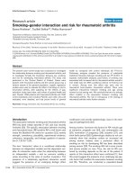
Figure 1
Cytokine networks and cellular interactions in cartilage destruction in rheumatoid arthritis. This scheme represents the progressive destruction of
the cartilage associated with the invading synovial pannus in rheumatoid arthritis. As a result of immune cell interactions involving T and B
lymphocytes, monocyte/macrophages, and dendritic cells, several different cytokines are produced in the inflamed synovium as a result of the influx
of inflammatory cells from the circulation and synovial cell hyperplasia. The upregulation of proinflammatory cytokines produced primarily in the
synovium, but also by chondrocytes, results in the upregulation of cartilage-degrading enzymes, of the matrix metalloproteinase (MMP) and ADAM
with thrombospondin-1 domains (ADAMTS) families, at the cartilage–pannus junction. Chemokines, nitric oxide (NO), and prostaglandins (PGs)
also contribute to the inflammation and tissue catabolism. SDF, stromal cell-derived factor 1; TNF, tumor necrosis factor; TGF, transforming growth
factor; IFN, interferon; Treg, regulatory T lymphocytes; Th, T helper cells.
showing that transgenic or dysregulated overexpression of
the TNF-α in causes polyarthritis in mice, chondrocytes were
found to express the human transgene [69]. When
backcrossed with arthritis-susceptible DBA/1 mice, a more
severe, erosive arthritis developed during successive genera-
tions [70]. Because few chondrocytes remained in older mice
with advanced arthritis and the extracellular matrix of the
cartilage was relatively preserved, it was proposed that the
chondrocytes may die early in the life of the mice by TNF-α-
driven apoptosis before significant proteoglycan degradation
can occur [70]. The higher potency of IL-1 compared with
TNF-α in driving cartilage erosion is supported by studies
showing that blockade of IL-1 is more effective than TNF-α
neutralization in CIA mice [71] and that IL-1 is a secondary
mediator in TNF-α transgenic mice [72]. Later studies in the
human RA/SCID (severe combined immunodeficiency) mouse
chimera indicated that TNF-α is a key molecule in the
inflammatory changes that occur in the rheumatoid synovium,
whereas cartilage damage occurs independently of this
cytokine [73]. Despite these findings in animal models, anti-
TNF therapy in patients with RA has been more successful in
preventing cartilage and bone destruction. This could be
related to the pharmacokinetic properties of IL-1Ra. It has
been suggested that alternative approaches for targeting IL-
1, including the use of soluble receptors and neutralizing
antibodies, need to be tested [67,74]. Supporting the
concept that IL-1 drives cartilage destruction are the findings
of a recent study by Schett’s group in which crossing the
arthritic human TNF transgenic (hTNFtg) mice with mice
deficient in IL-1α and IL-1β protected against cartilage
erosion without affecting synovial inflammation [75].
Cytokine networks
IL-1 and TNF-α can also induce chondrocytes to produce
several other proinflammatory cytokines, including IL-6, leukemia
inhibitory factor (LIF), IL-17, and IL-18, and chemokines
[76,77] (Fig. 1). IL-6 seems to perform a dual function by
increasing products that downregulate inflammation such as
IL-1Ra, soluble TNF receptor (sTNFR), and TIMPs, while also
enhancing immune cell function and inflammation [41,78].
The inhibition of proteoglycan synthesis and other chondro-
cyte responses in vitro require the soluble IL-6 receptor α
(sIL-6Rα), which permits the synergistic stimulation of MMP
expression by IL-1 and IL-6 [79]. IL-6 blockade is under
current investigation in animal models and clinical trials
[80,81]. The use of the IL-6 gene promoter as an inducible
adenoviral gene delivery system proposed for the local
treatment of arthritis would presumably target cartilage
destruction as well as inflammation [82]. Other members of
the IL-6 family that act through receptors that heterodimerize
with gp130 may also modulate chondrocyte function. IL-11
shares several actions of IL-6, including the stimulation of
TIMP production without affecting MMP production [79] and
may actually inhibit cartilage destruction [83]. Leukemia
inhibitory factor (LIF), similarly to the other chondrocyte-
derived autocrine factors described above, may participate in
a positive feedback loop by increasing the production of IL-6
by chondrocytes. Oncostatin M (OSM), which is a product of
macrophages and activated T cells, can act alone or
synergistically with IL-1 to stimulate the production of MMPs
and aggrecanases by chondrocytes [38,79,84]. Direct
evidence supporting a role for OSM in contributing to
cartilage loss in inflammatory arthritis is provided by studies in
animal models [85,86].
IL-17A, one of at least six family members, is primarily a
product of T helper type 17 (Th17) cells, a newly described
subset of T cells, which is a potent inducer of catabolic
responses in chondrocytes by itself or in synergy with other
cytokines [87,88]. IL-17 can drive T-cell-dependent erosive
arthritis in the TNF-deficient and IL-1Ra knockout mice, and
treatment of mice with CIA or AIA with neutralizing IL-17
antibody effectively inhibits cartilage destruction in those
models of RA [89-92].
The IL-1R/Toll-like receptor (TLR) superfamily of receptors
has a key role in innate immunity and inflammation. Studies in
arthritis induced with streptococcal cell wall showed that joint
inflammation and cartilage proteoglycan loss is predominantly
dependent on TLR-2 signaling [93]. Human articular
chondrocytes can express TLR-1, TLR-2, and TLR-4, and
activation of TLR-2 by IL-1, TNF-α, peptidoglycans, lipopoly-
saccharide, or fibronectin fragments increases the production
of MMPs, nitric oxide (NO), prostaglandin E (PGE), and
VEGF [94-96]. In arthritis mediated by immune complex,
TLR-4 regulates early-onset inflammation and cartilage
destruction by IL-10-mediated upregulation of Fcγ receptor
expression and enhanced production of cytokines [97].
Because the IL-18 receptor shares homology with IL-1RI and
has a TLR signaling domain, therapeutic strategies similar to
those for targeting IL-1 signaling have been explored [78,98].
In animal models, IL-18, by means of TLR-2, promotes joint
inflammation in a partly TNF-α-dependent manner and
induces IL-1-driven cartilage destruction [99]. IL-18 has
effects similar to IL-1 in human chondrocytes, and stimulates
chondrocyte apoptosis, although studies do not suggest a
pivotal role in cartilage destruction in RA [100-102]. Of the
other members of the IL-1 family recently identified by DNA
database searches, IL-1F8 seems to be capable of
stimulating the production of IL-6, IL-8, and NO by human
chondrocytes, but at 100-fold to 1,000-fold higher
concentrations than that of IL-1 [103]. IL-32, a recently
discovered cytokine that induces TNF-α, IL-1β, IL-6, and
chemokines and is expressed in the synovia of patients with
RA, contributes to TNF-α-dependent inflammation and a loss
of cartilage proteoglycan [104].
IL-4, IL-10, and IL-13 are generally classified as inhibitory or
modulatory cytokines because they are able to inhibit many of
the cartilage catabolic processes induced by proinflammatory
cytokines [105]. Their therapeutic application has been
proposed to restore the cytokine balance in RA [106,107].
Available online />Page 5 of 13
(page number not for citation purposes)
The efficacy of IL-4, IL-10, and IL-13 in retarding cartilage
damage may be related, in part, to their stimulatory effects on
IL-1Ra production [108,109]. Despite the capacity of IL-4 to
inhibit the effects of proinflammatory cytokines on chondro-
cyte function [110,111], differential effects have been
observed in mice, depending on the model used [112,113].
Gene transfer of IL-10 in combination with IL-1Ra inhibits
cartilage destruction by a mechanism involving activin, a
TGF-β family member [114]. IL-10 is part of the response
induced by immunomodulatory neuropeptides that have
recently been shown to inhibit inflammation and cartilage and
bone destruction by downregulating the Th1-driven immune
response and upregulating IL-10/TGF-β-producing regulatory
T (Treg) lymphocytes [115]. IL-13 decreases the breakdown
of collagen and proteoglycans by inhibiting IL-1- and OSM-
induced MMP-3 and MMP-13 expression [116]. Local gene
transfer of IL-13 inhibits chondrocyte death and MMP-
mediated cartilage degradation despite enhanced inflamma-
tion in the immune-complex arthritis model [117].
Mediators and mechanisms in the responses
of chondrocytes to inflammatory cytokines
In addition to inducing the synthesis of MMPs and other
proteinases by chondrocytes, IL-1 and TNF-α upregulate the
production of NO by means of inducible nitric oxide
synthetase (iNOS, or NOS2), and that of PGE
2
by stimulating
the expression or activities of cyclooxygenase (COX)-2,
microsomal PGE synthase-1 (mPGES-1), and soluble
phospholipase A2 (sPLA2). Although PGE
2
and NO have
been well characterized as proinflammatory mediators, there
is evidence of crosstalk between them in the regulation of
chondrocyte function (reviewed in [118]). COX-2 is also
involved in the chondrocyte response to high shear stress,
associated with decreased antioxidant capacity and
increased apoptosis [119]. In the production of prosta-
glandins, mPGES-1, which is induced by IL-1 in chondro-
cytes, is a major player [120,121]. In addition to opposing the
induction of COX-2, iNOS, and MMPs and the suppression
of aggrecan synthesis by IL-1, activators of the peroxisome
proliferator-activated receptor γ (PPAR-γ), including the endo-
genous ligand 15-deoxy-Δ
12,14
-prostaglandin J
2
(PGJ
2
), inhibit
the IL-1-induced expression of mPGES-1 [122,123]. Recent
evidence indicates that PPAR-α agonists may protect
chondrocytes against IL-1-induced responses by increasing
the expression of IL-1Ra [124].
Adipokines, which were originally identified as products of
adipocytes, have recently been shown to have roles in
cartilage metabolism [125]. White adipose tissue has been
proposed as a major source of both proinflammatory and anti-
inflammatory cytokines, including IL-Ra and IL-10 [126].
Leptin expression is enhanced during acute inflammation,
correlating negatively with inflammatory markers in RA sera
[127], and has been proposed to serve as a link between the
neuroendocrine and immune systems [128]. The elevated
expression of leptin in OA cartilage and in osteophytes, and
its capacity to stimulate insulin-like growth factor (IGF)-1 and
TGF-β1 synthesis, suggest a role for this adipokine in
anabolic responses of chondrocytes [129]. Leptin synergizes
with IL-1 or interferon-γ to increase NO production in
chondrocytes [130], and leptin deficiency attenuates
inflammatory processes in experimental arthritis [131]. It has
been proposed that the dysregulated balance between leptin
and other adipokines, such as adiponectin, promotes
destructive inflammatory processes [132].
Several additional mediators that affect chondrocyte metabo-
lism have been described. The IL-1-induced SOCS3
(suppressor of cytokine signaling 3) acts as a negative
feedback regulator during desensitization toward IGF-1 in the
absence of NO by inhibiting the phosphorylation of insulin
receptor substrate (IRS)-1 [133]. Recent evidence indicates
that RAGE, the receptor for advanced glycation end products
(AGEs), interacts preferentially with S100A4, a member of the
S100 family of calcium-binding proteins, in chondrocytes and
stimulates MMP-13 production through the phosphorylation of
Pyk2, mitogen-activated protein kinases, and NF-κB [134].
The fibroblast activation protein α (FAP-α), a membrane serine
proteinase, which colocalizes in synovium with MMP-1 and
MMP-13 and is induced by IL-1 and OSM in chondrocytes,
may have a role in collagen degradation [135,136]. Many of
these proteins may be activated during the chondrocyte
response to abnormal stimuli and may serve as endogenous
mediators of cellular responses to stress and inflammation.
Signaling mechanisms, gene transcription,
and genome analyses
Signal transduction molecules and transcription factors
activated by inflammatory mediators in chondrocytes and
synovial cells have been studied to identify potential
therapeutic targets. For example, NF-κB is a ‘master switch’
of the inflammatory cascade [137], and the signaling
intermediates in the p38 and JNK pathways have also been
targeted for future therapeutic development [138]. In addition
to NF-κB, members of the CCAAT-enhancer-binding protein
(C/EBP), Ets, and activator protein (AP)-1 families are
important for the regulation of gene expression by IL-1 and
TNF-α [43,139-142] and have been localized in rheumatoid
tissues [143,144]. The JAK/Stat3 signaling pathway is
important for signaling by gp130 cytokines [145]. Cytokine-
induced transcription factors also suppress the expression of
several genes associated with the differentiated chondrocyte
phenotype, including type II collagen (COL2A1), aggrecan,
and CD-RAP [146-148]. Chondrocyte-specific transcription
factors, including Sox9 (which regulates cartilage formation
during development [139]), have not been studied in the
context of cartilage metabolism in RA. Genomic and
proteomic analyses that have been performed in cytokine-
treated chondrocytes, in cartilage from patients with
osteoarthritis, and in rheumatoid synovium have provided
some insights into novel mechanisms that might govern
chondrocyte responses in RA [149-154]. So far, more than
Arthritis Research & Therapy Vol 9 No 5 Otero and Goldring
Page 6 of 13
(page number not for citation purposes)
1,000 differentially expressed transcripts have been identified
in cartilage derived from patients with arthritis [155].
Chemokines
The role of chemokines in RA synovium, where they are
involved in neutrophil activation, chemotaxis, and angio-
genesis, is well established, but their potential contribution to
cartilage metabolism has been recognized only recently
[156-159]. IL-8, probably the most potent and abundant
chemotactic agent in RA synovial fluids, and other chemo-
kines, such as monocyte chemoattractant protein (MCP)-1
and RANTES, are produced primarily by the synovium and
serve as indicators of synovitis. Chondrocytes, when
activated by IL-1 and TNF-α, express several chemokines,
including IL-8, MCP-1, and MCP-4, macrophage inflammatory
protein (MIP)-1α, MIP-1β, RANTES, and GROα, as well as
receptors that enable responses to some of these
chemokines, and may feedback regulate synovial cell
responses [160,161]. High levels of stromal cell-derived
factor 1 (SDF-1) are detected in RA synovial fluids, and its
receptor, CXCR4, is expressed by chondrocytes but not
synovial fibroblasts, suggesting a direct influence of this
chemokine on cartilage damage [162]. Microarray studies
have elucidated several chemokines that are inducible in
chondrocytes by fibronectin fragments and cytokines [154].
Adhesion molecules and angiogenesis
In addition to the requirement of chemokines for the recruit-
ment of T lymphocytes and other inflammatory cells to the
subsynovial lining, adhesion receptors must be available on
synovial blood vessels for binding the circulating leukocytes
and other cell types with which they interact in the inflamed
tissue, including macrophages, dendritic cells, and fibro-
blasts. The principal families of adhesion molecules involved
are the selectins, the integrins, the cadherins, and variants of
the immunoglobulin supergene family. Although these
molecules are common to different inflammatory sites, many
of the prominent adhesion proteins expressed in the inflamed
rheumatoid synovium are also expressed in cartilage. For
example, vascular cell adhesion molecule (VCAM)-1 and
intercellular adhesion molecule (ICAM)-1, which are members
of the immunoglobulin family, are expressed by human
articular chondrocytes as well as synovial and endothelial
cells, although their function on chondrocytes may not be
significant unless damage to the matrix permits cell–cell
interactions [163]. VCAM-1, as well as VEGF, fibroblast
growth factor (FGF), and TNF-α, contributes to angiogenesis
during synovitis and to the activation of chondrocytes during
cartilage degradation [164,165]. VEGF expression is
upregulated by inflammatory cytokines in both chondrocytes
and synovial cells and by hypoxia [166,167], and Vegfb
knockout mice are protected against synovial angiogenesis in
the CIA and AIA models [168].
Several members of the integrin family are expressed by
chondrocytes. The α1β1 and α5β1 integrins function as
receptors for fragments of collagen and fibronectin, respec-
tively. The stimulation of α5β1 integrin by integrin-activating
antibodies or fibronectin fragments results in increased MMP
production and requires reactive oxygen species [169]. In
contrast, the discoidin domain receptor-2 specifically
increases MMP-13 production by recognizing intact type II
collagen fibrils that have been denuded by proteoglycans, as
occurs in osteoarthritis [170,171], but its role in RA has not
been determined. Specific roles for the hyaluronan receptor,
CD44, in cell–matrix interactions in cartilage have also been
identified [172]. CD44 expression is upregulated on
chondrocytes in articular cartilage and synoviocytes from
patients with RA [173,174]. Hyaluronan binding to CD44
increases MMP-13 and NO production by chondrocytes
[175]. Furthermore, induction of MMP-specific cleavage of
type II collagen and NO production by the heparin-binding
fragment of fibronectin is mediated by CD44 [176].
Cadherins are adhesion molecules that mediate cell–cell
adhesion by binding a cadherin of the same cell type on an
adjacent cell. The recent identification of cadherin-11 as a
key adhesion molecule, which regulates the formation of the
synovial lining during development and the synoviocyte
function postnatally, has provided the opportunity to examine
its role in inflammatory joint disease [177]. Cadherin-11
deficiency, or treatment with cadherin-11 antibody or a
cadherin-11 fusion protein, decreased synovial inflammation
and decreased cartilage erosion in an animal model of
arthritis. Furthermore, cadherin-11 facilitated synoviocyte
invasion into cartilage-like extracellular matrix in an in vitro
model, suggesting that this molecule could serve as a
specific target for therapy against cartilage destruction in
inflammatory arthritis [178].
Bone-related factors
The potent induction by IL-17 of receptor activator of NF-κB
ligand (RANKL), which is produced by synoviocytes and
T cells in RA synovium [179] and mediates osteoclast
differentiation and activity, may partly account for the capacity
of IL-17 to induce bone destruction in an IL-1-independent
manner and bypass the requirement for TNF in the
development of inflammatory arthritis [88]. Both RANKL and
its receptor RANK, a member of the TNF receptor family, are
expressed in adult articular chondrocytes [180], but a direct
action in cartilage has not yet been identified. Although
RANKL deficiency blocks bone destruction without direct
effects on cartilage destruction in inflammatory models, it is
possible that indirect cartilage-protective effects may occur
through interference with the degradation of subchondral
bone [179,181,182].
Wnt signaling, through the canonical β-catenin pathway and
activation of T-cell factor (TCF)/Lef transcription factors,
functions in a cell-autonomous manner to induce osteoblast
differentiation and suppress chondrocyte differentiation in
early osteo-chondroprogenitors [183]. During chondro-
Available online />Page 7 of 13
(page number not for citation purposes)
genesis, Wnt/β-catenin acts at two stages, at low levels to
promote chondroprogenitor differentiation and later at high
levels to promote chondrocyte hypertrophic differentiation
and subsequent endochondral ossification [183,184].
Because ectopic Wnt/β-catenin signaling leads to enhanced
ossification and the suppression of cartilage formation during
skeletal development, the disruption of Wnt signaling in adult
cartilage would be expected to have pathological
consequences. For example, activation of β-catenin in mature
cartilage cells stimulates hypertrophy, matrix mineralization,
and expression of VEGF, ADAMTS5, MMP-13, and several
other MMPs [184]. A recent study showed limited expression
of β-catenin in joint tissues of patients with RA, but high
expression of the inhibitor of Wnt/β-catenin signaling, DKK-1,
in the inflamed synovium, especially in the synoviocytes and
synovial microvessels, and in cartilage adjacent to inflam-
matory tissue [185]. This study also showed expression of
DKK-1 in a TNF-α-dependent manner in TNF transgenic mice
and blockade of RANKL-dependent bone resorption by the
administration of DKK-1 antibody, as a result of upregulation
of the RANKL inhibitor osteoprotegerin [185] (reviewed in
[186]).
Conclusion
Significant advances have been made in recent years that
have contributed to our understanding of the cellular
interactions in the RA joint involving macrophages, T and B
lymphocytes, and synovial fibroblasts. Laboratory investi-
gations in vitro and in vivo have resulted in new findings
about the role of the chondrocyte in remodeling the cartilage
matrix in the RA joint. Although the mediators involved in
immunomodulation and synovial cell function, including
cytokines, chemokines, and adhesion molecules, have
primary roles in the inflammatory and catabolic processes in
the joint, they may also promote cartilage damage, directly or
indirectly. Despite the clinical success of anti-TNF therapy for
RA, there is still a need for therapeutic strategies that prevent
the extensive cartilage and bone loss. Recent work that has
identified novel molecules and mechanisms, as well as
providing new understanding of the contributions of known
mediators, offers the possibility of developing new therapies
for targeting cartilage destruction in inflammatory joint
disease.
Competing interests
The authors declare that they have no competing interests.
References
1. Tak PP, Bresnihan B: The pathogenesis and prevention of joint
damage in rheumatoid arthritis: advances from synovial
biopsy and tissue analysis. Arthritis Rheum 2000, 43:2619-
2633.
2. Firestein GS: Evolving concepts of rheumatoid arthritis. Nature
2003, 423:356-361.
3. Meyer LH, Franssen L, Pap T: The role of mesenchymal cells in
the pathophysiology of inflammatory arthritis. Best Pract Res
Clin Rheumatol 2006, 20:969-981.
4. Knedla A, Neumann E, Muller-Ladner U: Developments in the
synovial biology field 2006. Arthritis Res Ther 2007, 9:209.
5. Szekanecz Z, Koch AE: Macrophages and their products in
rheumatoid arthritis. Curr Opin Rheumatol 2007, 19:289-295.
6. Gravallese EM, Harada Y, Wang J-T, Gorn A, Thornhill T, Goldring
SR: Identification of cell types responsible for bone resorption
in rheumatoid arthritis and juvenile rheumatoid arthritis. Am J
Pathol 1998, 152:943-951.
7. Pettit AR, Walsh NC, Manning C, Goldring SR, Gravallese EM:
RANKL protein is expressed at the pannus–bone interface at
sites of articular bone erosion in rheumatoid arthritis.
Rheumatology (Oxford) 2006, 45:1068-1076.
8. Mulherin D, Fitzgerald O, Bresnihan B: Clinical improvement and
radiological deterioration in rheumatoid arthritis: evidence
that the pathogenesis of synovial inflammation and articular
erosion may differ. Br J Rheumatol 1996, 35:1263-1268.
9. Kirwan J, Byron M, Watt I: The relationship between soft tissue
swelling, joint space narrowing and erosive damage in hand
X-rays of patients with rheumatoid arthritis. Rheumatology
(Oxford) 2001, 40:297-301.
10. van den Berg WB, van Riel PL: Uncoupling of inflammation and
destruction in rheumatoid arthritis: myth or reality? Arthritis
Rheum 2005, 52:995-999.
11. Graudal N: The natural history and prognosis of rheumatoid
arthritis: association of radiographic outcome with process
variables, joint motion and immune proteins. Scand J Rheuma-
tol Suppl 2004:1-38.
12. van Lent PL, Grevers L, Lubberts E, de Vries TJ, Nabbe KC,
Verbeek S, Oppers B, Sloetjes A, Blom AB, van den Berg WB:
Fc
γγ
receptors directly mediate cartilage, but not bone,
destruction in murine antigen-induced arthritis: uncoupling of
cartilage damage from bone erosion and joint inflammation.
Arthritis Rheum 2006, 54:3868-3877.
13. Verstappen SM, Poole AR, Ionescu M, King LE, Abrahamowicz M,
Hofman DM, Bijlsma JW, Lafeber FP: Radiographic joint
damage in rheumatoid arthritis is associated with differences
in cartilage turnover and can be predicted by serum biomark-
ers: an evaluation from 1 to 4 years after diagnosis. Arthritis
Res Ther 2006, 8:R31.
14. Goldring SR, Goldring MB: Rheumatoid arthritis and other
inflammatory joint pathologies. In Dynamics of Bone and Carti-
lage Metabolism. 2nd edition. Edited by Seibel MJ, Robins SP,
Bilezikian JP. San Diego: Academic Press; 2006:843-869.
15. Charni-Ben Tabassi N, Garnero P: Monitoring cartilage
turnover. Curr Rheumatol Rep 2007, 9:16-24.
16. Verzijl N, DeGroot J, Thorpe SR, Bank RA, Shaw JN, Lyons TJ,
Bijlsma JW, Lafeber FP, Baynes JW, TeKoppele JM: Effect of col-
lagen turnover on the accumulation of advanced glycation
end products. J Biol Chem 2000, 275:39027-39031.
17. Maroudas A, Bayliss MT, Uchitel-Kaushansky N, Schneiderman R,
Gilav E: Aggrecan turnover in human articular cartilage: use of
aspartic acid racemization as a marker of molecular age. Arch
Biochem Biophys 1998, 350:61-71.
18. Shikhman AR, Brinson DC, Lotz MK: Distinct pathways regulate
facilitated glucose transport in human articular chondrocytes
during anabolic and catabolic responses. Am J Physiol
Endocrinol Metab 2004, 286:E980-E985.
19. Mobasheri A, Richardson S, Mobasheri R, Shakibaei M, Hoyland
JA: Hypoxia inducible factor-1 and facilitative glucose trans-
porters GLUT1 and GLUT3: putative molecular components of
the oxygen and glucose sensing apparatus in articular chon-
drocytes. Histol Histopathol 2005, 20:1327-1338.
20. Wilkins RJ, Browning JA, Ellory JC: Surviving in a matrix: mem-
brane transport in articular chondrocytes. J Membr Biol 2000,
177:95-108.
21. Pufe T, Kurz B, Petersen W, Varoga D, Mentlein R, Kulow S,
Lemke A, Tillmann B: The influence of biomechanical parame-
Arthritis Research & Therapy Vol 9 No 5 Otero and Goldring
Page 8 of 13
(page number not for citation purposes)
This review is part of a series on
Cells of the synovium in rheumatoid arthritis
edited by Gary Firestein.
Other articles in this series can be found at
/>review-series.asp?series=ar_Cells
ters on the expression of VEGF and endostatin in the bone
and joint system. Ann Anat 2005, 187:461-472.
22. Lin C, McGough R, Aswad B, Block JA, Terek R: Hypoxia
induces HIF-1
αα
and VEGF expression in chondrosarcoma
cells and chondrocytes. J Orthop Res 2004, 22:1175-1181.
23. McNulty AL, Stabler TV, Vail TP, McDaniel GE, Kraus VB: Dehy-
droascorbate transport in human chondrocytes is regulated
by hypoxia and is a physiologically relevant source of ascor-
bic acid in the joint. Arthritis Rheum 2005, 52:2676-2685.
24. Robins JC, Akeno N, Mukherjee A, Dalal RR, Aronow BJ,
Koopman P, Clemens TL: Hypoxia induces chondrocyte-spe-
cific gene expression in mesenchymal cells in association
with transcriptional activation of Sox9. Bone 2005, 37:313-
322.
25. Kobayashi I, Ziff M: Electron microscopic studies of the carti-
lage–pannus junction in rheumatoid arthritis. Arthritis Rheum
1975, 18:475-483.
26. Woolley DE, Crossley MJ, Evanson JM: Collagenase at sites of
cartilage erosion in the rheumatoid joint. Arthritis Rheum 1977,
20:1231-1239.
27. Edwards JCW: Fibroblast biology. Development and differenti-
ation of synovial fibroblasts in arthritis. Arthritis Res 2000, 2:
344-347.
28. Kimura H, Tateishi HJ, Ziff M: Surface ultrastructure of rheuma-
toid articular cartilage. Arthritis Rheum 1977, 20:1085-1098.
29. Dodge GR, Poole AR: Immunohistochemical detection and
immunochemical analysis of type II collagen degradation in
human normal, rheumatoid, and osteoarthritic articular carti-
lages and in explants of bovine articular cartilage cultured
with interleukin 1. J Clin Invest 1989, 83:647-661.
30. Mitchell NS, Shepard N: Changes in proteoglycan and collagen
in cartilage in rheumatoid arthritis. J Bone Joint Surg 1978,
60A:349-354.
31. Dodge GR, Pidoux I, Poole AR: The degradation of type II colla-
gen in rheumatoid arthritis: an immunoelectron microscopic
study. Matrix 1991, 11:330-338.
32. Murphy G, Lee MH: What are the roles of metalloproteinases
in cartilage and bone damage? Ann Rheum Dis 2005, 64
(Suppl 4):iv44-iv47.
33. Hembry RM, Bagga MR, Reynolds JJ, Hamblen DL: Immunolo-
calisation studies on six matrix metalloproteinases and their
inhibitors, TIMP-1 and TIMP-2, in synovia from patients with
osteo- and rheumatoid arthritis. Ann Rheum Dis 1995, 54:25-
32.
34. Tetlow LC, Woolley DE: Comparative immunolocalization
studies of collagenase 1 and collagenase 3 production in the
rheumatoid lesion, and by human chondrocytes and synovio-
cytes in vitro. Br J Rheumatol 1998, 37:64-70.
35. Cunnane G, FitzGerald O, Hummel KM, Youssef PP, Gay RE, Gay
S, Bresnihan B: Synovial tissue protease gene expression and
joint erosions in early rheumatoid arthritis. Arthritis Rheum
2001, 44:1744-1753.
36. Woolley DE, Tetlow LC: Observations on the microenviron-
mental nature of cartilage degradation in rheumatoid arthritis.
Ann Rheum Dis 1997, 56:151-161.
37. Ainola MM, Mandelin JA, Liljestrom MP, Li TF, Hukkanen MV,
Konttinen YT: Pannus invasion and cartilage degradation in
rheumatoid arthritis: involvement of MMP-3 and interleukin-
1
ββ
. Clin Exp Rheumatol 2005, 23:644-650.
38. Barksby HE, Milner JM, Patterson AM, Peake NJ, Hui W, Robson
T, Lakey R, Middleton J, Cawston TE, Richards CD, et al.: Matrix
metalloproteinase 10 promotion of collagenolysis via procol-
lagenase activation: implications for cartilage degradation in
arthritis. Arthritis Rheum 2006, 54:3244-3253.
39. Rutkauskaite E, Volkmer D, Shigeyama Y, Schedel J, Pap G,
Muller-Ladner U, Meinecke I, Alexander D, Gay RE, Drynda S, et
al.: Retroviral gene transfer of an antisense construct against
membrane type 1 matrix metalloproteinase reduces the inva-
siveness of rheumatoid arthritis synovial fibroblasts. Arthritis
Rheum 2005, 52:2010-2014.
40. Kevorkian L, Young DA, Darrah C, Donell ST, Shepstone L, Porter
S, Brockbank SM, Edwards DR, Parker AE, Clark IM: Expression
profiling of metalloproteinases and their inhibitors in carti-
lage. Arthritis Rheum 2004, 50:131-141.
41. Cawston TE, Wilson AJ: Understanding the role of tissue
degrading enzymes and their inhibitors in development and
disease. Best Pract Res Clin Rheumatol 2006, 20:983-1002.
42. Patel IR, Attur MG, Patel RN, Stuchin SA, Abagyan RA, Abramson
SB, Amin AR: TNF-
αα
convertase enzyme from human arthritis-
affected cartilage: isolation of cDNA by differential display,
expression of the active enzyme, and regulation of TNF-
αα
. J
Immunol 1998, 160:4570-4579.
43. Burrage PS, Huntington JT, Sporn MB, Brinckerhoff CE: Regula-
tion of matrix metalloproteinase gene expression by a
retinoid X receptor-specific ligand. Arthritis Rheum 2007, 56:
892-904.
44. Overall CM, Blobel CP: In search of partners: linking extracel-
lular proteases to substrates. Nat Rev Mol Cell Biol 2007, 8:
245-257.
45. Arner EC: Aggrecanase-mediated cartilage degradation. Curr
Opin Pharmacol 2002, 2:322-329.
46. Plaas A, Osborn B, Yoshihara Y, Bai Y, Bloom T, Nelson F, Mikecz
K, Sandy JD: Aggrecanolysis in human osteoarthritis: confocal
localization and biochemical characterization of ADAMTS5–
hyaluronan complexes in articular cartilages. Osteoarthritis
Cartilage 2007, 15:719-734.
47. Sandy JD: A contentious issue finds some clarity: on the inde-
pendent and complementary roles of aggrecanase activity
and MMP activity in human joint aggrecanolysis. Osteoarthritis
Cartilage 2006, 14:95-100.
48. Stanton H, Rogerson FM, East CJ, Golub SB, Lawlor KE, Meeker
CT, Little CB, Last K, Farmer PJ, Campbell IK, et al.: ADAMTS5 is
the major aggrecanase in mouse cartilage in vivo and in vitro.
Nature 2005, 434:648-652.
49. Glasson SS, Askew R, Sheppard B, Carito B, Blanchet T, Ma HL,
Flannery CR, Peluso D, Kanki K, Yang Z, et al.: Deletion of active
ADAMTS5 prevents cartilage degradation in a murine model
of osteoarthritis. Nature 2005, 434:644-648.
50. Kashiwagi M, Tortorella M, Nagase H, Brew K: TIMP-3 is a
potent inhibitor of aggrecanase 1 (ADAM-TS4) and aggre-
canase 2 (ADAM-TS5). J Biol Chem 2001, 276:12501-12504.
51. Hui W, Cawston T, Rowan AD: Transforming growth factor
ββ
1
and insulin-like growth factor 1 block collagen degradation
induced by oncostatin M in combination with tumour necrosis
factor
αα
from bovine cartilage. Ann Rheum Dis 2003, 62:172-174.
52. El Mabrouk M, Qureshi HY, Li WQ, Sylvester J, Zafarullah M:
Interleukin-4 antagonizes oncostatin M and transforming
growth factor
ββ
-induced responses in articular chondrocytes.
J Cell Biochem 2007, in press [epub ahead of print, June 1].
53. Yamanishi Y, Firestein GS: Pathogenesis of rheumatoid arthri-
tis: the role of synoviocytes. Rheum Dis Clin North Am 2001,
27:355-371.
54. Nagase H, Kashiwagi M: Aggrecanases and cartilage matrix
degradation. Arthritis Res Ther 2003, 5:94-103.
55. Hou WS, Li W, Keyszer G, Weber E, Levy R, Klein MJ, Gravallese
EM, Goldring SR, Bromme D: Comparison of cathepsins K and
S expression within the rheumatoid and osteoarthritic syn-
ovium. Arthritis Rheum 2002, 46:663-674.
56. Salminen-Mankonen HJ, Morko J, Vuorio E: Role of cathepsin K
in normal joints and in the development of arthritis. Curr Drug
Targets 2007, 8:315-323.
57. Fraser A, Fearon U, Billinghurst RC, Ionescu M, Reece R, Barwick
T, Emery P, Poole AR, Veale DJ: Turnover of type II collagen
and aggrecan in cartilage matrix at the onset of inflammatory
arthritis in humans: relationship to mediators of systemic and
local inflammation. Arthritis Rheum 2003, 48:3085-3095.
58. Lindqvist E, Eberhardt K, Bendtzen K, Heinegard D, Saxne T:
Prognostic laboratory markers of joint damage in rheumatoid
arthritis. Ann Rheum Dis 2005, 64:196-201.
59. Baeten D, Steenbakkers PG, Rijnders AM, Boots AM, Veys EM,
De Keyser F: Detection of major histocompatibility complex/
human cartilage gp-39 complexes in rheumatoid arthritis syn-
ovitis as a specific and independent histologic marker. Arthri-
tis Rheum 2004, 50:444-451.
60. van Bilsen JH, van Dongen H, Lard LR, van der Voort EI, Elferink
DG, Bakker AM, Miltenburg AM, Huizinga TW, de Vries RR, Toes
RE: Functional regulatory immune responses against human
cartilage glycoprotein-39 in health vs. proinflammatory
responses in rheumatoid arthritis. Proc Natl Acad Sci USA
2004, 101:17180-17185.
61. Ling H, Recklies AD: The chitinase 3-like protein human carti-
lage glycoprotein 39 inhibits cellular responses to the inflam-
matory cytokines interleukin-1 and tumour necrosis factor-
αα
.
Biochem J 2004, 380:651-659.
Available online />Page 9 of 13
(page number not for citation purposes)
62. Recklies AD, Ling H, White C, Bernier SM: Inflammatory
cytokines induce production of CHI3L1 by articular chondro-
cytes. J Biol Chem 2005, 280:41213-41221.
63. Sekine T, Masuko-Hongo K, Matsui T, Asahara H, Takigawa M,
Nishioka K, Kato T: Recognition of YKL-39, a human cartilage
related protein, as a target antigen in patients with rheuma-
toid arthritis. Ann Rheum Dis 2001, 60:49-54.
64. Knorr T, Obermayr F, Bartnik E, Zien A, Aigner T: YKL-39 (chiti-
nase 3-like protein 2), but not YKL-40 (chitinase 3-like protein
1), is up regulated in osteoarthritic chondrocytes. Ann Rheum
Dis 2003, 62:995-998.
65. Saito S, Kondo S, Mishima S, Ishiguro N, Hasegawa Y, Sandell
LJ, Iwata H: Analysis of cartilage-derived retinoic-acid-sensi-
tive protein (CD-RAP) in synovial fluid from patients with
osteoarthritis and rheumatoid arthritis. J Bone Joint Surg Br
2002, 84:1066-1069.
66. Dayer JM: The process of identifying and understanding
cytokines: from basic studies to treating rheumatic diseases.
Best Pract Res Clin Rheumatol 2004, 18:31-45.
67. van den Berg WB: Animal models of arthritis. What have we
learned? J Rheumatol Suppl 2005, 72:7-9.
68. Horai R, Saijo S, Tanioka J, Nakae S, Sudo K, Okahara A, Ikuse T,
Asano M, Iwakura Y: Development of chronic inflammatory
arthropathy resembling rheumatoid arthritis in interleukin 1
receptor antagonist-deficient mice. J Exp Med 2000, 191:313-
320.
69. Keffer J, Probert L, Cazlaris H, Georgopoulos S, Kaslaris E, Kious-
sis D, Kollias G: Transgenic mice expressing human tumour
necrosis factor: a predictive genetic model of arthritis. EMBO
J 1991, 10:4025-4031.
70. Butler DM, Malfait AM, Mason LJ, Warden PJ, Kollias G, Maini RN,
Feldmann M, Brennan FM: DBA/1 mice expressing the human
TNF-
αα
transgene develop a severe, erosive arthritis: charac-
terization of the cytokine cascade and cellular composition. J
Immunol 1997, 159:2867-2876.
71. Joosten LA, Helsen MM, van de Loo FA, van den Berg WB: Anti-
cytokine treatment of established type II collagen-induced
arthritis in DBA/1 mice. A comparative study using anti-TNF
αα
,
anti-IL-1
αα
/
ββ
, and IL-1Ra. Arthritis Rheum 1996, 39:797-809.
72. Probert L, Plows D, Kontogeorgos G, Kollias G: The type I inter-
leukin-1 receptor acts in series with tumor necrosis factor
(TNF) to induce arthritis in TNF-transgenic mice. Eur J
Immunol 1995, 25:1794-1797.
73. Matsuno H, Yudoh K, Katayama R, Nakazawa F, Uzuki M, Sawai T,
Yonezawa T, Saeki Y, Panayi GS, Pitzalis C, et al.: The role of
TNF-
αα
in the pathogenesis of inflammation and joint destruc-
tion in rheumatoid arthritis (RA): a study using a human RA/
SCID mouse chimera. Rheumatology (Oxford) 2002, 41:329-
337.
74. Burger D, Dayer JM, Palmer G, Gabay C: Is IL-1 a good thera-
peutic target in the treatment of arthritis? Best Pract Res Clin
Rheumatol 2006, 20:879-896.
75. Zwerina J, Redlich K, Polzer K, Joosten L, Kroenke G, Distler J,
Hess A, Pundt N, Pap T, Hoffmann O, et al.: TNF-induced struc-
tural joint damage is mediated by IL-1. Proc Natl Acad Sci
USA 2007, 104:11742-11747.
76. Goldring SR, Goldring MB: The role of cytokines in cartilage
matrix degeneration in osteoarthritis. Clin Orthop Relat Res
2004, 427(Suppl):S27-S36.
77. Aida Y, Maeno M, Suzuki N, Namba A, Motohashi M, Matsumoto
M, Makimura M, Matsumura H: The effect of IL-1
ββ
on the
expression of inflammatory cytokines and their receptors in
human chondrocytes. Life Sci 2006, 79:764-771.
78. Connell L, McInnes IB: New cytokine targets in inflammatory
rheumatic diseases. Best Pract Res Clin Rheumatol 2006, 20:
865-878.
79. Rowan AD, Koshy PJ, Shingleton WD, Degnan BA, Heath JK, Ver-
nallis AB, Spaull JR, Life PF, Hudson K, Cawston TE: Synergistic
effects of glycoprotein 130 binding cytokines in combination
with interleukin-1 on cartilage collagen breakdown. Arthritis
Rheum 2001, 44:1620-1632.
80. Choy E: Clinical experience with inhibition of interleukin-6.
Rheum Dis Clin North Am 2004, 30:405-415.
81. Nishimoto N, Kishimoto T: Interleukin 6: from bench to bedside.
Nat Clin Pract Rheumatol 2006, 2:619-626.
82. van de Loo FA, de Hooge AS, Smeets RL, Bakker AC, Bennink
MB, Arntz OJ, Joosten LA, van Beuningen HM, van der Kraan PK,
Varley AW, et al.: An inflammation-inducible adenoviral
expression system for local treatment of the arthritic joint.
Gene Ther 2004, 11:581-590.
83. Sack U, Sehm B, Kahlenberg F, Murr A, Lehmann J, Tannapfel A,
Uberla K, Moessner A, Dietrich A, Emmrich F, et al.: Investigation
of arthritic joint destruction by a novel fibroblast-based
model. Ann NY Acad Sci 2005, 1051:291-298.
84. Koshy PJ, Lundy CJ, Rowan AD, Porter S, Edwards DR, Hogan A,
Clark IM, Cawston TE: The modulation of matrix metallopro-
teinase and ADAM gene expression in human chondrocytes
by interleukin-1 and oncostatin M: a time-course study using
real-time quantitative reverse transcription-polymerase chain
reaction. Arthritis Rheum 2002, 46:961-967.
85. Plater-Zyberk C, Buckton J, Thompson S, Spaull J, Zanders E,
Papworth J, Life PF: Amelioration of arthritis in two murine
models using antibodies to oncostatin M. Arthritis Rheum
2001, 44:2697-2702.
86. Rowan AD, Hui W, Cawston TE, Richards CD: Adenoviral gene
transfer of interleukin-1 in combination with oncostatin M
induces significant joint damage in a murine model. Am J
Pathol 2003, 162:1975-1984.
87. Koshy PJ, Henderson N, Logan C, Life PF, Cawston TE, Rowan
AD: Interleukin 17 induces cartilage collagen breakdown:
novel synergistic effects in combination with proinflammatory
cytokines. Ann Rheum Dis 2002, 61:704-713.
88. Lubberts E, Koenders MI, van den Berg WB: The role of T cell
interleukin-17 in conducting destructive arthritis: lessons
from animal models. Arthritis Res Ther 2005, 7:29-37.
89. Lubberts E, Koenders MI, Oppers-Walgreen B, van den Bersse-
laar L, Coenen-de Roo CJ, Joosten LA, van den Berg WB: Treat-
ment with a neutralizing anti-murine interleukin-17 antibody
after the onset of collagen-induced arthritis reduces joint
inflammation, cartilage destruction, and bone erosion. Arthritis
Rheum 2004, 50:650-659.
90. Koenders MI, Lubberts E, Oppers-Walgreen B, van den Bersse-
laar L, Helsen MM, Di Padova FE, Boots AM, Gram H, Joosten LA,
van den Berg WB: Blocking of interleukin-17 during reactiva-
tion of experimental arthritis prevents joint inflammation and
bone erosion by decreasing RANKL and interleukin-1. Am J
Pathol 2005, 167:141-149.
91. Koenders MI, Joosten LA, van den Berg WB: Potential new
targets in arthritis therapy: interleukin (IL)-17 and its relation
to tumour necrosis factor and IL-1 in experimental arthritis.
Ann Rheum Dis 2006, 65 (Suppl 3):iii29-iii33.
92. Koenders MI, Lubberts E, van de Loo FA, Oppers-Walgreen B,
van den Bersselaar L, Helsen MM, Kolls JK, Di Padova FE,
Joosten LA, van den Berg WB: Interleukin-17 acts indepen-
dently of TNF-
αα
under arthritic conditions. J Immunol 2006,
176:6262-6269.
93. Joosten LA, Koenders MI, Smeets RL, Heuvelmans-Jacobs M,
Helsen MM, Takeda K, Akira S, Lubberts E, van de Loo FA, van
den Berg WB: Toll-like receptor 2 pathway drives streptococ-
cal cell wall-induced joint inflammation: critical role of myeloid
differentiation factor 88. J Immunol 2003, 171:6145-6153.
94. Su SL, Tsai CD, Lee CH, Salter DM, Lee HS: Expression and
regulation of Toll-like receptor 2 by IL-1
ββ
and fibronectin frag-
ments in human articular chondrocytes. Osteoarthritis Carti-
lage 2005, 13:879-886.
95. Kim HA, Cho ML, Choi HY, Yoon CS, Jhun JY, Oh HJ, Kim HY:
The catabolic pathway mediated by Toll-like receptors in
human osteoarthritic chondrocytes. Arthritis Rheum 2006, 54:
2152-2163.
96. Varoga D, Paulsen F, Mentlein R, Fay J, Kurz B, Schutz R, Wruck
C, Goldring MB, Pufe T: TLR-2-mediated induction of vascular
endothelial growth factor (VEGF) in cartilage in septic joint
disease. J Pathol 2006, 210:315-324.
97. van Lent PL, Blom AB, Grevers L, Sloetjes A, van den Berg WB:
Toll-like receptor 4 induced Fc
γγ
R expression potentiates early
onset of joint inflammation and cartilage destruction during
immune complex arthritis: Toll-like receptor 4 largely regu-
lates Fc
γγ
R expression by interleukin 10. Ann Rheum Dis 2007,
66:334-340.
98. Asquith DL, McInnes IB: Emerging cytokine targets in rheuma-
toid arthritis. Curr Opin Rheumatol 2007, 19:246-251.
99. Joosten LA, Smeets RL, Koenders MI, van den Bersselaar LA,
Helsen MM, Oppers-Walgreen B, Lubberts E, Iwakura Y, van de
Loo FA, van den Berg WB: Interleukin-18 promotes joint
Arthritis Research & Therapy Vol 9 No 5 Otero and Goldring
Page 10 of 13
(page number not for citation purposes)
inflammation and induces interleukin-1-driven cartilage
destruction. Am J Pathol 2004, 165:959-967.
100. Matsui K, Tsutsui H, Nakanishi K: Pathophysiological roles for
IL-18 in inflammatory arthritis. Expert Opin Ther Targets 2003,
7:701-724.
101. Dai SM, Shan ZZ, Nishioka K, Yudoh K: Implication of inter-
leukin 18 in production of matrix metalloproteinases in articu-
lar chondrocytes in arthritis: direct effect on chondrocytes
may not be pivotal. Ann Rheum Dis 2005, 64:735-742.
102. John T, Kohl B, Mobasheri A, Ertel W, Shakibaei M: Interleukin-
18 induces apoptosis in human articular chondrocytes. Histol
Histopathol 2007, 22:469-482.
103. Magne D, Palmer G, Barton JL, Mezin F, Talabot-Ayer D, Bas S,
Duffy T, Noger M, Guerne PA, Nicklin MJ, et al.: The new IL-1
family member IL-1F8 stimulates production of inflammatory
mediators by synovial fibroblasts and articular chondrocytes.
Arthritis Res Ther 2006, 8:R80.
104. Joosten LA, Netea MG, Kim SH, Yoon DY, Oppers-Walgreen B,
Radstake TR, Barrera P, van de Loo FA, Dinarello CA, van den
Berg WB: IL-32, a proinflammatory cytokine in rheumatoid
arthritis. Proc Natl Acad Sci USA 2006, 103:3298-3303.
105. Bessis N, Boissier MC: Novel pro-inflammatory interleukins:
potential therapeutic targets in rheumatoid arthritis. Joint
Bone Spine 2001, 68:477-481.
106. van de Loo FA, Smeets RL, van den Berg WB: Gene therapy in
animal models of rheumatoid arthritis: are we ready for the
patients? Arthritis Res Ther 2004, 6:183-196.
107. van de Loo FA, Geurts J, van den Berg WB: Gene therapy works
in animal models of rheumatoid arthritis … so what! Curr
Rheumatol Rep 2006, 8:386-393.
108. Palmer G, Guerne PA, Mezin F, Maret M, Guicheux J, Goldring MB,
Gabay C: Production of interleukin-1 receptor antagonist by
human articular chondrocytes. Arthritis Res 2002, 4:226-231.
109. Fernandes JC, Martel-Pelletier J, Pelletier JP: The role of
cytokines in osteoarthritis pathophysiology. Biorheology 2002,
39:237-246.
110. Chowdhury TT, Bader DL, Lee DA: Anti-inflammatory effects of
IL-4 and dynamic compression in IL-1
ββ
stimulated chondro-
cytes. Biochem Biophys Res Commun 2006, 339:241-247.
111. Schuerwegh AJ, Dombrecht EJ, Stevens WJ, Van Offel JF, Bridts
CH, De Clerck LS: Influence of pro-inflammatory (IL-1
αα
, IL-6,
TNF-
αα
, IFN-
γγ
) and anti-inflammatory (IL-4) cytokines on chon-
drocyte function. Osteoarthritis Cartilage 2003, 11:681-687.
112. Nandakumar KS, Holmdahl R: Arthritis induced with cartilage-
specific antibodies is IL-4-dependent. Eur J Immunol 2006, 36:
1608-1618.
113. Ho SH, Hahn W, Lee HJ, Kim DS, Jeong JG, Kim S, Yu SS, Jeon
ES, Kim JM: Protection against collagen-induced arthritis by
electrotransfer of an expression plasmid for the interleukin-4.
Biochem Biophys Res Commun 2004, 321:759-766.
114. Neumann E, Judex M, Kullmann F, Grifka J, Robbins PD, Pap T,
Gay RE, Evans CH, Gay S, Scholmerich J, et al.: Inhibition of
cartilage destruction by double gene transfer of IL-1Ra and
IL-10 involves the activin pathway. Gene Ther 2002, 9:1508-
1519.
115. Gonzalez-Rey E, Chorny A, Varela N, O’Valle F, Delgado M: Ther-
apeutic effect of urocortin on collagen-induced arthritis by
down-regulation of inflammatory and Th1 responses and
induction of regulatory T cells. Arthritis Rheum 2007, 56:531-
543.
116. Cleaver CS, Rowan AD, Cawston TE: Interleukin 13 blocks the
release of collagen from bovine nasal cartilage treated with
proinflammatory cytokines. Ann Rheum Dis 2001, 60:150-157.
117. Nabbe KC, van Lent PL, Holthuysen AE, Sloetjes AW, Koch AE,
Radstake TR, van den Berg WB: Local IL-13 gene transfer prior
to immune-complex arthritis inhibits chondrocyte death and
matrix-metalloproteinase-mediated cartilage matrix degrada-
tion despite enhanced joint inflammation. Arthritis Res Ther
2005, 7:R392-R401.
118. Goldring MB, Berenbaum F: The regulation of chondrocyte
function by proinflammatory mediators: prostaglandins and
nitric oxide. Clin Orthop Relat Res 2004, 427(Suppl):S37-S46.
119. Healy ZR, Lee NH, Gao X, Goldring MB, Talalay P, Kensler TW,
Konstantopoulos K: Divergent responses of chondrocytes and
endothelial cells to shear stress: cross-talk among COX-2, the
phase 2 response, and apoptosis. Proc Natl Acad Sci USA
2005.
120. Masuko-Hongo K, Berenbaum F, Humbert L, Salvat C, Goldring
MB, Thirion S: Up-regulation of microsomal prostaglandin E
synthase 1 in osteoarthritic human cartilage: critical roles of
the ERK-1/2 and p38 signaling pathways. Arthritis Rheum
2004, 50:2829-2838.
121. Whiteman M, Spencer JP, Zhu YZ, Armstrong JS, Schantz JT:
Peroxynitrite-modified collagen-II induces p38/ERK and NF-
κκ
B-dependent synthesis of prostaglandin E2 and nitric oxide
in chondrogenically differentiated mesenchymal progenitor
cells. Osteoarthritis Cartilage 2006, 14:460-470.
122. Li X, Afif H, Cheng S, Martel-Pelletier J, Pelletier JP, Ranger P,
Fahmi H: Expression and regulation of microsomal
prostaglandin E synthase-1 in human osteoarthritic cartilage
and chondrocytes. J Rheumatol 2005, 32:887-895.
123. Cheng S, Afif H, Martel-Pelletier J, Pelletier JP, Li X, Farrajota K,
Lavigne M, Fahmi H: Activation of peroxisome proliferator-acti-
vated receptor
γγ
inhibits interleukin-1
ββ
-induced membrane-
associated prostaglandin E2 synthase-1 expression in human
synovial fibroblasts by interfering with Egr-1. J Biol Chem
2004, 279:22057-22065.
124. Francois M, Richette P, Tsagris L, Fitting C, Lemay C, Benallaoua
M, Tahiri K, Corvol MT: Activation of the peroxisome prolifera-
tor-activated receptor
αα
pathway potentiates interleukin-1
receptor antagonist production in cytokine-treated chondro-
cytes. Arthritis Rheum 2006, 54:1233-1245.
125. Loeser RF: Systemic and local regulation of articular cartilage
metabolism: where does leptin fit in the puzzle? Arthritis
Rheum 2003, 48:3009-3012.
126. Dayer JM, Chicheportiche R, Juge-Aubry C, Meier C: Adipose
tissue has anti-inflammatory properties: focus on IL-1 recep-
tor antagonist (IL-1Ra). Ann N Y Acad Sci 2006, 1069:444-453.
127. Popa C, Netea MG, Radstake TR, van Riel PL, Barrera P, van der
Meer JW: Markers of inflammation are negatively correlated
with serum leptin in rheumatoid arthritis. Ann Rheum Dis
2005, 64:1195-1198.
128. Otero M, Lago R, Gomez R, Dieguez C, Lago F, Gomez-Reino J,
Gualillo O: Towards a pro-inflammatory and immunomodula-
tory emerging role of leptin. Rheumatology (Oxford) 2006, 45:
944-950.
129. Dumond H, Presle N, Terlain B, Mainard D, Loeuille D, Netter P,
Pottie P: Evidence for a key role of leptin in osteoarthritis.
Arthritis Rheum 2003, 48:3118-3129.
130. Otero M, Lago R, Lago F, Reino JJ, Gualillo O: Signalling
pathway involved in nitric oxide synthase type II activation in
chondrocytes: synergistic effect of leptin with interleukin-1.
Arthritis Res Ther 2005, 7:R581-R591.
131. Palmer G, Aurrand-Lions M, Contassot E, Talabot-Ayer D,
Ducrest-Gay D, Vesin C, Chobaz-Peclat V, Busso N, Gabay C:
Indirect effects of leptin receptor deficiency on lymphocyte
populations and immune response in db/db mice. J Immunol
2006, 177:2899-2907.
132. Lago F, Dieguez C, Gomez-Reino JJ, Gualillo O: The emerging
role of adipokines as mediators of inflammation and immune
responses. Cytokine Growth Factor Rev 2007, 18:313-325.
133. Smeets RL, Veenbergen S, Arntz OJ, Bennink MB, Joosten LA,
van den Berg WB, van de Loo FA: A novel role for suppressor
of cytokine signaling 3 in cartilage destruction via induction of
chondrocyte desensitization toward insulin-like growth factor.
Arthritis Rheum 2006, 54:1518-1528.
134. Yammani RR, Carlson CS, Bresnick AR, Loeser RF: Increase in
production of matrix metalloproteinase 13 by human articular
chondrocytes due to stimulation with S100A4: role of the
receptor for advanced glycation end products. Arthritis Rheum
2006, 54:2901-2911.
135. Bauer S, Jendro MC, Wadle A, Kleber S, Stenner F, Dinser R,
Reich A, Faccin E, Godde S, Dinges H, et al.: Fibroblast activa-
tion protein is expressed by rheumatoid myofibroblast-like
synoviocytes. Arthritis Res Ther 2006, 8:R171.
136. Milner JM, Kevorkian L, Young DA, Jones D, Wait R, Donell ST,
Barksby E, Patterson AM, Middleton J, Cravatt BF, et al.: Fibrob-
last activation protein
αα
is expressed by chondrocytes follow-
ing a pro-inflammatory stimulus and is elevated in
osteoarthritis. Arthritis Res Ther 2006, 8:R23.
137. Firestein GS: NF-
κκ
B: Holy Grail for rheumatoid arthritis? Arthri-
tis Rheum 2004, 50:2381-2386.
138. Vincenti MP, Brinckerhoff CE: Transcriptional regulation of col-
lagenase (MMP-1, MMP-13) genes in arthritis: integration of
Available online />Page 11 of 13
(page number not for citation purposes)
complex signaling pathways for the recruitment of gene-spe-
cific transcription factors. Arthritis Res 2002, 4:157-164.
139. Imamura T, Imamura C, Iwamoto Y, Sandell LJ: Transcriptional
co-activators CREB-binding protein/p300 increase chondro-
cyte Cd-rap gene expression by multiple mechanisms includ-
ing sequestration of the repressor CCAAT/enhancer-binding
protein. J Biol Chem 2005, 280:16625-16634.
140. Grall FT, Prall WC, Wei W, Gu X, Cho JY, Choy BK, Zerbini LF,
Inan MS, Goldring SR, Gravallese EM, et al.: The Ets transcrip-
tion factor ESE-1 mediates induction of the COX-2 gene by
LPS in monocytes. FEBS J 2005, 272:1676-1687.
141. Raymond L, Eck S, Mollmark J, Hays E, Tomek I, Kantor S, Elliott
S, Vincenti M: Interleukin-1
ββ
induction of matrix metallopro-
teinase-1 transcription in chondrocytes requires ERK-depen-
dent activation of CCAAT enhancer-binding protein-
ββ
. J Cell
Physiol 2006, 207:683-688.
142. Rannou F, Francois M, Corvol MT, Berenbaum F: Cartilage
breakdown in rheumatoid arthritis. Joint Bone Spine 2006, 73:
29-36.
143. Grall F, Gu X, Tan L, Cho J-Y, Inan MS, Pettit A, Thamrongsak U,
Choy BK, Manning C, Akbarali Y, et al.: Responses to the pro-
inflammatory cytokines interleukin-1 and tumor necrosis
factor
αα
in cells derived from rheumatoid synovium and other
joint tissues involve NF
κκ
B-mediated induction of the Ets tran-
scription factor ESE-1. Arthritis Rheum 2003, 48:1249-1260.
144. Benito MJ, Murphy E, Murphy EP, van den Berg WB, FitzGerald
O, Bresnihan B: Increased synovial tissue NF-
κκ
B1 expression
at sites adjacent to the cartilage–pannus junction in rheuma-
toid arthritis. Arthritis Rheum 2004, 50:1781-1787.
145. Katoh M: STAT3-induced WNT5A signaling loop in embryonic
stem cells, adult normal tissues, chronic persistent inflamma-
tion, rheumatoid arthritis and cancer [review]. Int J Mol Med
2007, 19:273-278.
146. Okazaki K, Li J, Yu H, Fukui N, Sandell LJ: CCAAT/Enhancer-
binding proteins
ββ
and
δδ
mediate the repression of gene tran-
scription of cartilage-derived retinoic acid-sensitive protein
induced by interleukin-1
ββ
. J Biol Chem 2002, 277:31526-31533.
147. Tan L, Peng H, Osaki M, Choy BK, Auron PE, Sandell LJ, Goldring
MB: Egr-1 mediates transcriptional repression of COL2A1
promoter activity by interleukin-1
ββ
. J Biol Chem 2003, 278:
17688-17700.
148. Seguin CA, Bernier SM: TNF
αα
suppresses link protein and type
II collagen expression in chondrocytes: role of MEK1/2 and
NF-
κκ
B signaling pathways. J Cell Physiol 2003, 197:356-369.
149. Heller RA, Schena M, Chai A, Shalon D, Bedilion T, Gilmore J,
Woolley DE, Davis RW: Discovery and analysis of inflammatory
disease-related genes using cDNA microarrays. Proc Natl
Acad Sci USA 1997, 94:2150-2155.
150. Vincenti MP, Brinckerhoff CE: Early response genes induced in
chondrocytes stimulated with the inflammatory cytokine inter-
leukin-1
ββ
. Arthritis Res 2001, 3:381-388.
151. Barksby HE, Hui W, Wappler I, Peters HH, Milner JM, Richards
CD, Cawston TE, Rowan AD: Interleukin-1 in combination with
oncostatin M up-regulates multiple genes in chondrocytes:
implications for cartilage destruction and repair. Arthritis
Rheum 2006, 54:540-550.
152. Aigner T, Fundel K, Saas J, Gebhard PM, Haag J, Weiss T, Zien A,
Obermayr F, Zimmer R, Bartnik E: Large-scale gene expression
profiling reveals major pathogenetic pathways of cartilage
degeneration in osteoarthritis. Arthritis Rheum 2006, 54:3533-
3544.
153. Sato T, Konomi K, Yamasaki S, Aratani S, Tsuchimochi K, Yok-
ouchi M, Masuko-Hongo K, Yagishita N, Nakamura H, Komiya S,
et al.: Comparative analysis of gene expression profiles in
intact and damaged regions of human osteoarthritic cartilage.
Arthritis Rheum 2006, 54:808-817.
154. Pulai JI, Chen H, Im HJ, Kumar S, Hanning C, Hegde PS, Loeser
RF: NF-
κκ
B mediates the stimulation of cytokine and chemo-
kine expression by human articular chondrocytes in response
to fibronectin fragments. J Immunol 2005, 174:5781-5788.
155. Attur MG, Dave MN, Akamatsu M, Katoh M, Amin AR: A system
biology approach to bioinformatics and functional genomics
in complex human diseases: arthritis. Curr Issues Mol Biol
2002, 4:129-146.
156. Goldring MB, Goldring SR: Role of cytokines and chemokines
in cartilage and bone destruction in arthritis. Curr Opin
Orthopaed 2002, 13:351-362.
157. Koch AE: Chemokines and their receptors in rheumatoid
arthritis: future targets? Arthritis Rheum 2005, 52:710-721.
158. Vergunst CE, van de Sande MG, Lebre MC, Tak PP: The role of
chemokines in rheumatoid arthritis and osteoarthritis. Scand
J Rheumatol 2005, 34:415-425.
159. Nakamura H, Masuko K, Yudoh K, Kato T, Nishioka K: Effects of
celecoxib on human chondrocytes – enhanced production of
chemokines. Clin Exp Rheumatol 2007, 25:11-16.
160. Borzi RM, Mazzetti I, Marcu KB, Facchini A: Chemokines in carti-
lage degradation. Clin Orthop Relat Res 2004, 427(Suppl):
S53-S61.
161. Iwamoto T, Okamoto H, Iikuni N, Takeuchi M, Toyama Y, Tomatsu
T, Kamatani N, Momohara S: Monocyte chemoattractant
protein-4 (MCP-4)/CCL13 is highly expressed in cartilage
from patients with rheumatoid arthritis. Rheumatology (Oxford)
2006, 45:421-424.
162. Kanbe K, Takemura T, Takeuchi K, Chen Q, Takagishi K, Inoue K:
Synovectomy reduces stromal-cell-derived factor-1 (SDF-1)
which is involved in the destruction of cartilage in osteoarthri-
tis and rheumatoid arthritis. J Bone Joint Surg Br 2004, 86:
296-300.
163. Green DM, Noble PC, Ahuero JS, Birdsall HH: Cellular events
leading to chondrocyte death after cartilage impact injury.
Arthritis Rheum 2006, 54:1509-1517.
164. Fearon U, Griosios K, Fraser A, Reece R, Emery P, Jones PF,
Veale DJ: Angiopoietins, growth factors, and vascular mor-
phology in early arthritis. J Rheumatol 2003, 30:260-268.
165. Szekanecz Z, Gaspar L, Koch AE: Angiogenesis in rheumatoid
arthritis. Front Biosci 2005, 10:1739-1753.
166. Honorati MC, Neri S, Cattini L, Facchini A: Interleukin-17, a reg-
ulator of angiogenic factor release by synovial fibroblasts.
Osteoarthritis Cartilage 2005, 14:345-352.
167. Fearon U, Mullan R, Markham T, Connolly M, Sullivan S, Poole AR,
FitzGerald O, Bresnihan B, Veale DJ: Oncostatin M induces
angiogenesis and cartilage degradation in rheumatoid arthri-
tis synovial tissue and human cartilage cocultures. Arthritis
Rheum 2006, 54:3152-3162.
168. Mould AW, Tonks ID, Cahill MM, Pettit AR, Thomas R, Hayward
NK, Kay GF: Vegfb gene knockout mice display reduced
pathology and synovial angiogenesis in both antigen-induced
and collagen-induced models of arthritis. Arthritis Rheum
2003, 48:2660-2669.
169. Del Carlo M, Schwartz D, Erickson EA, Loeser RF: Endogenous
production of reactive oxygen species is required for stimula-
tion of human articular chondrocyte matrix metalloproteinase
production by fibronectin fragments. Free Radic Biol Med
2007, 42:1350-1358.
170. Xu L, Peng H, Wu D, Hu K, Goldring MB, Olsen BR, Li Y: Activa-
tion of the discoidin domain receptor 2 induces expression of
matrix metalloproteinase 13 associated with osteoarthritis in
mice. J Biol Chem 2005, 280:548-555.
171. Hu K, Xu L, Cao L, Flahiff CM, Brussiau J, Ho K, Setton LA, Youn
I, Guilak F, Olsen BR, et al.: Pathogenesis of osteoarthritis-like
changes in the joints of mice deficient in type IX collagen.
Arthritis Rheum 2006, 54:2891-2900.
172. Knudson CB, Knudson W: Hyaluronan and CD44: modulators
of chondrocyte metabolism. Clin Orthop Relat Res 2004, 427
(Suppl):S152-S162.
173. Takagi T, Okamoto R, Suzuki K, Hayashi T, Sato M, Kurosaka N,
Koshino T: Up-regulation of CD44 in rheumatoid chondro-
cytes. Scand J Rheumatol 2001, 30:110-113.
174. Naor D, Nedvetzki S: CD44 in rheumatoid arthritis. Arthritis Res
Ther 2003, 5:105-115.
175. Ohno S, Im HJ, Knudson CB, Knudson W: Hyaluronan oligosac-
charides induce matrix metalloproteinase 13 via transcrip-
tional activation of NF
κκ
B and p38 MAP kinase in articular
chondrocytes. J Biol Chem 2006, 281:17952-17960.
176. Yasuda T, Kakinuma T, Julovi SM, Yoshida M, Hiramitsu T,
Akiyoshi M, Nakamura T: COOH-terminal heparin-binding
fibronectin fragment induces nitric oxide production in
rheumatoid cartilage through CD44. Rheumatology (Oxford)
2004, 43:1116-1120.
177. Valencia X, Higgins JM, Kiener HP, Lee DM, Podrebarac TA,
Dascher CC, Watts GF, Mizoguchi E, Simmons B, Patel DD, et
al.: Cadherin-11 provides specific cellular adhesion between
fibroblast-like synoviocytes. J Exp Med 2004, 200:1673-1679.
178. Lee DM, Kiener HP, Agarwal SK, Noss EH, Watts GF, Chisaka O,
Arthritis Research & Therapy Vol 9 No 5 Otero and Goldring
Page 12 of 13
(page number not for citation purposes)
Takeichi M, Brenner MB: Cadherin-11 in synovial lining forma-
tion and pathology in arthritis. Science 2007, 315:1006-1010.
179. Kong YY, Yoshida H, Sarosi I, Tan HL, Timms E, Capparelli C,
Morony S, Oliveira-dos-Santos AJ, Van G, Itie A, et al.: OPGL is a
key regulator of osteoclastogenesis, lymphocyte develop-
ment and lymph-node organogenesis. Nature 1999, 397:315-
323.
180. Komuro H, Olee T, Kuhn K, Quach J, Brinson DC, Shikhman A,
Valbracht J, Creighton-Achermann L, Lotz M: The osteoprote-
gerin/receptor activator of nuclear factor
κκ
B/receptor activa-
tor of nuclear factor
κκ
B ligand system in cartilage. Arthritis
Rheum 2001, 44:2768-2776.
181. Pettit AR, Ji H, von Stechow D, Muller R, Goldring SR, Choi Y,
Benoist C, Gravallese EM: TRANCE/RANKL knockout mice are
protected from bone erosion in a serum transfer model of
arthritis. Am J Pathol 2001, 159:1689-1699.
182. Zwerina J, Hayer S, Redlich K, Bobacz K, Kollias G, Smolen JS,
Schett G: Activation of p38 MAPK is a key step in tumor
necrosis factor-mediated inflammatory bone destruction.
Arthritis Rheum 2006, 54:463-472.
183. Day TF, Guo X, Garrett-Beal L, Yang Y: Wnt/
ββ
-catenin signaling
in mesenchymal progenitors controls osteoblast and chon-
drocyte differentiation during vertebrate skeletogenesis. Dev
Cell 2005, 8:739-750.
184. Tamamura Y, Otani T, Kanatani N, Koyama E, Kitagaki J, Komori T,
Yamada Y, Costantini F, Wakisaka S, Pacifici M, et al.: Develop-
mental regulation of Wnt/
ββ
-catenin signals is required for
growth plate assembly, cartilage integrity, and endochondral
ossification. J Biol Chem 2005, 280:19185-19195.
185. Diarra D, Stolina M, Polzer K, Zwerina J, Ominsky MS, Dwyer D,
Korb A, Smolen J, Hoffmann M, Scheinecker C, et al.: Dickkopf-1
is a master regulator of joint remodeling. Nat Med 2007, 13:
156-163.
186. Goldring SR, Goldring MB: Eating bone or adding it: the Wnt
pathway decides. Nat Med 2007, 13:133-134.
Available online />Page 13 of 13
(page number not for citation purposes)