Enzymes in the Environment: Activity, Ecology and Applications - Chapter 5 ppt
Bạn đang xem bản rút gọn của tài liệu. Xem và tải ngay bản đầy đủ của tài liệu tại đây (477.6 KB, 27 trang )
5
Enzymes in the Arbuscular
Mycorrhizal Symbiosis
Jose
´
Manuel Garcı
´
a-Garrido, Juan Antonio Ocampo, and Inmaculada
Garcı
´
a-Romera
Estacio
´
n Experimental del Zaidı
´
n, CSIC, Granada, Spain
I. INTRODUCTION
Terrestrial fungi can adopt different life strategies to exploit nutrient sources. They grow
as saprotrophs on simple or complex organic substrates, or they can establish a nutritional
relationship with higher plants, either as biotrophs or as necrotrophs. Mycorrhizal associa-
tions are the most important mutualistic biotrophic interactions (1). Over 80% of vascular
flowering plants are capable of entering into symbiotic associations with arbuscular mycor-
rhizal (AM) fungi (2).
The fungi that form these associations are members of the zygomycetes, and the
current classification places them all into one order, Glomales (3). They are strictly depen-
dent on their host plant to complete their life cycle, whereas other mycorrhizal fungi, such
as ericoid fungi, can be grown in pure culture (4–6). The AM association is a relatively
nonspecific, highly compatible, long-lasting mutuality from which both partners derive
benefit. The plant supplies the fungus with carbon, on which it is entirely dependent. The
fungal contribution is more complex. Although it is clear that the fungi assist the plant
with the acquisition of phosphate and other mineral nutrients from the soil, AM fungi also
may influence the plant’s resistance to invading pathogens (7). In addition to its ecological
significance, the association also may have applications in agriculture. This is particularly
important for developing more sustainable systems (8) because mycorrhizae create an
intimate link between the soil and the plant and may be manipulated to improve plant
nutrition efficiency and soil conservation.
The interaction begins when fungal hyphae arising from spores in the soil and on
adjacent colonized roots or hyphae contact the root surface. Here they differentiate to
form appressoria and penetrate the root. The formation of appressoria is one of the first
morphological signs that recognition between the plant and the fungus has occurred. Once
inside the root, the fungus may grow both inter- and intracellularly throughout the cortex,
but AM fungi do not invade the vascular or the meristematic regions. The types of internal
structures that develop depend on the plant/fungal combination and may include intracellu-
lar differentiated hyphae called arbuscules and/or intracellular coils (9). Wall-like mate-
rial containing proteins and polysaccharides is deposited by the continuous host plas-
Copyright © 2002 Marcel Dekker, Inc.
malemma against the wall of the fungus, forming an interfacial matrix or apoplast (10).
Although the fungal hypha penetrates the cortical cell wall to form arbuscules within the
cell, it does not penetrate the plant plasma membrane, which extends to surround
the arbuscule (11). Arbuscules die after a few days encased in host cell wall material.
The senescence of arbuscules does not affect the development of the residual mycelia,
which continue to grow and form arbuscules in other parenchymal cells. The complex
interaction at the cellular and molecular level that has resulted in a functional AM symbio-
sis must be based on highly evolved physiological and genetic coordination between fun-
gus and host.
The variety of factors that act immediately before and after contact of an AM fungus
with a root surface and might influence the success of root colonization is quite broad.
However, fungal development within the host may be modulated by the ability of fungus
and host to produce enzymes. The purpose of this chapter is to discuss the role of enzymes
in the penetration and development of the fungus inside the plant root.
II. ENZYMATIC MECHANISMS OF PENETRATION AND FORMATION
OF THE SYMBIOSIS
The mechanisms by which endomycorrhizal fungi enter and spread through host tissues
are unknown. Different steps in the infection process (e.g., formation of entry points, inter-
and intracellular colonization) necessitate the growth of hyphae along the middle lamella
or through cell walls of the host root. Only localized changes in wall texture have been
observed as endomycorrhizal fungi penetrate epidermal cells or develop through the mid-
dle lamella of parenchymal tissue, suggesting that wall-degrading enzyme activities within
host tissues are very limited (12).
Biotrophic fungi usually are thought to penetrate host tissues mechanically. It has
been calculated that high pressure can be generated by appressoria of Magnaporte grisea
(a nonmycorrhizal fungus) at the penetration point (13). This mechanical pressure allows
the fungus to perforate the host wall through formation of a penetration peg. Some wall
components, such as melanin, are considered to play an important role in the increase of
hydrostatic pressure since they act to trap solutes within the appressoria, causing water
to be absorbed because of the increasing osmotic gradient (13).
Most phytopathogenic fungi and bacteria are known to produce enzymes that de-
grade pectin, cellulolytic, and hemicellulolytic substances (14). These hydrolytic enzymes
play a fundamental role in pathogenesis (15,16). Polygalacturonase plays multiple roles
during infection; this enzyme allows the fungus to colonize the host tissues and to obtain
nutrients from the degradation of complex pectic substrates. Concomitantly, polygalactur-
onases can produce oligogalacturonides, which elicit plant defense response (17).
Many of the enzymes that degrade pectic, cellulolytic, and hemicellulolytic sub-
stances are produced by the plants themselves, including the fruits, epicotyls, cotyledons,
and other growing tissues (18–20). Research is scarce on these enzymes in plant roots,
and on their mode of action in the process of penetration and development of symbiotic
microorganisms (21). Infection of roots by other mutualistic microorganisms, such as Rhi-
zobium and Azospirillum species, appears to be mediated by cell wall–hydrolyzing en-
zymes (22–24).
The observation that arbuscular mycorrhizal (AM) fungi penetrate the plant cell wall
at the site of contact during the establishment of intracellular symbiosis (25) indicates that
Copyright © 2002 Marcel Dekker, Inc.
hydrolytic enzymes may be involved in the AM colonization process. However, since AM
fungi have not yet been cultured axenically in the absence of plant roots, it is difficult to
confirm the production of hydrolytic enzymes by AM fungi or their possible participation
in the colonization of roots. This is because of the very low levels of enzyme produced,
as occurs with the other mutualistic microorganisms (24). Investigations have demon-
strated the production of pectinase, cellulase, and xyloglucanase (5,6,26–32) from the
external hyphae and the mycorrhizal roots. It seems that mycorrhizal fungi colonize the
root tissues of their host plant by a combination of mechanical and enzymatic mechanisms
(33,34). A very weak and localized production of hydrolytic enzymes by AM fungi might
ensure that viability of the host is maintained and defense responses are not triggered,
allowing compatibility between plant and fungi (17).
The primary (growing) cell walls of plants are rigid yet dynamic structures com-
posed of roughly equal quantities (around 30% for each) of cellulose, hemicellulosic, and
pectic polysaccharides, plus about 10% glycoproteins (hydroxyproline-rich glycoproteins
and enzymes) and a small proportion of phenolic compounds (35,36). The cell wall com-
prises a crystalline microfibrillar array of cellulose embedded in an amorphous mass of
pectic and hemicellulose materials. The AM fungi hydrolyze these cellular complexes in
a very organized manner to make their entry into the root cortical cells (37). The mode
of action of some of the important enzymes and the role of these enzymes in the penetration
of the fungus inside the plant root are discussed later.
A. Cellulases
Cellulose is the best known of all plant cell wall polysaccharides. It is particularly abundant
in secondary cell walls and accounts for 20%–30% of the dry mass of most primary cell
walls (38). Chemically, cellulose is a linear β-4-linked d-glucan that provides the mechani-
cal strength of plant cell walls. Cellulose self-associates by intermolecular hydrogen bond-
ing to form microfibrils of at least 36 glucan chains and becomes strongly associated with
hemicellulose in the cell wall. Indeed, it has been suggested that the diameter of the cellu-
lose microfibril may be determined, at least in part, by the binding of hemicellulose during
cellulose synthesis, which prevents combining of small microfibrils into larger bun-
dles (39).
Cellulases comprise a number of extracellular β-1,4-glucanases. Endohydrolases
randomly disrupt internal linkages throughout β-1,4-glucan chains, producing glucose,
cellobiose, and high-molecular-weight fractions. Exohydrolases or β-1,4-cellobiohydro-
lases act only on the exposed ends of β-1,4-glucan chains releasing the disaccharide cello-
biose (17). β-Glucosidase and cellobiohydrolase also are part of the cellulase complex of
some microorganisms. Because of its crystalline nature, native cellulose is degraded
slowly. Plant pathologists generally have thought that cellulases are not particularly impor-
tant in pathogenesis since extensive cellulose degradation typically occurs only late in
infection, if at all. However, when the major endoglucanase genes of the phytopathogenic
bacteria Pseudomonas solanacearum and Xanthomonas campestris pv. compestris were
disrupted, virulence decreased (14).
Extracts of arbuscular mycorrhizal fungus (AMF) spores and external mycelium of
G. mosseae have been shown to have endo- and exoglucanase activities (27). The enzyme
activities in spores and external mycelium indicated which types of enzymes are found
in mycorrhizae during root colonization. Endo- and exoglucanase activities increased in
plants colonized by AMF when G. mosseae was in its logarithmic stage of growth (40). No
Copyright © 2002 Marcel Dekker, Inc.
relationship was found between number of vesicles and endo- and exoglucanase activities,
although the maximum hydrolytic activities coincided with the beginning of entry point
formation and arbuscule development (40).
Endoglucanases are present in noncolonized roots during growth and development
(41). Several electrophoretic bands of endoglucanase activity observed in colonized plants
had the same mobility as in noncolonized plants; however, some of these bands were
present at earlier stages of plant growth in mycorrhizal plants than in nonmycorrhizal
plants (27). The presence of bands different from those observed in nonmycorrhizal roots
or external mycelia suggests that some of this activity may be induced by the fungus in
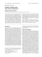
the plant (Fig. 1). These findings indicate that endoglucanases produced by either the plant
or the AM fungus may be involved in the process of host wall degradation. Some of the
endoglucanase activity can be attributed to the extramatrical phase of the AM fungi since
at least one of the endoglucanase activities found in the external mycelium and in the
mycorrhizal root extracts showed the same electrophoretic mobility (42,43) (Fig. 1). En-
doglucanase (EC 3.2.1.4) was purified from roots of onion (Allium cepa) colonized by G.
mosseae. The endoglucanase has a relative molecular weight of about 27 kD and behaves
as a monomer in its native form (44).
B. Pectinases
Pectins and related polysaccharides provide a protective material between plant cells. The
term pectin encompasses a complex group of polysaccharides, some of which may be
structural domains of larger, more complex molecules. The classic pectin fraction from
Figure 1 Nondenaturing polyacrylamide gradient gel electrophoresis of cellulase on 4–12% acryl-
amide. Lane 1, extract from non-AM onion roots; lane 2, extracts from AM onion roots; lane 3,
extracts from external mycelium of Glomus mosseae.
Copyright © 2002 Marcel Dekker, Inc.
oat seedlings contains 23% galacturonic acid and earlier it was thought that pectin con-
sisted solely of α-d-1,4-linked galacturonic acid residues. Today, all evidence suggests
that other sugars are covalently attached to the polygalacturonide backbone and that other
sugars may even form an integral part of the main chain (45,46).
Pectinolysis is carried out by a complex of enzymes (pectinases), which include
endo- and exopectate lyase (PL), endo- and exopolygalacturonase (PG), and pectin meth-
ylesterase (PME).
Degradation of pectin was reported for a sterile ericoid mycelium isolated from
Calluna vulgaris by Perotto and associates (4) and Cairney and Burke (47). A wide range
of ericoid fungi from different geographic regions was capable of growing on pectin as
a sole carbon source. Ericoid fungi seem to use polygalacturonase during their saprotrophic
life.
Attempts to demonstrate pectinase in extracts from AM tissues have not been suc-
cessful (48). However, catabolic repression experiments by Garcı
´
a-Romera and colleagues
(49) showed that pectolytic enzymes may be involved in the process of root colonization
by AM fungi. The spores and external mycelium of G. mosseae possess a complex of
pectinolytic (pectin esterase [PE], pectin lyase [PL], pectatolyase [PNL], polygalacturo-
nase [PG], and polymethylgalacturonase [PMG]) activities (26). The production of hy-
drolytic enzymes was studied during the process of penetration and development by G.
mosseae in plant roots (29). The PE activity was consistently higher throughout the process
of root colonization in plants inoculated with AM fungi than in controls. PE is thought
to facilitate the action of the other pectinase enzymes (50). PMG and PNL (pectinolytic)
activities were higher during the logarithmic stage of AM development in plants inoculated
with the fungus than in nonmycorrhizal plants. The increase in fungal structures that pene-
trate the cell wall during the logarithmic stage of root colonization may explain the in-
crease in PMG and PNL activities at this time. However, PG and PL (pectolytic) activities
in AM plants were similar to those in controls throughout the experiment (29). The lack
of differences in these degradative enzymes is not, however, conclusive evidence that they
do not participate in the colonization process. It may indicate that PG and PL are involved
during other stages of development (i.e., appressoria formation and penetration) in view
of the presence of these enzymes in the extracts of spores and external mycelium of AM
fungi (26).
The simultaneous presence of polygalacturonase produced by the fungus and of
pectins secreted by the plant in the interfacial matrix suggests that the fungus might use
pectins as a food source (31), as suggested by Dexheimer and coworkers (51).
Wall-degrading pectic enzymes uniquely associated with the interface of fine arbus-
cule branches may contribute to the interference of wall formation of the host plant. Active
H
ϩ
adenosine triphosphatase (ATPase) on fungal and plant membranes bordering the inter-
face suggest that protons accumulate in the interfacial matrix and the resulting change in
pH also could contribute to wall loosening (52).
C. Hemicellulases
Hemicelluloses are an integral part of all plant cell walls and form about 25% of the total
dry weight of annuals and up to 40% in woody species. Hemicelluloses consist of chains
of sugars in nonfibrillar organization that are linked to cellulose microfibrils by weak
hydrogen bonds. In dicot primary walls, the major hemicelluloses are neutral xyloglucans
and acidic arabinoxylans; in monocots they are acidic arabinoxylans and neutral β-(1-3,1-
Copyright © 2002 Marcel Dekker, Inc.
4)-glucans(53).Xyloglucansareβ-1,4-glucanswithsidechainsthatcanhydrogenbond
tocellulosemicrofibrils,cross-linkingthemandrestrainingcellexpansion.Inadditionto
astructuralrole,xyloglucanscanbehydrolyzedbyhydrolyticenzymes,andtheoligosac-
charidesproducedmayactassignalmolecules(15,54).
Theplantcellwallcontainsglucanasesandglycosidasesthathydrolyzexyloglucan
intomonosaccharides.Endo-β-1,4-glucanaseactivityisresponsibleforthefirststepof
degradationwherebythexyloglucanisendohydrolyzedintolargefragmentsandexo-1,4-
glucanaseactivityliberateslow-molecular-weightfractionsfromtheendsoflongpolysac-
charidechains(41).Theproductionofhemicellulolyticenzymeshasbeenobservednot
onlyinparasitesbutalsoinmutualisticmicroorganismssuchasRhizobiumspecies(24)
andarbuscularmycorrhiza(28).
Endoxyloglucanaseactivityincreasesduringgrowthanddevelopmentofroots(55).
Thisactivitywasconsistentlyhigheratthebeginningofcolonizationandthelogarithmic
stageofdevelopmentofmycorrhizalfungus(55).Theincreaseinfungalstructuresthat
penetratethecellwallduringthelogarithmicstageofrootcolonizationmayexplainthe
increaseinthedifferentactivitiesatthistime(56).Theevolutionofendoxyloglucanase
activitiesinplantsparalleledthechangesintheexternalmycelium.Therewere,however,
bandsofxyloglucanaseactivityinnonmycorrhizalrootsthatwereabsentinmycorrhizal
roots;thatmaysuggestqualitativeinhibitionbythefungusofsomeplantactivity.Inhibi-
tionofplantproteinsynthesisbyAMfungihasbeenobservedinseveralplant–AMfungi
associations(57,58).
III.ENZYMESINTHEPHYSIOLOGYOFTHEASSOCIATION
A.PhosphorusUptake
Itnowisestablishedthatmycorrhizalcolonizationcanenhancetheuptakefromsoilof
solubleinorganicPbyplantroots(59).Althoughparticularlyimportantinlow-Psoils,
anincreasedrateofPuptakecanoccuroverarangeofsoilPlevelsevenwhenmycorrhizal
growthresponsesnolongeroccur.TheenhancedPuptakebymycorrhizalplantsismost
likelytheresultoftheexternalfungalhyphae’sactingasanextensionoftherootsystem,
therebyprovidingamoreefficient(moreextensiveandbetterdistributed)absorbingsur-
faceforuptakeofnutrientsfromthesoilandfortranslocationtothehostroot(60).External
hyphaeofAMfungimustabsorborthophosphate(Pi)byactivetransport(59,61).They
haveanactiveH
ϩ
-ATPaseintheplasmamembranethatwouldbecapableofgenerating
therequiredproton-motiveforcetodriveH
ϩ
-phosphatecotransport,andPcertainlyis
accumulatedtohighconcentration(62).
Polyphosphate(poly-P)isamajorPreserveinmanyfungianditaccumulatesin
vacuolesofAMfungi(63).Transferofmycorrhizalrootsfromlow-tohigh-Pmedia
resultsinarapidaccumulationofpoly-P(64).Enzymesofpoly-Psynthesishavebeen
foundinmycorrhizaltissue(63,65).Polyphosphatekinase,whichcatalyzesthetransfer
oftheterminalphosphatefromATPtopoly-P,wasdetectedinbothexternalhyphaeand
mycorrhizalrootsbutnotinuninfectedroots,indicatingthatpoly-Pcanbesynthesized
onlybythefungalcomponentofthemycorrhiza.
AlthoughitnowseemslikelythatPistranslocatedbyprotoplasmicstreaminginto
theintraradicalhyphaeaspoly-P(66),littleisyetknownofthebiochemicalmechanisms
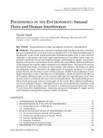
involved.Thetransportthroughthehyphaeandunloadingstepswithinthearbusculemay
belinkedtopoly-Pmetabolism(Fig.2).Highproportionoflong-chainpoly-Ptototal
Copyright © 2002 Marcel Dekker, Inc.
Figure 2 Enzymes involved in P transport in AM roots.
poly-P was observed in the external hyphae, and short-chain poly-P was higher in the
internal hyphae (67). Long-chain poly-P seems to be more efficient in transporting Pi from
the extraradical to the intraradical part of the fungi. Activity of enzymes of polyphosphate
breakdown (exopolyphosphatase and endopolyphosphatase) is greater in mycorrhizal roots
than in uninfected roots (65). Both enzymes have been detected in extracts of internal
hyphae, but not those of external hyphae. The long-chain poly-P may be partly hydrolyzed
into short-chain poly-P with endopolyphosphatase. Depolymerized short-chain poly-P
may be hydrolyzed further with exopolyphosphatase to liberate Pi (67). Alternatively, the
reaction catalyzed by polyphosphate kinase is readily reversible, so there is also the possi-
bility that poly-P could be hydrolyzed, liberating ATP (63). The Pi (or ATP) so released
in the arbuscule then would be transferred into the host (66).
Copyright © 2002 Marcel Dekker, Inc.
The presence of intense ATPase activity indicates there is a carrier that mediates
active transport mechanisms for Pi uptake at the host plasmalemma. ATPase activity has
been observed in both plant and fungal plasma membranes and in the interfacial matrix
associated with young arbuscules that decreased with senescence of arbuscules (68). A
lack of H
ϩ
-ATPase activity in the host periarbuscular membrane surrounding nonfunc-
tional arbuscules has been reported (69).
Other enzymes also have been implicated in P metabolism. Mycorrhizal-specific
alkaline phosphatase is located in the vacuole of extraradical and intraradical hyphae (70–
72). Maximum activity occurs while infections are young (100% arbuscular), coinciding
with the start of the mycorrhizal growth response, but disappears with degeneration and
collapse of the arbuscule. This enzyme appears to be of fungal origin. However, the role
of alkaline phosphatase in Pi metabolism is still unknown (59,71).
The amount of P in soil available to plants is small, about 1% to 5% of the total P
content. This finding has led to the suggestion that AM fungi are capable of utilizing
insoluble P sources. Organic phosphates in soil may be utilized by plants through the
action of phosphatases. Phosphatase activity in soil may originate from the plant roots or
from microorganisms (73,74). High levels of acid and alkaline phosphatases have been
found in the roots (70) and rhizosphere (75,76) of plants colonized by AM fungi. This
increase in phosphatase activity would result in Pi’s being liberated from organic phos-
phates immediately adjacent to the cell surface to be captured by the uptake mechanisms
of mycorrhizal fungi. Some results have shown exudation of phosphatase by the external
hypha and efficient hydrolysis of phytate-P by the phosphatase of mycorrhizal hyphae (76).
Acid phosphatase activity release was visually shown as a red-colored ‘‘hyphal print’’
on filter paper treated with napthyl phosphate and Fast Red TR (diazotized 2-amino-5
chlorotoluene 1,5-naphthalene disulphonate) (77). However, other results indicated that
the role of fungal phosphatases in P uptake from organic P is not clear: (1) extracellular
phosphatase activity of mycorrhizal roots was stimulated in the presence of easily hy-
drolyzed substrates (76) but repressed by nonhydrolyzable forms of organic P (Po) (78),
(2) no effect of mycorrhiza on specific activity of phosphatase was detected for clover
grown in soil amended with
32
P-labeled organic matter (79), (3) the production of phospha-
tase varied with the choice of host plant and fungal endophyte (75,78), and no relationship
between the level of AM colonization and phosphatase activity in different wheat cultivars
has been found (80,81), (4) the addition of P fertilizer and CaCO
3
to soils decreased AM
colonization but increased phosphatase activity in the plant rhizosphere (82); and (5) soil
microorganims can mineralize organic P and AM hyphae may use Pi derived from their
activity (83). Thus the results obtained are conflicting despite much effort (84).
One of the most important factors involved in controlling AM colonization of roots
is soil and plant P. High P concentrations inhibit mycorrhizal colonization (60,85). The
activity of mycorrhiza-specific alkaline phosphatases of Glomus species declines at high
P levels (86,87). These observations suggest these enzymes would be involved in the
regulation of mycorrhizal colonization of roots by P content of plants (85). However, high
soil P concentration decreased AM colonization of roots by Gigaspora species but did
not affect alkaline phosphatase activity (72). Thus the mechanism whereby the internal
P content of the host regulates mycorrhizal infection is not clear.
B. Nitrogen Metabolism
Mycorrhizal plants sometimes improved nodulation and N fixation (88), an effect that
may be due to enhanced P uptake (89). However, AM contribute to the N nutrition of the
Copyright © 2002 Marcel Dekker, Inc.
host by assimilation of soil nitrogen (N). The plant growth response to AM colonization
may be greater in the presence of NH
4
than NO
3
(59).
Ammonium N and nitrate N have different pathways for metabolism, cation-carbox-
ylate storage, and pH regulation, and hence they have rather different biochemical and
physiological implications for the host (89). Nitrate is reduced, first to nitrite by nitrate
reductase, then to ammonium by nitrite reductase. Ammonium N, once inside the cell,
becomes directly incorporated into the various pathways for amino acid synthesis. Assimi-
lation may be by glutamate dehydrogenase (GDH), or via glutamine synthetase (GS) and
glutamate synthase (GOGAT) with the formation of glutamate. GS activity is increased
in mycorrhizal root systems, partly as a result of a contribution from the fungi themselves;
activity has been detected in fungal tissue separated from mycorrhizal roots (90). Improved
P nutrition in the plants resulted in only a small increase in activity, confirming that the
fungi have an important contribution and that GS is not related to P nutrition. In contrast,
GDH activity showed no direct relationship with colonization (90). This limited evidence
suggests that the fungi may have the capacity to assimilate NH
ϩ
4
and, in consequence, N
is likely to be transferred from fungi to plants in organic form.
Nitrate reductase activity has been detected in isolated spores of AM fungi (91,92).
There are suggestions that either the AM fungi increase the nitrate reductase activity in
the host plant (regardless of the P content) or the AM fungi have enzymatic activity per
se (93). The fungal nitrate reductase messenger ribonucleic acid (mRNA) was detected
in arbuscules but not in vesicles by in situ hybridization (94). The observation that AM
fungi possess the gene coding for assimilatory nitrate reductase does not rule out the
possibility that plant root cells mainly reduce nitrate in the AM symbiosis (95). The plant
colonized by different AM fungi showed different nitrate reductase activity (93). Nitrate
reductase and glutamine synthetase decreased with the age of mycorrhizal plants (96).
Nitrite formation catalyzed by nitrate reductase was mainly reduced nicotinamide-adenine
dinucleotide phosphate–(NADPH)-dependent in roots of AM colonized plants but not in
those nonmycorrhizal plants, a finding consistent with the fact that the nitrate reductases
of fungi preferentially utilized NADPH as the reductant (94). These investigations suggest
that the fungus in AM-colonized root performs nitrate uptake and nitrate reduction to
some degree. Because of its toxicity, the nitrite formed probably is not exported from the
fungal to the plant cells. Other enzymes of nitrate assimilation have been described to
occur in AM fungi (90). Thus nitrite reductase, glutamate synthetase, and glutamate syn-
thase may transform nitrite, and N compounds (e.g., ammonium, glutamine, glutamate)
probably are transferred from arbuscules to host cells.
C. Carbohydrate Assimilation
It is commonly accepted that the AM fungi are obligate symbionts and that carbohydrates
are transferred from autotroph to heterotroph. It is likely that the fungus obtains the bulk
of its carbon from host sugars; short-term
14
CO
2
labeling experiments have shown transport
of photosynthate from the host to the fungus (97). Most (70% to 90%) of the
14
C label
present in both the roots and mycelium was in the form of soluble carbohydrates. The
carbohydrates, predominantly sucrose, are delivered to the apoplast by the host cell. Then
sucrose is hydrolyzed in the apoplastic interface by an acid invertase of plant origin, and
the resulting hexoses are absorbed by the fungus (59), and used for trehalose synthesis.
Trehalose has been shown to accumulate in both spores and external hyphae of AM fungi
(66,98,99). Trehalose was detected in roots of colonized plants but not of control plants
(56). Polyphosphates may serve in phosphorylation for the active transport of carbon skele-
Copyright © 2002 Marcel Dekker, Inc.
tons into the arbuscule from the host either through the ATP produced by degradative
polyphosphate kinase action or through a direct phosphorylation of sugars by enzymes of
the polyphosphate glucokinase type (64). On the other hand, trehalase has been found in
plants (100), and this enzyme increased upon mycorrhizal colonization (101). The biologi-
cal function of plant trehalases is unknown, but they might be involved in the degradation
of trehalose released from senescent AM fungus.
The possible metabolic pathways of carbon utilization in AM are largely uninvesti-
gated. Dehydrogenases indicative of glycolysis are found in hyphae, vesicles, arbuscules,
and spore germ tubes (102). From this, MacDonald and Lewis (102) have inferred that
AM fungi employ the Embden–Meyerhof–Parnas glycolytic scheme, the hexose mono-
phosphate shunt (or pentose phosphate cycle), and the tricarboxylic acid cycle.
A cyanide-insensitive respiratory pathway has been noted in AM roots (103). Such
a pathway of electron transport to oxygen has been established in the sheath tissue of
ectomycorrhizal roots (104). This pathway is not coupled to oxidative phosphorylation
and may operate when oxidative phosphorylation is reduced by adenosine diphosphate
(ADP) limitations. It is likely that the operation of such a pathway would increase the
overall utilization of carbohydrates in mycorrhizal tissues (66).
IV. ENZYMES IMPLICATED IN THE HOST DEFENSE RESPONSE
TO ARBUSCULAR MYCORRHIZAL FUNGAL COLONIZATION
Arbuscular mycorrhizal fungal penetration and establishment in the host roots involve a
complex sequence of events and intracellular modifications that influence root colonization
(25). Genotype and environmental factors influence the infection intensity or even the
host compatibility and/or resistance (33,105).
The key to understanding the phenomenon of compatibility is to study recognition
mechanisms and molecules involved in early stages of the AM interaction. In this sense,
the formation of appressoria is one of the first morphological signs that recognition be-
tween the plant and the fungus has occurred. Some authors suggest that plant defense
reactions may occur only after appresorium formation when the fungus has changed its
state from saprophytic to infective (106).
Although AM fungi are considered as biotrophic microorganisms and biotrophs gen-
erally exhibit a high degree of host specificity, most AM fungi that have been studied
show little or no specificity and are not thought to induce typical defense responses in host
plants. Nevertheless, some plant resistance markers have been investigated in compatible
symbiotic AM fungus–root interactions, and the early activation of certain plant defense
genes has been shown (105). Since the plant host can elicit a weak defense response to the
invading fungus, this may be a natural mechanism to control the number and/or location of
infections. Furthermore, some phenomena of suppression of defense responses have been
demonstrated in mycorrhizal roots (107,108). Whether this suppression is systemic or
restricted to the infected area or whether products of symbiosis-related plant genes sup-
press the defense genes directly or through activation of fungal-derived suppressors re-
mains to be elucidated. So far, it is not known how the induction/suppression of mecha-
nisms associated with plant resistance could participate in the phenomenon of
compatibility between plant roots and AM fungi. The investigation of early events and
molecules involved in fungal–plant interactions is crucial for a better understanding of
symbiosis.
Copyright © 2002 Marcel Dekker, Inc.
In the following sections, some results obtained from studying the enzymatic activi-
ties produced by the host are reviewed and their contribution in the induction/suppression
of mechanisms associated with plant resistance and in the control of intraradical fungal
growth and maintenance of the symbiotic status is discussed. For ease of discussion the
defense-related activities are divided into three classes based on their role in defense re-
sponse. The first class involves hydrolases such as chitinases and β-1,3-glucanases that
act directly as potent inhibitors of fungal growth. The second class involves enzymes
related to oxidative stress such as catalases and peroxidases, and the third class consists
of key enzymes that catalyze core reactions in phenylpropanoid metabolism.
A. Chitinases and -1,3-Glucanases
The initiation of chitinase and other hydrolase activities is predominantly one of the coor-
dinated and widespread mechanisms of plant defense against pathogen attack. There is
good evidence that the action of the endohydrolases leads to detrimental effects, such as
the inhibition of hyphal growth by invading fungi, as well as the probable release of
signaling molecules (β-glucans and chitin/chitosan oligomers) that activate defense genes
in the plants (109,110).
Most research into chitinase enzyme activity in plant roots colonized by the AM
fungi has focused on the measurements of enzymatic activity during the different phases
of mycorrhizal development. Several authors have shown that roots of infected plants
contain enhanced levels of endochitinase activity at early stages of AM development.
These results have been obtained with different combinations of plant and fungus (111–
114). The peak of chitinase activity is followed by a period in which the enzyme activity
is generally repressed to levels that are below those in nonmycorrhizal roots and that
coincide with the extensive fungal development within roots (111–114). A similar result
has been observed for β-1,3-glucanases. In bean and tomato mycorrhizal roots, β-1,3-
glucanase activity was suppressed during the phase of rapid colonization (113,114).
Greater suppression of chitinase activity was observed in soybean roots under low
P concentration and coincided with the period of maximum intraradical growth rate of
the fungus (115). A correlation between the rate of suppression of chitinase activity in
the inoculated roots and the infectivity of fungal isolates was observed (116). The maxi-
mum level of suppression was found in roots of soybean plants infected with the more
infective isolate of Glomus intraradices (116).
In some particular plant–fungal combinations, the initial increase of chitinase activ-
ity was not followed by suppression, and higher levels of activity persisted (117). In some
cases, no changes in chitinase and β-1,3-glucanase activities were detected between inocu-
lated and noninoculated plants at all stages of mycorrhiza development (118–119).
Corroborating the biochemical data, differential gene expression of acidic and basic
forms of chitinase and β-1,3-glucanase has been observed during mycorrhiza formation in
different plant–fungal combinations (108,113,120,121). Studies of in situ localization of
transcripts of bean endochitinases and β-1,3-glucanases in mycorrhizal roots showed that
mRNAs accumulated predominantly in the vascular cylinder (121). Nevertheless, the accu-
mulation of chitinase and β-1,3-glucanase transcripts has been observed around a number
of cortical cells containing arbuscules (120,121), suggesting that the encoded enzyme might
be involved in the control of intraradical fungal growth. The accumulation of β-1,3-gluca-
nase mRNA in cells containing arbuscules was modulated by P concentration. The higher
level of mRNA accumulation was obtained at a low level of P concentration (121).
Copyright © 2002 Marcel Dekker, Inc.
ThepatternsofenzymeactivityandmRNAaccumulationsuggestthatchitinases
andβ-1,3-glucanasesmightbepartoftheearlydefenseresponsebytheplanttotheinvad-
ingfungus,whichisthensuppressedassymbioticinteractionsdevelop.Inthiscontext,
planthydrolasesmaybeinvolvedintheregulationofAMdevelopment.Nevertheless,
someexperimentaldatarevealedthatitisnotlikelythatplantchitinasesandglucanases
areessentialtothecontrolofthegrowthofAMfungi.Transgenicplantsconstitutively
expressinghighlevelsofdifferentacidicformsoftobaccoPRs(includingchitinasesand
β-1,3-glucanases)becamenormallycolonizedbytheAMfungi(122,123).Thefactthat
chitinasesandβ-1,3-glucanasesinducedbytheAMsymbioticfungiorbyconstitutive
geneexpression,donotpreventrootcolonizationsuggeststhattheyareineffectivein
controllingfungaldevelopment.ThelowenzymaticaffinityforAMfungalcomponents
orinaccessibilityoftheseenzymestofungalcellwallcomponentsmaycausethisineffec-
tiveness(112).
Conversely,specificacidicformsofchitinaseandβ-1,3-glucanaseareactivatedin
severalplantscolonizedbyAMfungi.Thesesymbiotic,specificisoenzymeshavebeen
reportedinpea(124),tobacco(118),andtomato(125–127)rootsandaredifferentfrom
pathogen-inducedisoformsorconstitutiveenzymes.Inaddition,newchitosanaseisoforms
havebeenshowninpea(128)andtomato(126).Chitosanasesarehydrolyticenzymes
actingonchitosan,aderivativepartiallyorfullydeacetylatedofchitin(129).Interestingly,
themycorrhizal-relatedchitinaseisoformdescribedintomato-colonizedrootsappearedto
displaychitosanaseactivity.Thisbifunctionalcharacterwasnotfoundfortheconstitutive
enzymes,orinPhytophthorasp.–inducedchitinases(126).Mycorrhizal-specificplantchi-
tinasesarenotactiveinpathogen-infectedroots(118,124–125)orinRhizobiumsp.legume
symbiosis(130),indicatingadifferentialinductionandfunction.
Althoughtheprecisefunctionofplanthydrolaseactivitiesintheestablishmentof
AMsymbioticinteractionisstillunclear,theirstimulationseemstobeakeypointinthe
mechanismofrecognitionandsignalingbetweenplantrootsandAMfungi.Aregulatory
roleoftheseenzymesduringestablishmentofAMandotherrootsymbiosishasbeen
proposed.Stimulationofspecificplantchitinaseshasbeenreportedinsoybean/Rhizobium
sp.(131)andectomycorrhiza(132).Ithasbeenpostulatedthatchitinasesmaybeinvolved
intherecognitionoftherhizobialnodulationsignalsand,thus,intheregulationofthe
nodulationprocess(133).Thedatasuggestaspecificrolefortheseenzymes,onethat
couldberelatedintheAMsymbiosistothedetection,modification,and/orreleaseof
chitinorchitosanoligomersfromthefungalcellwallthatcanactassignalingcompounds
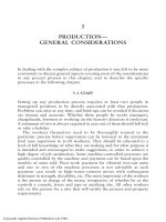
duringthedevelopmentofAM(Fig.3).Inthisprocessofsignalexchange,themodulation
of chitinase activity by substrate specificity could be important (126). Furthermore, a puta-
tive role in bioprotection against fungal pathogens has been proposed for the new mycor-
rhiza-specific hydrolases. In this sense, the additional acidic and basic β-1,3-glucanase
isoforms revealed during the interaction of Phytophtora parasitica and tomato plants pre-
inoculated with G. mosseae could be implicated in the protective effect caused by AM
symbiosis (127).
B. Catalases, Peroxidases, and Other Enzymes Related
to Oxidative Stress
One of the major and rapid processes in the response of plant cells to environmental
stresses is the generation of an oxidative burst, characterized by the release of active
oxygen species (AOS) (134). This rapid response has been characterized in the hypersensi-
Copyright © 2002 Marcel Dekker, Inc.
Figure 3 A speculative model showing possible participation of plant and fungal enzymes at early
stages of plant–AM fungal interactions.
Fungal elicitors released during or after appressorium formation may be coupled to active
plant receptors. In the process of release, perception of modification of these fungal signaling mole-
cules could involve/stimulate plant enzymes. Alternatively, these plant enzymes may play a role
facilitating changes in plasma membrane and cell wall architecture associated with early symbiotic
interactions. Additionally, fungal enzymes involved in plant cell wall modification could facilitate
the process of signal generation by cell wall architecture modification.
These initial reactions lead to the activation of a subset of elicitor-responsive genes, including
defense and symbiotic genes. Possibly, gene activation is a consequence of a complex mechanism
of signal transduction. The correct balance of the function of these induced genes is one of the keys
of AM fungal–plant compatibility.
Unfortunately, most of the components of the signal recognition and transduction are un-
known, and the assignment of components and sequence of events requires additional data.
tive response (HR) of plants to pathogens or elicitors (134,135). The predominant species
detected in plant–pathogen interactions are superoxide anion (O
2
Ϫ
), hydrogen peroxide
(H
2
O
2
), and the hydroxyl radical (OH
Ϫ
). The experimental data suggest different roles of
the AOS in plant defense response (134,136). They can contribute to cell wall protein
cross-linking and programmed host cell death during the hypersensitive response and may
Copyright © 2002 Marcel Dekker, Inc.
directlycontributetoreductionofpathogenviabilityandgrowth.Inaddition,theyhave
beenproposedasmediatorsinpathwaysleadingtodefense-relatedgeneexpression(136).
ThereleaseofAOSinsomeplant–pathogeninteractionscanresultindamageto
thehosttissues.Therefore,mechanismsthatlimitthedurationoftheoxidativeburstand
itstoxiceffectsarenecessarytominimizedamagetotheplantitself.Oneofthesemecha-
nismsistheactionofendogenousantioxidantenzymes,suchassuperoxidedismutases,
catalases,peroxidases,andglutathioneperoxidases,whicharecapableofneutralizingthe
AOS.
Duringtheestablishmentofacompatibleplant–fungusAMsymbiosis,thehost
plantshowedlittlereactionatthecytologicalleveltoappressoriumformationorinfection
hyphae.Occasionallysomethickeningwasobservedinepidermalcellwallsatthepoint
ofcontactwithappressoria(105),andonlyaresponsesimilartoHRhasbeendetected
inRiT-DNA–transformedrootsofalfalfacolonizedbyGigasporamargarita(137).Nev-
ertheless,recentstudies,usingthediaminobenzidine(DAB)stainingtechnique,revealed
thatabrownishstain,indicativeofH
2
O
2
accumulation,waspresentwithincorticalroot
cellsinthespaceoccupiedbyclumpedarbusculesandaroundhyphaltipsattemptingto
penetraterootsofMedicagotruncatulacolonizedbyG.intraradices(138).Theseresults
suggestthatalocallyrestrictedoxidativeburstcouldbeinvolvedintheresponseofthe
planttoAMformationanddevelopment.
Relativelyfewdataexistconcerningthepossibleparticipationofantioxidanten-
zymesintheplantresponsetoAMformation.Apeakofcellwall–boundperoxidasewas
observedduringtheinitialstagesoffungalpenetrationinleek(Alliumporrum)cells.Once
infectionwasestablished,theactivitydecreasedtothelevelsshowninnonmycorrhizal
plants(139).Inpotatoroots,theactivityofextracellularperoxidaserecoveredinroot
leachateswasnotstimulatedbyAMinfection;peroxidaseactivitypergramoffreshweight
wassignificantlyenhancedinAMroots(140).Whenpotatoplantsweregrownwithhigher
Psupply,extracellularperoxidaseactivityincreasedlinearlywithincreasingPsupply,
suggestingaroleofperoxidaseinlimitingAMinfectioninwell-P-nourishedplants(140).
Theanalysisofcatalaseandascorbateperoxidaseactivitiesduringtheearlystageof
tobacco–Glomusmosseaeinteractionrevealedtransientenhancementsofbothenzymatic
activitiesintheinoculatedplants(141).Theseincreasescoincidedwiththestageofappre-
ssoriaformationonrootsurfacesandtheappearanceofapeakofaccumulationoffree
salicylicacidininoculatedroots(141).Thesedataindicatetheactivationofcatalaseand
peroxidaseactivitiesinrootcellswherethefungusformingappressoriamightbepartof
theplantresponsetotheinvadingfungus.Theroleoftheseenzymesinthisresponse
couldberelatedtoactivationofadefensivemechanismortoaprocessofcellwallrepair
atthesiteofinfection(Fig.3).Alternatively,theearlyactivationofcatalaseandperoxidase
may play a role that facilitates changes in cell wall architecture associated with early
symbiotic interactions. This hypothesis has been proposed for the Rhizobium meliloti–
Medicago truncatula association, when a Rhizobium sp.–induced peroxidase gene is in-
duced rapidly and transiently by compatible R. meliloti, and Nod factor. The transcript is
localized to differentiating epidermal cells in the root zone that is subsequently infected
by Rhizobium sp. (142).
Other important enzymes that participate in the primary defense against the AOS
are the superoxide dismutases (SODs) that catalyze the disproportionation of the superox-
ide free radical (O
2
Ϫ
)toH
2
O
2
and O
2
. Data suggest that SOD acts as an antioxidant system
in the N
2
fixation process of nodules (143). Some changes in the isoenzymatic pattern
and SOD activity in several plant–AM fungal symbioses have been reported (144–148).
Copyright © 2002 Marcel Dekker, Inc.
The appearance of additional SOD isozymes in plant roots inoculated with G. mos-
seae has been reported in red clover (144,146) and onion plants (144). This new isozyme
was not detected in noninoculated plants nor in plants inoculated with G. intraradices;
thus SOD enzymes appearance was associated with specific fungal–plant interaction (144).
So far, it is not known what role SOD enzymatic activity plays during the period of AM
development; nevertheless some data suggest that the change in enzymatic activity was
related to differences between AM inoculated and noninoculated plants in stress situations,
including drought exposure (148) and plant senescence (144).
C. Enzymes That Catalyze Core Reactions
in the Phenylpropanoid Metabolism
Phenylpropanoid compounds include a variety of chemical formulas with a wide range
of biological roles in plant life cycles. The biochemical reactions and genetic regulation
of phenylpropanoid metabolism are complex, because many compounds are constitutive
or induced, depending on the plant species or tissues, and a broad variety of biotic and
abiotic stresses can induce their accumulation (149).
Among phenylpropanoid compounds, flavonoid and isoflavonoid compounds are
involved in diverse aspects of plant growth, development, and interactions with microor-
ganisms, mainly in defense responses with the liberation of phytoalexins (150) and in
Rhizobium sp.–legume interaction, when specific flavonoids act to initiate the symbi-
osis (151).
Flavonoids can stimulate spore germination, hyphal growth, and enhancements of
AM colonization by AM fungi (152). Some of these compounds have been isolated and
characterized. Their effect on different AM fungal species has been assayed, and the results
show that the AM fungal growth response to root flavonoids is not uniform (153).
In Medicago species, transient increases in different flavonoid/isoflavonoid com-
pounds were found, depending on the specific interaction of Medicago and fungal species
(107,111,154,155). Some of these compounds stimulated hyphal growth (154). Formono-
netin was found to accumulate in Medicago sativa roots in the presence of the fungus
Glomus intraradix, before fungal penetration and colonization (111). The analysis of enzy-
matic activities and accumulation of mRNA transcripts encoding enzymes of flavonoid/
isoflavonoid metabolism revealed that the changes in compound accumulation correlate
with increases in mRNA and enzyme activity. Increases in enzymatic activity and messen-
ger ribonucleic acid (mRNA) accumulation of phenylalanine ammonia lyase (PAL), chal-
cone synthase (CHS), and chalcone isomerase (CHI) were observed in Medicago species
colonized by AM fungi (107,155). Increases in PAL transcript accumulation also have
been detected in rice roots inoculated with G. mosseae (156). This increase was concomi-
tant with the accumulation of salicylic acid, a phenolic acid derived from phenylpropanoid
metabolism that has been implicated in plant defense responses (157–159). Nevertheless,
no changes in transcript level were observed in other mycorrhizal interactions, such as
those involving bean (113) or parsley (160).
The increase, followed by decline, in transcript accumulation and enzyme activity
of PAL and CHI in roots of alfalfa during infection by G. intraradices (107) has been
interpreted as a mechanism of activation/suppression of the defense reaction elicited by
G. intraradices in alfalfa (161). Although phenylpropanoid metabolism can be enhanced
in roots during symbiotic interactions, this is not a general phenomenon, and the extent,
timing, and enzymatic activities and compounds released appear to depend on the plant
Copyright © 2002 Marcel Dekker, Inc.
and fungal genotypes involved. In situ localization of transcripts encoding PAL and CHS
in mycorrhizal roots showed that the transcripts were discretely localized in cells con-
taining arbuscules (155). The expression of other gene encoding enzymes in the flavonoid/
isoflavonoids pathway, such as CHI or isoflavone reductase (IFR), was not significantly
affected in mycorrhizal roots (155).
Altogether, the available data suggest an activation of phenylpropanoid metabolism
in mycorrhizal roots, characterized by the weak, localized, and uncoordinated induction
of genes and accumulation of phytoalexin products, some of them at high levels (162).
Nevertheless, no evidence exists to support a specific role for flavonoid/isoflavonoid in
the AM symbiosis. Even though several of them can stimulate hyphal growth, some results
suggest that they are not necessarily signaling compounds involved in the AM symbi-
osis (163).
V. ENZYMES AS A METABOLIC ACTIVITY INDEX
Mycorrhizal colonization of plant roots has been evaluated by using nonvital staining
techniques. However, often there is no relationship between percentage mycorrhizal colo-
nization and the effectiveness of a particular fungus in plant growth (164). Several authors
have developed vital staining techniques to measure metabolic active fungal colonization
of roots.
The alkaline phosphatase enzyme in intraradical hyphae was found to be related to
the stimulation of the growth of the plants when colonized by AM fungi (165). Alkaline
phosphatase can be histochemically visualized in external and intracellular mycelium
(87,166). Fungal alkaline phosphatase activity diminished in plants growing under several
adverse conditions in spite of the constant level of mycorrhizal colonization assessed with
nonvital stains. Alkaline phosphatase has been proposed as a vital marker for root coloniza-
tion (71). However, the application of a fungicide, which inhibits P uptake, and transfer
to plants via hyphae do not affect alkaline phosphatase activity (167). More work needs
to be conducted on the fungal phosphatases in order to identify whether these enzymes
play a key role in the efficiency of the symbiosis.
MacDonald and Lewis (102) developed a histochemical technique to stain for succi-
nate dehydrogenase (SDH) activity in AM fungi. Several authors showed that this enzy-
matic activity of the fungus was depressed when herbicides (168) or fungicides (169,170)
were applied to mycorrhizal colonized plants, in spite of the small effect on the fungal
structures visualized by nonvital staining techniques. A decrease in SDH activity also was
observed (along with the formation of septa in the intraradical hypha) when mycorrhizal
plants were subjected to the presence of the antagonistic fungus Trichoderma koningii
(171). Some correlation between the frequency of SDH-active arbuscules and shoot mass
of plants has been found (86,172). However, the proportion of the AM mycelium with
SDH activity is not related to the effect of the fungus on plant growth (169,173,174).
Thus SDH activity appears to be a sensitive parameter for measures effects of environmen-
tal stress on the fungi but is not a sensitive parameter for measuring growth.
Other enzymes, such as malate dehydrogenase (MDH) (175) and colonization-
specific phosphatase (IPS) (176), have been proposed as markers of the fungal metabolic
activity. A very high correlation coefficient has been found between the intensity of the
fungal MDH electrophoretic bands and the AM colonization of roots measured by the
concentration of glucosamine (175,177). The activity of this MDH was inhibited also in
Copyright © 2002 Marcel Dekker, Inc.
the presence of fungicides. Conversely, some IPS (probably a neutral phosphatase) was
detected in mycorrhizal plants. The activity increased as the colonization rate increased
and decreased at the stationary phase of the host growth when the colonization rate was
still high (176).
VI. ENZYMES IN AM FUNGI IDENTIFICATION
The assessments of the biodiversity of AM fungi in ecosystems have relied on the isolation
of the resting spores from soils. This approach does not necessarily supply useful informa-
tion about functional ecological characteristics since the fungus that colonizes roots might
not produce spores under certain conditions. One plant can be colonized by several AM
fungi at the same time, but the intraradical mycelium of the different species of AM
fungi shows little morphological variation (164). Molecular techniques have been used to
identify AM fungi (178). However, with isozyme techniques it is possible to measure the
metabolically active mycorrhizae; this is not yet possible with DNA-based techniques.
AM fungi can be identified within roots by differences in the mobility of specific fungal
enzymes on polyacrylamide gel electrophoresis (179–181). This method has been used
to identify and quantify endophytes within a root in a competition experiment with differ-
ent AM isolates (182). The staining intensity of esterase, glutamate oxaloacetate transami-
nase, and peptidase (measured as peak height on a densitometer trace) was correlated with
the biomass of the fungus in the root sample, and so this offers a method of quantifying
the contribution of a single fungus to a mixed colonization (183).
The use of MDH and esterase enzymes allows characterization of different AM fungi
(184). The grouping of Gigaspora sp. isolates provided by the SSU sequence analysis was
similar to the grouping of the isozyme profile of MDH (185).
VII. CONCLUSIONS
Fungi that form AM associations have not been cultured in the absence of host roots.
Most of the enzymatic studies on AM symbiosis have been performed with extracts of
mycorrhizal roots containing both plant and fungal enzymes (43). The small differences
in hydrolytic enzyme activities between mycorrhizal and nonmycorrhizal plants, together
with the low rate of production of cell wall hydrolytic enzymes, suggest that AM fungi
penetrate the root surface mostly by mechanical force. Appressoria with well-melanized
walls produce hyphae that tend to progress by growing between root epidermal cells rather
than by crossing their outer walls. Once inside the roots, many AM fungi produce intercel-
lular hyphae that run within huge air channels (186), and then cross the wall of cortical
cells to become intracellular, producing penetration pegs and causing only limited and
subtle changes in the structure of the host wall (187). These slight modifications suggest
that they may produce at this stage very limited or localized amounts of hydrolytic en-
zymes. AM fungi seem to colonize the root tissues of their host plant by means of a
combination of mechanical and enzymatic mechanisms. Very weak and localized produc-
tion of enzymes might ensure that viability of the host is maintained, defense responses
are not triggered, and a high degree of compatibility is reached.
Some plant defense responses have been shown to be activated in compatible AM
fungus–root interactions. The role of these plant defense compounds could be to control
Copyright © 2002 Marcel Dekker, Inc.
intraradical fungal growth and maintenance of the symbiotic status. Plant defense chemi-
cals and enzymes may be involved in the perception, modification, and/or release of fungal
cell wall fragments that can act as signaling compounds during the process of recognition
and formation of AM symbiosis.
The enhanced growth of plants colonized by AM fungi results from improved uptake
of soil P (59). Enzymes are involved in P transport from the fungus to the host plant, but
it is not clear whether AM enzymes are involved in P mobilization and fungal/plant uptake
from soil. AM fungi use P from the soluble fraction of soil (59). However, there are
indications of P mineralization from organic fractions of soil by fungal phosphatases that
may represent another source of P uptake in P-deficient soils (76,77). The increase of N
uptake by AM symbiosis has been attributed to better P nutrition of plants (164). Neverthe-
less, enzymes implicated in assimilation of ammonium N and nitrate N that have been
found in arbuscular fungi that contribute to N nutrition of the host plant by assimilation
of soil N regardless of P effect (90).
In spite of the importance of AM symbiosis to nutrient movement and soil conserva-
tion, very few studies on the impact of the fungal enzymes on soil have been carried out
(188). There is a clear need to extend studies on the ecological role of AM enzymes in soil.
REFERENCES
1. JL Harley. Introduction: The state of art. In: JR Norris, DJ Read, AK Varma, eds. Methods
in Microbiology. Vol 21. London: Academic Press, 1991, pp 1–24.
2. R Law, DH Lewis. Biotic environments and the maintenance of sex-some evidence from
mutualistic symbioses. Biol J Linn Soc 20:249–276, 1983.
3. JB Morton, GL Benny. Revised classification of arbuscular mycorrhizal fungi (zygomycetes):
A new order, Glomales, two new suborders, Glomineae and Gigasporaceae, with an amenda-
tion of Glomaceae. Mycotaxonomy 37:471–491, 1990.
4. S Perotto, V Bettini, P Bonfante. The biology of mycorrhiza in the Ericaceae. I. The isolation
of the endophyte and synthesis of mycorrhizas in aseptic cultures. New Phytol 72:371–379,
1993.
5. S Perotto, R Peretto, A Schubert, A Varma, P Bonfante. Ericoid mycorrhizal fungi: Cellular
and molecular basis of their interactions with host plant. Can J Bot 73:557–568, 1995.
6. A Varma, P Bonfante. Utilization of cell wall-related carbohydrates by ericoid mycorrhizal
endophytes. Symbiosis 16:301–313, 1994.
7. KK Newsham, AH Fitter, AR Watterson. Arbuscular mycorrhiza protect an annual grass
from root pathogenic fungi in the field. J Ecol 83:991–1000, 1995.
8. RP Schreiner, GJ Bethlenfalvay. Mycorrhizal interactions in sustainable agriculture. Crit Rev
Biotechnol 15:271–287, 1995.
9. FA Smith, SE Smith. Structural diversity in (vesicular)-arbuscular mycorrhizal symbioses.
New Phytol 137:373–388, 1997.
10. P Bonfante-Fasolo. Ultrastructural analysis reveals the complex interactions between root
cells and arbuscular mycorrhizal fungi. In: S Gianinazzi, H Schu
¨
epp, eds. Impact of Arbuscu-
lar Mycorrhizas on Sustainable Agriculture. Basel, Switzerland: Birkaha
¨
user Verlag, 1984,
pp. 73–87.
11. MJ Harrison. Development of the arbuscular mycorrhizal symbiosis. Curr Opin Plant Biol
1:360–365, 1998.
12. V Gianinazzi-Pearson, S Gianinazzi. Protein and protein activities in endomycorrhizal symbi-
oses. In: A Varma, B Hock, eds. Mycorrhiza: Structure, Function, Molecular Biology and
Biotechnology. Berlin, Heidelberg, New York: Springer-Verlag, 1995, pp 251–267.
Copyright © 2002 Marcel Dekker, Inc.
13. RJ Howard, MA Ferrari. Role of melanin in appressorium function. Exp Mycol 13:403–418,
1989.
14. JD Walton. Deconstructing the cell wall. Plant Physiol 104:1113–1118, 1994.
15. T Hayashi. Xyloglucans in the primary cell wall. Annu Rev Plant Physiol Plant Mol Biol
40:139–168, 1989.
16. MG Hahn, P Bucheli, F Cervone, SH Doares, RA O’Neill, A Darvill, P Arbesheim. The
role of cell wall constituents in plant-pathogen interactions. In: E Nester, T Kosuge, eds.
Plant-Microbe Interactions. New York: McGraw Hill, 1989, pp 131–181.
17. A Varma. Hydrolytic enzymes from arbuscular mycorrhizae: the current status. In: A Varma,
B Hock, eds. Mycorrhiza: Structure, Function, Molecular Biology and Biotechnology. Berlin,
Heidelberg, New York: Springer-Verlag, 1999, pp 372–389.
18. SC Fry, RC Smith, KF Renwick, DJ Martin, SK Hodge, KG Matthews. Xyloglucan endotrans-
glycosylase, a new wall-loosening enzyme activity from plants. Biochem J 282:821–828,
1992.
19. G Maclachlan, C Brady. Multiple forms of 1,4-β-glucanase in ripening tomato fruits include
a xyloglucanase activatable by xyloglucan oligosaccharides. Aust J Plant Physiol 19:137–
146, 1992.
20. NC Carpita. Structure and biogenesis of the cell walls of grasses. Annu Rev Plant Physiol
Plant Mol Biol 47:445–476, 1996.
21. A Collmer, DW Bauer, SW He, M Lindeberg, S Kelemu, P Rodrı
´
guez-Palenzuela, TJ Burr,
AK Chatterjee. Pectic enzyme production and bacterial plant pathogenicity. In: H Hennecke,
DPS Verma, eds. Advances in Molecular Genetics of Plant-Microbe Interactions. Vol. 1.
Dordrecht, The Netherlands: Kluwer Academic Publishers, 1991, pp 65–72.
22. M Umali-Garcia, DH Hubbell, MH Gaskins, FB Dazzo. Association of Azospirilum with
grass roots. Appl Environ Microbiol 39:219–226, 1980.
23. PF Mateos, JI Jimenez-Zurdo, J Chen, AS Squartini, SK Haack, E Martinez-Molina, DH
Hubbell, FB Dazzo. Cell-associated pectinolytic and cellulolytic enzymes in Rhizobium le-
guminosarum biovar. trifoli. Appl Environ Microbiol 58:1816–1822, 1992.
24. JI Jimene
´
z-Zurdo, PF Mateos, FB Dazzo, E Martı
´
nez-Molina. Cell-bound cellulase and po-
lygalacturonase production by Rhizobium and Bradyrhizobium species. Soil Biol Biochem
28:917–921, 1996.
25. P Bonfante-Fasolo, S Perotto. Plants and endomycorrhizal fungi: The cellular and molecular
basis of their interaction. In: DPS Verma, ed. Molecular Signals in Plant-Microbe Interac-
tions. Boca Raton, Florida: CRC, 1992, pp 445–470.
26. I Garcı
´
a-Romera, JM Garcı
´
a-Garrido, JA Ocampo. Pectolytic enzymes in the vesicular-
arbuscular mycorrhizal fungus Glomus mosseae. FEMS Microbiol Lett 78:343–346,
1991.
27. JM Garcı
´
a-Garrido, I Garcı
´
a-Romera, JA Ocampo. Cellulase production by the vesicular-
arbuscular fungus Glomus mosseae (Nicol. and Gerd.) Gerd. and Trappe. New Phytol 121:
221–226, 1992.
28. I Garcı
´
a-Romera, JM Garcı
´
a-Garrido, E Martinez-Molina, JA Ocampo. Possible influence
of hydrolytic enzymes on vesicular arbuscular mycorrhizal infection of alfalfa. Soil Biol
Biochem 22:149–152, 1990.
29. I Garcı
´
a-Romera, JM Garcı
´
a-Garrido, JA Ocampo. Pectinase activity in vesicular-arbuscular
mycorrhiza during colonization of lettuce. Symbiosis 12:189–198, 1991.
30. JM Garcı
´
a-Garrido, I Garcı
´
a-Romera, JA Ocampo. Cellulase activity in lettuce and onion
plants colonized by the vesicular-arbuscular mycorrhizal fungus Glomus mosseae. Soil Biol
Biochem 25:503–504, 1992.
31. R Peretto, V Bettini, F Favaron, P Alghisi, P Bonfante. Polygalacturonase activity and local-
ization in arbuscular mycorrhizal roots of Allium porrum. Mycorrhiza 5:157–164, 1995.
32. S Perotto, JD Coisson, I Perugini, V Cometti, P Bonfante. Production of pectin-degrading
enzymes by ericoid mycorrhizal fungi. New Phytol 135:151–162, 1997.
Copyright © 2002 Marcel Dekker, Inc.
33. P Bonfante, S Perotto. Strategies of arbuscular mycorrhizal fungi when infecting host plants.
New Phytol 130:3–21, 1995.
34. V Gianinazzi-Pearson, E Dumas-Gaudot, G Armelle, A Tahiri, S Gianinazzi. Cellular and
molecular defence-related root responses to invasion by arbuscular mycorrhizal fungi. New
Phytol 133:45–58, 1996.
35. P Bonfante. The role of the cell wall in mycorrhizal association. In: S Scannerini, D Smith,
P Bonfante-Fasolo, V Gianinazzi-Pearson, eds. NATO ASI series, series H, vol. 17. New
York: Berlin, Heidelberg, Springer, 1988, pp 219–236.
36. DA Brummell, CC Coralie, AB Bennett. Plant endo-1,4-β-D-glucanases: Structure, proper-
ties and physiological function. Am Chem Soc Symp Ser 566:100–129, 1994.
37. M Saito. Enzyme activities of the internal hyphae of Gigaspora margarita isolated from
onion root compared with those of the germinated spores. In: C Azco
´
n-Aguilar, JM Barea,
eds. Mycorrhizas in Integrated Systems from Genes to Plant Development. Brussels. Luxem-
bourg: Official Publications of the European Communities, 1996, pp 260–262.
38. NC Carpita, DM Gibeaut. Structural models of primary cell walls in flowering plants: Consis-
tency of molecular structure with the physical properties of the walls during growth. Plant
J 3:1–30, 1993.
39. RL Fischer, AB Bennett. Role of cell wall hydrolases in fruit ripening. Annu Rev Plant
Physiol Plant Mol Biol 42:675–703, 1991.
40. JM Garcı
´
a-Garrido, MN Cabello, I Garcı
´
a-Romera, JA Ocampo. Endoglucanase activity in
lettuce plants colonized with the vesicular arbuscular mycorrhizal fungus Glomus fascicula-
tum. Soil Biol Biochem 24:955–959, 1992.
41. SC Fry. Polysaccharide-modifying enzymes in the plant cell wall. Annu Rev Plant Physiol
Plant Mol Biol 46:497–520, 1995.
42. I Garcı
´
a-Romera, JM Garcı
´
a-Garrido, E Martinez-Molina, JA Ocampo. Production of pecto-
lytic enzymes in lettuce root colonized with Glomus mosseae. Soil Biol Biochem 23:597–
601, 1991.
43. I Garcı
´
a-Romera, JM Garcı
´
a-Garrido, A Rejo
´
n-Palomares, JA Ocampo. Enzimatic mecha-
nisms of penetration and development of arbuscular mycorrhizal fungi in plants. In: SG Pan-
dalai, ed. Recent Research Developments in Soil Biology and Biochemistry. Thuandrum,
India: Research Signpost, 1997, pp 121–136.
44. JM Garcı
´
a-Garrido, I Garcı
´
a-Romera, MD Parra-Garcı
´
a, JA Ocampo. Purification of an ar-
buscular mycorrhizal endoglucanase form onion root colonized by Glomus mosseae. Soil
Biol Biochem 28:1443–1449, 1996.
45. PM Dey, JB Harborne. Plant biochemistry. London: Academic Press, 1995.
46. JSG Reid. Carbohydrate metabolism: Structural carbohydrate. In: PM Dey, JB Harborne,
eds. Plant Biochemistry. London: Academic Press, 1995, pp 205–235.
47. JWG Cairney, RM Burke. Plant cell wall-degrading enzymes in ericoid and ectomycorrhizal
fungi. In: C. Azco
´
n-Aguilar, JM Barea, eds. Mycorrhizas in Integrated Systems from Genes
to Plant Development. Brussels. Luxembourg: Official Publications of the European Commu-
nities, 1996, pp 218–221.
48. AJ Anderson. Mycorrhizae-host specificity and recognition. Am Phytopathol Soc 78:375–
378, 1988.
49. I Garcı
´
a-Romera, JM Garcı
´
a-Garrido, JA Ocampo. Hydrolytic enzymes in arbuscular mycor-
rhiza. In: C Azco
´
n-Aguilar, JM Barea, eds. Mycorrhizas in Integrated Systems from Genes
to Plant Development. Brussels. Luxembourg: Official Publications of the European Commu-
nities, 1996, pp 234–237.
50. F Stutzenberger. Pectinase production. In: J Lederberg, ed. Encyclopedia of Microbiology.
Vol III, San Diego: Academic Press, 1992, pp 327–337.
51. J Dexheimer, S Gianinazzi, V Gianinazzi-Pearson. Ultrastructural cytochemistry of the host-
fungus interfaces in the endomycorrhizal association Glomus mosseae/Allium cepa. Z Pfanz
92:191–206, 1979.
Copyright © 2002 Marcel Dekker, Inc.
52. A Gollotte, M-C Lemoine, V Gianinazzi-Pearson. Morphofuntional integration and cellular
compatibility between endomycorrhizal symbionts. In: KG Mukerji,ed.ConceptsinMycorrhi-
zal Research. Dordrecht, The Netherlands: Kluwer Academic Publisher, 1996, pp 91–111.
53. SC Fry. Cellulases, hemicellulases and auxin-stimulated growth: A possible relationship.
Physiol Plant 75:532–536, 1989.
54. SC Fry. The structure and functions of xyloglucan. J Exp Bot 40:1–11, 1989.
55. A Rejo
´
n-Palomares, JM Garcı
´
a-Garrido, JA Ocampo, I Garcı
´
a-Romera. Presence of xyloglu-
can-hydrolyzing glucanases (xyloglucanases) in arbuscular mycorrhizal symbiosis. Symbio-
sis 21:249–261, 1996.
56. A Schubert, P Wyss. Trehalase activity in mycorrhizal and nonmycorrhizal root of leek and
soybean. Mycorrhiza 6:401–404, 1995.
57. JM Garcı
´
a-Garrido, N Toro, JA Ocampo. Presence of specific polypeptides in onion roots
colonized by Glomus mosseae. Mycorrhiza 2:175–177, 1993.
58. E Dumas-Gaudot, P Guillaume, A Tahiri-Alaoui, V Gianinazzi-Pearson, S Gianinazzi.
Changes in polypeptide patterns in tobacco roots colonized by two Glomus species. Mycor-
rhiza 4:215–221, 1994.
59. SE Smith, DJ Read. Mycorrhizal symbiosis. 2nd ed. New York: Academic Press. 1997.
60. PB Tinker. Effects of vesicular-arbuscular mycorrhizas on higher plants. Symp Soc Exp Biol
29:325–328, 1975.
61. MJ Harrison, ML Van Buuren. A phosphate transporter from the mycorrhizal fungus Glomus
versiforme. Nature 378:626–629, 1995.
62. J Lei, G Becard, JG Catford, Y Piche. Root factors stimulate
32
P uptake and plasmalemma
ATPase activity in a vesicular-arbuscular fungus, Gigaspora margarita. New Phytol 118:
289–294, 1991.
63. RE Beever, DJW Burn. Phosphorus uptake storage and utilization by fungi. Adv Bot Res
8:128–219, 1980.
64. JA Callow, LCM Capaccio, G Parish, PB Tinker. Detection and estimation of polyphosphate
in vesicular-arbuscular mycorrhizas. New Phytol 80:125–134, 1978.
65. LCM Capaccio, JA Callow. The enzymes of polyphosphate metabolism in vesicular-arbuscu-
lar mycorrhizas. New Phytol 91:81–91, 1982.
66. KM Cooper. Physiology of VA mycorrhizal association. In: CL Powell, DJ Bagyaraj, eds.
VA Mycorrhizae. Boca Raton, Florida: CRC, 1984, pp 155–203.
67. MZ Solaiman, T Ezawa, T Kojima, M Saito. Polyphosphates in intraradical and extraradical
hyphae of an arbuscular mycorrhizal fungus Gigaspora margarita. Appl Environ Microbiol
65:5604, 1999.
68. V Gianinazzi-Pearson, SE Smith, S Gianinazzi, FA Smith. Enzymatic studies on the metabo-
lism of vesicular-arbuscular mycorrhizas. V. Is H
ϩ
-ATPase a component of ATP-hydrolysing
enzyme activities in plant-fungus interfaces? New Phytol 117:61–64, 1991.
69. V Gianinazzi-Pearson, A Gollote, J Leherminier, B Tisserant, P Franken, E Dumas-Gaudot,
MC Lemoine, D Van Tuinen, S Gianinazzi. Cellular and molecular approaches in the charac-
terization of symbiotic events in functional arbuscular mycorrhizal associations. Can J Bot
73:526–532, 1995.
70. S Gianinazzi, V Gianinazzi-Pearson, J Dexheimer. Enzymatic studies on the metabolism of
vesicular-arbuscular mycorrhizas. III. Ultrastructural localisation of acid and alkaline phos-
phatases in onion roots infected by Glomus mosseae (Nicol. and Gerd.). New Phytol 82:
127–132, 1979.
71. V Gianinazzi-Pearson, E Dumas-Gaudot, S Gianinazzi. Proteins and proteins activities in
endomycorrhizal symbioses. In: A Varma, B Hock, eds. Mycorrhiza. 2nd ed. Berlin:
Springer-Verlag, 1999, pp 255–272.
72. CL Boddington, JC Dodd. Evidence that differences in phosphate metabolism in mycorrhizas
formed by species of Glomus and Gigaspora might be related to their life-cycle strategies.
New Phytol 142:531–538, 1999.
Copyright © 2002 Marcel Dekker, Inc.
73. B Dinkelaker, H Marschner. In vivo demonstration of acid phosphatase activity in the rhizo-
sphere of soil-grown plants. Plant Soil 144:199–205, 1992.
74. JC Tarafdar, N Classen. Organic phosphorus compounds as a phosphorus source for higher
plants through the activity of phosphatases produced by plant roots and microorganisms.
Biol Fertil Soils 5:308–312, 1988.
75. JC Dodd, CC Burton, RG Burns, P Jeffries. Phosphatase activity associated with the roots
and the rhizosphere of plants infected with vesicular-arbuscular mycorrhizal fungi. New Phy-
tol 107:163–172, 1987.
76. JC Tarafdar, H Marschner. Phosphatase activity in the rhizosphere of VA-mycorrhizal wheat
supplied with inorganic and organic phosphorus. Soil Biol Biochem 26:387–395, 1994.
77. JC Tarafdar. Visual demonstration of in vivo acid phosphatase activity of VA mycorrhizal
fungi. Curr Sci 69:541–543, 1995.
78. R Azco
´
n, F Borie, JM Barea. Exocellular phosphatase activity of lavender and wheat
roots as affected by phytate and mycorrhizal inoculation. In: S Gianinazzi, V Gianinazzi-
Pearson, A Trouvelot, eds. Les Mycorrhizes: Biologie et Utilization. Dijon: INRA, 1982, pp
83–85.
79. E Joner, I Jakobsen. Uptake of
32
P from labelled organic matter by mycorrhizal and non-
mycorrhizal subterranean clover (T. subterraneum L.). Plant Soil 172:221–227, 1995.
80. R Rubio, E Moraga, F Borie. Acid phosphatase activity and vesicular-arbuscular mycorrhizal
infection associated with roots of four wheat cultivars. J Plant Nutr 13:585–598, 1990.
81. F Borie, R Rubio, I Martinez, C Castillo. Mycorrhizal and phosphatase activities of two wheat
cultivars at different developmental stages. Second International Conference on Mycorrhiza,
Uppsala, 1998, p 31.
82. R Rubio, F Borie, E Moraga, E Albornoz. Efecto del encalado sobre algunos para
´
metros
biolo
´
gicos y rendimiento de trebol rosado (Trifolium pratense L) en un suelo con alto
contenido de aluminio. Agric Tec 52:394–397, 1992.
83. E Joner, J Magid, TS Gahoonia, I Jakobsen. Phosphorus depletion and activity of phospha-
tases in the rhizosphere of mycorrhizal and non-mycorrhizal cucumber (Cucumis sativus L.).
Soil Biol Biochem 27:1145–1151, 1995.
84. I Jakobsen. Transport of phosphorus and carbon in arbuscular mycorrhizas. In: A Varma, B
Hock, eds. Mycorrhiza. 2nd ed. Berlin: Springer-Verlag, 1999, pp 305–332.
85. SE Smith, V Gianinazzi-Pearson, R Koide, JWG Cairney. Nutrient transport in mycorrhizas:
Structure, physiology and consequences for efficiency of the symbiosis. In AD Robson, N
Malajczuk, LK Abbot, eds. Management of Mycorrhizas in Agriculture, Horticulture and
Forestry, Dordrecht, The Netherlands: Kluwer Academic, 1994, pp 103–113.
86. JP Guillemin, MO Orozco, V Gianinazzi-Pearson, S Gianinazzi. Influence of phosphate fertil-
ization on fungal alkaline phosphatase and succinate dehydrogenase activies in arbuscular
mycorrhiza of soybean and pineapple. Agric Ecosyst Environ 53:63–69, 1995.
87. B Tisserant, S Gianinazzi, V Gianinazzi-Pearson. Relationships between lateral root order,
arbuscular mycorrhiza development, and the physiological state of the symbiotic fungus in
Platanus acerifolia. Can J Bot 74:1947–1955, 1996.
88. JM Barea, C Azcon-Aguilar. Mycorrhizas and their significance in nondulating nitrogen-
fixing plants. In: NC Brady, ed. Advances in Agronomy 36, London: Academic Press, 1983,
pp 1–54.
89. SE Smith. Mycorrhizas of autotrophic plants. Biol Rev 55:475–510, 1980.
90. SE Smith, SBT John, FA Smith, DJ Nicholas. Activity of glutamine synthetase and glutamate
dehydrogenase in Trifolium subterraneum L and Allium cepa L.: Effects of mycorrhizal infec-
tion and phosphate nutrition. New Phytol 99:211–227, 1985.
91. I Ho, JM Trappe. Nitrate reducing capacity of two vesicular arbuscular mycorrhizal fungi.
Mycologia 67:886–888, 1975.
92. P Sundaresan, NV Rata, P Gunasera, M Lakshman. Studies on nitrate reduction by VAM
fungal spores. Curr Sci 57:211–227, 1985.
Copyright © 2002 Marcel Dekker, Inc.
93. JM Ruiz-Lozano, R Azco
´
n. Mycorrhizal colonization and drought stress as factors affecting
nitrate reductase activity in lettuce plants. Agric Ecosyst Environ 60:175–181, 1996.
94. M Kaldorf, E Schmelzer, H Bothe. Expression of maize and fungal nitrate reductase genes
in arbuscular mycorrhiza. Mol Plant Microbe Interact 11:439–448, 1998.
95. M Kaldorf, W Zimmer, H Bothe. Genetic evidence for the occurrence of assimilatory nitrate
reductase in arbuscular mycorrhizal and other fungi. Mycorrhiza 5:23–28, 1994.
96. R Azcon, RM Tobar. Activity of nitrate reductase and glutamine synthase in shoot and root
of mycorrhizal Allium cepa: effect of drought stress. Plant Sci 133:1–8, 1998.
97. DI Bevege, GD Bowen, MF Skinner. Comparative carbohydrate physiology of ecto- and
endomycorrhizas. In: FE Sanders, B Mosse, P Tinker eds. Endomycorrhizas. London: Aca-
demic Press, 1975, pp 149–174.
98. F Amijee, DP Stribley. Soluble carbohydrates of vesicular-arbuscular mycorrhizal fungi. Mi-
cologist 21:20–21, 1987.
99. G Becard, LW Doner, DB Rolin, DD Douds, PE Pfeffer. Identification and quantification
of trehalose in vesicular-arbuscular mycorrhizal fungi in vivo
13
C NMR and HPLC analyses.
New Phytol 118:547–552, 1991.
100. J Muller, T Boller, A Wiemken. Trehalose and trehalase in plants: Recent developments.
Plant Sci 112:1–9, 1995.
101. L Schellenbaum, J Muller, T Boller, A Wiemken, H Schuepp. Effects of drought on non-
mycorrhizal and mycorrhizal maize: Changes in the pools of non-structural carbohydrates,
in the activities of invertase and trehalase, and in the pools of amino acids and imino acids.
New Phytol 138:59–66, 1998.
102. RM MacDonald, M Lewis. The occurrence of some acid phosphatases and dehydrogenases
in the vesicular-arbuscular mycorrhizal fungus Glomus mosseae. New Phytol 80:135–141,
1978.
103. RK Antibus, JM Trappe, AE Linkins. Cyanide resistant respiration in Salix nigra endomycor-
rhizae. Can J Bot 58:14–20, 1980.
104. JL Harley, CC McCready, RT Wedding. Control of respiration of beech mycorrhizas during
ageing. New Phytol 78:147, 1977.
105. V Gianinazzi-Pearson, E Dumas-Gaudot, A Gollotte, A Tahiri-Alaoui, S Gianinazzi. Cellular
and molecular defense-related root responses to invasion by arbuscular mycorrhizal fungi.
New Phytol 133:45–57, 1996.
106. M Giovannetti, C Sbrana, C Logi. Early processes involved in host recognition by arbuscular
mycorrhizal fungi. New Phytol 127:703–709, 1994.
107. H Volpin, DA Phillips, Y Okon, Y Kapulnik. Suppression of an isoflavonoid phytoalexin
defense response in mycorrhizal alfalfa roots. Plant Physiol 108:1449–1454, 1995.
108. R David, H Itzhaki, I Ginzberg, Y Gafni, G Galili, Y Kapulnik. Suppression of tobacco basic
chitinase gene expression in response to colonization by the arbuscular mycorrhizal fungus
Glomus intraradices. Mol Plant Microbe Interact 11:489–497, 1998.
109. DB Collinge, KM Kragh, JD Mikkelsen, KK Nielsen, U Rasmussen, K Vad. Plant chitinase.
Plant J 3:31–40, 1993.
110. LS Graham, NB Steklen. Plant chitinases. Can J Bot 72:1057–1083, 1994.
111. H Volpin, Y Elkind, Y Okon, Y Kapulnik. A vesicular arbuscular mycorrhizal fungus
(Glomus intraradix) induces a defense response in alfalfa roots. Plant Physiol 104:683–689,
1994.
112. P Spanu, T Boller, A Ludwig, A Wiemken, A Faccio, P Bonfante-Fasolo. Chitinase in roots
of mycorrhizal Allium porrum: Regulation and localization. Planta 177:447–455, 1989.
113. MR Lambais, MC Mehdy. Suppression of endochitinase, β-1,3-endoglucanase, and chalcone
isomerase expression in bean vesicular-arbuscular mycorrhizal roots under different soil
phosphate conditions. Mol Plant Microbe Interac 6:75–83, 1993.
114. H Vierheilig, M Alt, U Mohr, T Boller, A Wiemken. Ethylene biosynthesis and activities of
chitinase and β-1,3-glucanase in the roots of host and non-host plants of vesicular-arbuscular
Copyright © 2002 Marcel Dekker, Inc.
mycorrhizal fungi after inoculation with Glomus mosseae. J Plant Physiol 143:337–343,
1994.
115. MR Lambais, MC Mehdy. Differential expression of defense-related genes in arbuscular
mycorrhiza. Can J Bot 73:533–540, 1995.
116. MR Lambais. Expression of plant defense genes during the development of vesicular-
arbuscular mycorrhiza. PhD Austin: Division of Biological Sciences, The University of Texas
at Austin, 1993.
117. HW Dehne, F Scho
¨
nbeck. The influence of endotrophic mycorrhiza on plant diseases. 3.
Chitinase-activity and ornithine-cycle. J Plant Dis Proteins 85:666–678, 1978.
118. E Dumas-Gaudot, V Furlan, J Grenier, A Asselin. New acidic chitinase isoform induced in
tobacco roots by vesicular-arbuscular mycorrhizal fungi. Mycorrhiza 1:133–136, 1992.
119. U Mohr, J Lange, T Boller, A Wiemken, R Vo
¨
geli-Lange. Plant defence genes are induced in
the pathogenic interaction between bean roots and Fusarium solani, but not in the symbiotic
interaction with the arbuscular mycorrhizal fungus Glomus mosseae. New Phytol 138:589–
598, 1998.
120. KA Blee, AJ Anderson. Defense-related transcript accumulation in Phaseolus vulgaris L.
colonized by the arbuscular mycorrhizal fungus Glomus intraradices Schenk & Smith. Plant
Physiol 110:675–688, 1996.
121. MR Lambais, MC Mehdy. Spatial distribution of chitinases and β-1,3-glucanase transcripts
in bean arbuscular mycorrhizal roots under low and high soil phosphate conditions. New
Phytol 140:33–42, 1998.
122. H Vierheilig, M Alt, JM Neuhaus, T Boller, A Wiemken. Colonization of transgenic Nicoti-
ana silvestris plants, expressing different forms of Nicotiana tabacum chitinase, by the root
pathogen Rhizoctonia solani and by the mycorrhizal symbiont Glomus mosseae. Mol Plant
Microbe Interact 6:261–264, 1993.
123. H Vierheilig, M Alt, J Lange, M Gut-Rella, A Wiemken, T Boller. Colonization of
transgenics tobacco constitutively expressing pathogenesis-related proteins by vesicular-
arbuscular mycorrhizal fungus Glomus mosseae. Appl Environ Microbiol 61:3031–3034,
1995.
124. B Dassi, E Dumas-Gaudot, A Asselin, C Richard, S Gianinazzi. Chitinase and β-1,3-gluca-
nase isoforms expressed in pea roots inoculated with arbuscular mycorrhizal or pathogenic
fungi. Eur J Plant Pathol 102:105–108, 1996.
125. MJ Pozo, E Dumas-Gaudot, S Slezack, C Cordier, A Asselin, S Gianinazzi, V Gianinazzi-
Pearson, C Azco
´
n-Aguilar, JM Barea. Induction of new chitinase isoforms in tomato roots
during interactions with Glomus mosseae and/or Phytophthora nicotianae var, parasitica.
Agronomie 16:689–697, 1996.
126. MJ Pozo, C Azco
´
n-Aguilar, E Dumas-Gaudot, JM Barea. Chitosanase and chitinase activities
in tomato roots during interactions with arbuscular mycorrhizal fungi or Phytophthora para-
sitica. J Exp Bot 49:1729–1739, 1998.
127. MJ Pozo, C Azco
´
n-Aguilar, E Dumas-Gaudot, JM Barea. β-1,3-glucanase activities in tomato
roots inoculated with arbuscular mycorrhizal fungi and/or Phytophthora parasitica and their
possible involvement in bioprotection. Plant Sci 141:149–157, 1999.
128. E Dumas-Gaudot, J Grenier, V Furlan, A Asselin. Chitinase, chitosanase and β-1,3-glucanase
activities in Allium and Pisum roots colonized by Glomus species. Plant Sci 84:17–24, 1992.
129. RL Monaaghan, DE Eveleigh, RP Tewari, ET Reese. Chitosanase, a novel enzyme. Nature
New Biol 245:78–80, 1973.
130. S Slezack, B Dassi, E Dumas-Gaudot. Arbuscular mycorrhiza-induced chitinase isoforms.
In: RAA Muzzarelli, ed. Chitin Enzymologie. Vol. 2. Ancona, Italy: Atec Edizioni, pp. 339–
347, 1996.
131. C Staehelin, J Mu
¨
ller, RB Mellor, A Wiemken T Boller. Chitinase and peroxidase in effective
(fixϩ) and ineffective (fixϪ) soybean nodules. Planta 187:295–300, 1992.
132. C Albrecht, A Asselin, Y Piche
´
, F Lapeyrie. Comparison of Eucalyptus root chitinase patterns
Copyright © 2002 Marcel Dekker, Inc.
following inoculation by ectomycorrhizal or pathogenic fungi in vitro. In: B Fritig, M Le-
grand, eds. Mechanisms of plant defence responses, developments in plant pathology. Vol
2 Dordrecht, The Netherlands: Kluber Academic, 1993, p 380.
133. RB Mellor, DB Collinge. A simple model based on known plant defence reactions is suffi-
cient to explain most aspects of nodulation. J Exp Bot 46:1–18, 1995.
134. MC Mehdy. Active oxygen species in plant defense against pathogens. Plant Physiol 105:
467–472, 1994.
135. C Lamb, RA Dixon. The oxidative burst in plant disease resistance. Annu Rev Plant Physiol
Plant Mol Biol 48:251–275, 1997.
136. A Levine, R Tehnhaken, RA Dixon. H
2
O
2
from the oxidative burst orchestrates the plant
hypersensitive disease resistance response. Cell 79:583–593, 1994.
137. DD Douds, L Galvez, G Be
´
card, Y Kapulnik. Regulation of arbuscular mycorrhizal develop-
ment by plant host and fungus species in alfalfa. New Phytol 138:27–35, 1998.
138. P Salzer, H Corbie
`
re, T Boller. Hydrogen peroxide accumulation in Medicago truncatula
roots colonized by the arbuscular mycorrhiza-forming fungus Glomus mosseae. Planta 208:
319–325, 1999.
139. P Spanu, P Bonfante-Fasolo. Cell wall-bound peroxidase activity in roots of mycorrhizal
Allium porrum. New Phytol 109:119–124, 1988.
140. DAJ McArthur, NR Knowles. Resistance responses of potato to vesicular-arbuscular mycor-
rhizal fungi under varying abiotic phosphorus level. Plant Physiol 100:341–351, 1992.
141. I Blilou, P Bueno, JA Ocampo, JM Garcı
´
a-Garrido. Induction of catalase and ascorbate per-
oxidase activities in tobacco roots inoculated with the arbuscular mycorrhizal fungus Glomus
mosseae. Mycol Res 104:722–725, 2000.
142. D Cook, D Dreyer, D Bonnet, M Howell, E Nony, K VandenBosch. Transient induction of
a peroxidase gene in Medicago truncatula precedes infection by Rhizobium meliloti. Plant
Cell 7:43–55, 1995.
143. M Becana, FJ Paris, LM Sandalio, LA del Rio. Isoenzymes of superoxide dismutase in nod-
ules of Phaseolus vulgaris L., Pisum sativum L., and Vigna unguiculata (L.) Walp. Plant
Physiol 90:1286–1292, 1989.
144. J Martı
´
n, I Garcı
´
a-Romera, JA Ocampo, JM Palma. Superoxide dismutase and arbuscular
mycorrhizal fungi: Relationship between the isoenzyme pattern and the colonizing fungus.
Symbiosis 24:247–258, 1998.
145. J Arines, A Vilarin
˜
o, JM Palma. Involvement of the superoxide dismutase enzyme in the
mycorrhization process. Agric Sci Fin 3:303–307, 1994.
146. JM Palma, MA Longa, LA del Rio, J Arines. Superoxide dismutase in vesicular arbuscular-
mycorrhizal red clover plants. Physiol Plant 87:77–83, 1993.
147. J Arines, M Quintela, A Vilarin
˜
o, JM Palma. Protein patterns and superoxide dismutase
activity in non-mycorrhizal and arbuscular mycorrhizal Pisum sativum L. plants. Plant Soil
166:37–45, 1994.
148. JM Ruiz-Lozano, R Azco
´
n, JM Palma. Superoxide dismutase activity in arbuscular mycorrhi-
zal Lactuca sativa plants subjected to drought stress. New Phytol 134:327–333, 1996.
149. RA Dixon, NL Paiva. Stress-induced phenylpropanoid metabolism. Plant Cell 7:1085–1097,
1995.
150. RA Dixon, AD Choudhary, K Dalkin, R Edwards, T Fahrendorf, G Gowri, MJ Harrison,
CJ Lamb, GJ Loake, CA Maxwell, J Orr, NL Paiva. Molecular biology of stress-induced
phenylpropanoid and isoflavonoid biosynthesis in alfalfa. In: HA Stafford, RK Ibrahim, eds.
Phenolic Metabolism in Plants. New York: Plenum Press, 1992, pp 91–138.
151. F Sa
´
nchez, JE Padilla, H Pere
´
z, M Lara. Control of nodulin genes in root-nodule development
and metabolism. Annu Rev Plant Physiol Plant Mol Biol 42:507–528, 1991.
152. V Gianinazzi-Pearson, B Branzanti, S Gianinazzi. In vitro enhancement of spore germination
and early hyphal growth of a vesicular-arbuscular mycorrhizal fungus by host root exudates
and plant flavonoids. Symbiosis 7:243–255, 1989.
Copyright © 2002 Marcel Dekker, Inc.