Musculoskeletal problems and injuries - part 6 pdf
Bạn đang xem bản rút gọn của tài liệu. Xem và tải ngay bản đầy đủ của tài liệu tại đây (535.25 KB, 31 trang )
7. Musculoskeletal Problems of Children 173
BC
E
SEVERITY OF
SPONDYLOLISTHESIS
SLIP ANGLE
D
C
B
A
5
1
2
3
4
90Њ
31Њ
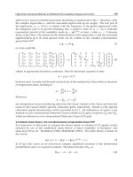
Fig. 7.10. Spondylolysis, and spondylolisthesis (right). (A)
Radiographic representation of an abnormal elongation (grey-
hound sign) of the pars interarticularis, or the “neck” of a scotty
dog (arrow). Other defects, such as sclerosis or lysis in the pars,
are best visualized in this “neck.” (From Lillegard and Kruse,
50
with permission.) (B) “Scotty dog.” A ϭ superior articular process
(ear); B ϭ pedicle (eye); C ϭ pars interarticularis (neck); D ϭ lam-
ina (body); E ϭ inferior articular process (front leg). (C) Severity of
spondylolisthesis and slip angle.
interarticularis) of L5. Sclerosis of the opposite pars may be
present. A standing spot lateral view of L5-S1 allows accurate
assessment of a possible slip. Scoliosis is commonly associated with
spondylolisthesis. Bone scans show increased activity on one or
both sides in symptomatic spondylolysis but are not routinely
required.
If asymptomatic, no treatment is required, and there is no need to
limit contact sports. For a mildly symptomatic patient, temporary
reduction of activity is all that is needed. If symptoms are alleviated,
progressive activity is permitted. Symptoms that are sudden in onset,
traumatically induced, or do not resolve with rest do heal—much as
any fracture would heal—after 10 to 12 weeks of immobilization in a
plastic body jacket or a Boston-type spinal orthosis. In general, once
symptoms resolve, the child can resume normal activities, although
advice regarding return to rigorous spine-bending athletic events (gym-
nastics, diving, downed lineman in football) is controversial (see
Chapter 10).
With spondylolisthesis, if slippage is less than 30% and symptoms
are minimal, treatment is conservative. With persistent pain unre-
sponsive to treatment or slippage more than 30% to 50%, spinal
fusion is recommended. Such fusion is generally at the L5-S1 level
and includes L4 if slippage is more than 50%.
44
Idiopathic Scoliosis
Idiopathic scoliosis is defined as lateral deviation of the spine of more
than 10 degrees (measured by the Cobb method),
45
with structural
change and without congenital anomalies of the vertebrae. It is inher-
ited in an autosomal-dominant manner with variable penetrance or a
multifactorial condition. It occurs in approximately 2% of the popu-
lation. Normally, only about one fifth to one sixth of this group
require treatment.
46
Scoliosis is a painless condition usually identified by shoulder,
scapular, or pelvic asymmetry during school screening or routine
physical examination. Forward bending (Adam’s) testing is done with
the child standing straight and bending forward with palms together
and knees straight. Truncal asymmetry, most commonly right rib
prominence, may be seen. Any limb length irregularity should be
noted and corrected by placing blocks under the short leg and level-
ing the pelvis prior to examination. Neurological examination is nor-
mal. Initial radiological evaluation consists of standing PA and lateral
spine films on a long cassette to include the pelvis. The curve is meas-
ured using the Cobb method
45
(Fig. 7.11). If a structural curve of 10
174 Mark D. Bracker et al.
to 20 degrees is identified, orthopedic referral is recommended.
Painful scoliosis or an atypical curve pattern (apex left thoracic) is
indicative of possible underlying neurological problems, such as
syringomyelia or spinal cord lesion, and is probably not idiopathic
scoliosis.
The risk of curve progression is higher in young children, in those
with large curves or double curves, and in girls. Bracing is usually ini-
tiated for curves of more than 20 degrees with documented progres-
sion and growth remaining or for curves initially 30 degrees or more.
Curves of more than 45 to 50 degrees are usually not amenable to
bracing, so surgery is recommended, as the risk of continued progres-
sion after skeletal maturity is high in this group.
46
Scheuermann’s Disease
Scheuermann’s disease (juvenile kyphosis) is defined as an abnormal
increase in thoracic kyphosis (normal 20–40 degrees) during puberty
with at least 5 degrees of anterior wedging of at least three or more
adjacent vertebrae. It is to be distinguished from postural round back,
which is more flexible and lacks radiographical changes in the verte-
brae.
47
The etiology is unclear, but a familial incidence is noted in
30% to 48% of cases. It occurs in about 1% of the population and is
more common in boys.
Clinically, it is possible to distinguish two forms of juvenile kypho-
sis. Thoracic Scheuermann’s disease has an apex of the curve at T7–9,
7. Musculoskeletal Problems of Children 175
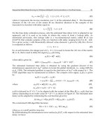
OBSERVATION
COBB ANGLE
0 - 25Њ 25Њ - 45Њ > 45Њ
BRACE SPINAL FUSION
Fig. 7.11. Measuring the Cobb angle and treatment of idiopathic
scoliosis.
176 Mark D. Bracker et al.
and thoracolumbar Scheuermann’s disease has an apex at T11-12.
Cosmetic deformity is often the chief complaint. Pain is usually
aching and occurs more commonly with the thoracolumbar form.
Radiographs should include standing posteroanterior and lateral
scoliosis films. Hyperextension lateral films help to determine the
flexibility of the curve. Radiographs show irregularity of the vertebral
endplates, anterior wedging of 5 degrees or more of three or more
adjacent vertebrae, Schmorl’s nodes, and increased kyphosis meas-
ured between T4 and T12 by the Cobb method.
Kyphosis may worsen during the growing period. Curves of 40 to
60 degrees may be treated by a trial of hyperextension exercises if
the curve is supple and demonstrates active correction. Curves of 60
to 75 degrees are treated with a Milwaukee brace or underarm
orthosis with a breastplate. Bracing is begun if the vertebral end plates
are not fused to the vertebral body, with full-time wearing for 6 to 12
months and then part-time (about 16 hours/day) for 6 months or until
the end plate fuses. Bracing is less effective for curves of more
than 65 to 75 degrees or after skeletal maturity. Surgery may be
indicated for cosmesis, progressive deformity despite bracing, or
intractable pain. No long-term cardiopulmonary problems have been
identified.
48,49
References
1. Karoll LA. Rotational deformities in the lower extremities. Curr Opin
Pediatr 1997;9:77–90.
2. Engel FM, Staheli LT. The natural history of torsion and other
factors influencing gait in early childhood. Clin Orthop 1974;99:
12–17.
3. Wells L. Common lower extremity problems in children, primary care.
Clin Office Pract 1996;23(2):299–303.
4. Brink DS, Levitsky DR. Cuneiform and cuboid wedge osteotomies for
correction of residual metatarsus adductus: a surgical review. J Foot
Ankle Surg 1995;34:371–8.
5. Fabray G, MacEwen GD, Shands AR Jr. Torsion of the femur: a follow-
up study in normal and abnormal conditions. J Bone Joint Surg
1973;55A:1726–38.
6. Kling TF, Hensinger RN. Angular and torsional deformities of the lower
limbs in children. Clin Orthop 1983;176:136–47.
7. Bruce RW Jr. Torsional and angular deformities. Pediatr Clin North Am
1996;43(4):867–81.
8. Mielke CH, Stevens PM. Hemiepiphyseal stapling for knee deformities
in children younger than 10 years: a preliminary report. J Pediatr Orthop
1996;16:423–9.
7. Musculoskeletal Problems of Children 177
9. Cummings RJ, Lovell WW. Operative treatment of congenital idiopathic
club foot. J Bone Joint Surg 1988;70A:1108–12.
10. Ponseti IV. Congenital clubfoot, the results of treatment. J Bone Joint
Surg 1963;45A:261–9.
11. Cowell H. Talocalcaneal coalition and new causes of peroneal spastic
flatfoot. Clin Orthop 1972;85:16–22.
12. Hoffinger SA. Evaluation and management of pediatric foot deformities.
Pediatr Clin North Am 1996;43:1091–111.
13. Paulos L, Samuelson KM. Pes cavovarus: review of a surgical approach
using selective soft tissue procedures. J Bone Joint Surg 1980;62A:
942–53.
14. Wenger DR, Mauldin D, Speck G, Morgan D, Lieber R. Corrective shoes
and inserts as treatment for flexible flatfoot in infants and children.
J Bone Joint Surg 1989;71A:800–10.
15. Steward M. Miscellaneous afflictions of the foot. In: Campbell’s opera-
tive orthopedics. St. Louis: Mosby, 1980;1703.
16. Salter RB, Zaltz C. Anatomic investigation of the mechanism of injury
and pathologic anatomy of “pulled elbow” in young children. Clin
Orthop 1971;77:134.
17. Southmayd W, Ehrlich MB. Idiopathic subluxation of the radial head.
Clin Orthop 1976;121:271.
18. Hardinse K. The etiology of transient synovitis of the hip in childhood.
J Bone Joint Surg 1970;52B:100–7.
19. Del Beccaro M, Champoux A, Bockers T, Mendelman P. Septic arthritis
versus transient synovitis of the hip: The value of screening laboratory
tests. Ann Emerg Med 1992;21(12):1418–22.
20. Roy DR. Current concepts in Legg-Calve-Perthes Disease. Pediatr Ann
1999;28(12):748–52.
21. Busch M, Morrisy R. Slipped capital femoral epiphysis. Orthop Clin
North Am 1987;18:637–47.
22. Fahey JJ, O’Brien ET. Acute slipped capital femoral epiphysis: review of
the literature and report of ten cases. J Bone Joint Surg 1965;47A: 1105–27.
23. Kallio PE, Paterson DC, Foster BK, Lequene GW. Classification in
slipped capital femoral epiphysis. Clin Orthop 1993;294:196–203.
24. Loder RT, Richards BS, Shapiro PS, Reznick LR, Aronsson DD. Acute
slipped capital femoral epiphysis: the importance of physical stability.
J Bone Joint Surg 1993;75A:1134–40.
25. Loder RT, Aronson DD, Greenfield ML. The epidemiology of bilateral
slipped capital femoral epiphysis. J Bone Joint Surg 1993;75A:1141–7.
26. Umas H, Liebling M, Moy L, Harmamati N, Macy N, Pritzker H. Slipped
capital femoral epiphysis: a physeal lesion diagnosed by MRI, with radi-
ographic and CT correlation. Skel Radiol 1998;27:139–44.
27. Committee on Quality Improvement and Subcommittee on
Developmental Dysplasia of the Hip. American Academy of Pediatrics:
clinical practice guideline: early detection of developmental dysplasia of
the hip. Pediatrics 2000;105(4):896–905.
28. Gerscovich EO. Radiologists’ guide to the imaging in the diagnosis and
treatment of developmental dysplasia of the hip. Skel Radiol 1997;
26:386–97.
178 Mark D. Bracker et al.
29. Rosendahl K, Markestad T, Lie RT. Ultrasound in the early diagnosis of
congenital dislocation of the hip: the significance of hip stability versus
acetabular morphology. Pediatr Radiol 1992;22:430–3.
30. Graf R. Hip semiography: how reliable? Sector scanning versus linear
scanning? Dynamic versus static examination? Clin Orthop 1992;281:
18–21.
31. Weinstein SL. Congenital hip dislocation: long-range problems, residual
signs and symptoms after successful treatment. Clin Orthop 1992;
281:69–74.
32. Harris IE, Dickens R, Menelaus MB. Use of the Pavlic harness for
hip displacements: when to abandon treatment. Clin Orthop 1992;
281:29–33.
33. Gruppo R, Glueck CJ, Wall E, Roy D, Wang P. Legg-Perthes disease in
three siblings, two heterozygous and one homozygous for the factor V
Leiden mutation. J Pediatr 1998;132(5):885–8.
34. Catterall A. The natural history of Perthes’ disease. J Bone Joint Surg
1971;53B:37–53.
35. Gershuni DH. Preliminary evaluation and prognosis in Legg-Calvé-
Perthes disease. Clin Orthop 1980;150:16–22.
36. Herring JA. The treatment of Legg-Calve-Perthes disease. J Bone Joint
Surg 1994;76A(3):448–57.
37. McAndrew MP. Weinstein SL. A long-term follow-up of Legg-Calve-
Perthes disease. J Bone Joint Surg 1984;66A(6):860–9.
38. Osgood RB. Lesions of the tibial tubercle occurring during adolescence.
Boston Med J 1903;148:114–17.
39. Sever JW. Apophysitis of the os calcis. NY Med J 1912;95:
1025–9.
40. Obedian RS, Grelsamer RP. Osteochondritis dissecans of the distal femur
and patella. Clin Sports Med 1997;16:157–74.
41. De Smet AA. Omer AI, Graf BK. Untreated osteochondritis dissecans of
the femoral condyles: prediction of patient outcome using radiographic and
MR findings. Skel Radiol 1997;26:463–7.
42. Wiltse LL, Newman PH, Macnab I. Classification of spondylolysis and
spondylolisthesis. Clin Orthop 1976;117:23–9.
43. Hensinger RN. Spondylolysis and spondylolisthesis in children and ado-
lescents. J Bone Joint Surg 1989;71A:1098–107.
44. Boxall D, Bradford DS, Winter RB, Moe JH. Management of severe
spondylolisthesis in children and adolescents. J Bone Joint Surg
1979;61:479–95.
45. Sorensen KH. Scheuermann’s juvenile kyphosis. Copenhagen:
Munksgaard, 1964.
46. Hensinger RN, Greene TL, Hunter LY. Back pain and vertebral changes
simulating Scheuermann’s kyphosis. Spine 1982;6:341–2.
47. Bradford DS. Juvenile kyphosis. In: Bradford DS, Lonstein JE, Moe JH,
et al, eds. Moe’s textbook of scoliosis and other spinal deformities, 2nd
ed. Philadelphia: WB Saunders, 1987;347–68.
48. Cobb J. Outline for study of scoliosis. AAOS Instruct Course Lect
1948;5:261–275, Ann Arbor: J. W. Edwards.
49. Moe JH, Byrd JA III. Idiopathic scoliosis. In: Bradford DS, Lonstein JH,
Moe JH, et al, eds. Moe’s textbook of scoliosis and other spinal deformi-
ties, 2nd ed. Philadelphia: WB Saunders, 1987;191–232.
50. Lillegard W, Kruse R. In Taylor RB, eds. Family medicine: principles
and practice, 4th ed. New York: Springer-Verlag, 1993.
51. Taylor RB. Family medicine: principles and practice, 6
th
ed. New York:
Springer-Verlag, 2003.
52. Gerberg LF, Micheli LJ, Nontraumatic hip pain in active children: a crit-
ical differential. Phys Sports Med 1996;24:69–74.
53. Peck DM. Apophyseal injuries in the young athlete. Am Fam phys
1995;51:1891–5.
7. Musculoskeletal Problems of Children 179
Osteoporosis
Paula Cifuentes Henderson and
Richard P. Usatine
Osteoporosis is a major health concern affecting approximately 20
million people in the United States. It is responsible for more than 1.3
million fractures annually,
1
with $15 billion in direct financial expen-
ditures to treat these fractures.
2
The clinical consequences of an osteo-
porotic fracture include increased mortality, disability, and the need
for long-term nursing care. After a hip fracture the mortality rate of
patients 65 to 79 years old at 1 year is between 20% and 30%, and
these rates worsen with increased age.
3
Among those who survive,
50% won’t be able to work without some type of assistance. After a
collapsed osteoporotic vertebra, 30% of patients will experience
chronic disabling back pain and spinal deformity.
4,5
Osteoporotic frac-
tures have a profound impact on quality of life, decreasing the physi-
cal, functional, and psychological performance secondary to pain,
deformities, and inability to perform the activities of daily living
(ADL)
6
.
Osteoporosis is a disease characterized by low bone mass and
michroarchitectural deterioration of bone tissue leading to enhanced
bone fragility and a consequent increase in fracture risk.
7
It can be a
silent disease because it is often asymptomatic until a fracture occurs.
The lifetime risk of a 50-year-old white woman of having an osteo-
porotic fracture is 40%. Fractures secondary to osteoporosis are more
common in women than in men and in Caucasians and Asians than in
African Americans and Latinos.
8
These fractures most commonly
occur at the hip, vertebrae, and wrists.
Primary osteoporosis is related to aging and not associated with
chronic illness. Secondary osteoporosis is related to chronic conditions
that contribute to accelerated bone loss such as with hyperparathy-
roidism, malignancy, renal failure, and hyperthyroidism.
9
Assessment and Diagnosis
Risk Factor Assessment
Start with the medical history and ask questions about:
Menopause (surgical and natural)
Family history of osteoporosis (especially mother)
Exercise
Diet
Smoking
Alcohol intake
Other risk factors such as age, gender, ethnicity, and slender body habi-
tus can usually be observed without asking specific questions. The
physical exam includes the measurement of height and weight, and the
examination of the spine looking for any signs of deformity such as
kyphosis, scoliosis, and limited range of motion. Screening for sec-
ondary forms of osteoporosis may be helpful. Assess the patient’s risk
of falling by asking about a history of falls and a decrease in visual
acuity.
10,11
Genetic Issues
The prevalence of osteoporosis varies by sex, ethnicity, and race.
12
Decreased bone density is more common in women of Northern
European or Asian descent. Women and men experience age-related
decrease in bone mass density starting at midlife, but women experi-
ence more rapid bone loss after the menopause.
13
Genetic syndromes
like Turner’s (45,X0) syndrome patients have streak ovaries and
decreased estrogen production leading to the early development of
osteoporosis.
14
Endocrine Factors
Risk factors associated with decreased bone density include early
estrogen deficiency secondary to surgery or to early menopause,
hyperthyroidism, hyperparathyroidism, hypercortisolism, Addison’s
disease, and Cushing’s syndrome.
14
182 Paula Cifuentes Henderson and Richard P. Usatine
Medications
Chronic use of certain medications that affect the bone metabolism,
such as corticosteroids, exogenous thyroid hormone, gonadotropin-
releasing hormone (GnRH) analogues, anticoagulants, and anticon-
vulsants, increase the risk of osteoporosis and subsequent fractures.
15
Lifestyle
Excessive use of alcohol depresses osteoblastic function and increases
the risk of osteoporosis. Physical activity early in life contributes to
higher peak bone mass and reduces the risk of falls by approximately
25%.
16
Good nutrition with a balanced diet is necessary for the devel-
opment of healthy bones. Calcium and vitamin D are required for the
prevention and treatment of osteoporosis. There are data to support rec-
ommendations (found later in the chapter) for specific dietary calcium
intakes at various stages in life.
17,18
Patients at high risk also include
those who pursue thinness excessively, have a history of an eating dis-
order,
19
restrict their intake of dairy products, don’t consume enough
vegetables and fruits, and have a high intake of low-calcium/high-phos-
phorus beverages like sodas. These beverages have a negative effect on
calcium balance.
Laboratory Assessment
If the history and physical exam suggests secondary causes of osteo-
porosis, the physician should consider tests such as thyroid-stimulat-
ing hormone (TSH), parathyroid hormone (PTH), calcium, vitamin D,
urine N-teloptide, complete blood count (CBC), chem panel, cortisol,
erythrocyte sedimentation rate (ESR), or serum protein electrophore-
sis, based on the differential diagnosis.
20,21
Bone Densitometry Assessment
To prevent osteoporosis, the physician should attempt to establish
early detection of low bone mineral density (BMD). Currently there
is no accurate measure of bone strength, but BMD is the accepted
method to establish a diagnosis of osteoporosis and predict future
fracture risk.
22,23
The World Health Organization (WHO) defines
osteoporosis as a BMD 2.5 standard deviations (SDs) below the mean
for young white adult women. This definition does not apply to other
ethnic groups, men, or children.
7,24
The U.S. Preventive Services Task
Force suggests that the primary reason to screen postmenopausal
8. Osteoporosis 183
women is to check for a low BMD so that early intervention may be
initiated to slow the further decrease of the bone density.
25
The ulti-
mate goal is to prevent vertebral and hip fractures.
The most thoroughly studied and most widely used technique to
measure BMD is the dual-energy x-ray absortiometry (DEXA) scan.
This is considered to be the gold standard screening test to measure the
BMD of the hip and spine. It is less expensive and involves less radia-
tion exposure than the quantitative computed tomography (CT). Since
some patients don’t respond to therapy for osteoporosis, the BMD
results can also be used to follow them and evaluate their response to
treatment. Bone mass should be measured in postmenopausal women 1
to 2 years following the initiation of therapy.
The report of the DEXA provides a T score and a Z score. The T
score is defined as the number of SDs above or below the mean BMD
for sex- and race-matched young controls (not age matched). This
should be distinguished from a Z score, which is defined as the num-
ber of SDs above or below the average BMD comparing the patient
with the population adjusted for age, sex, and race. These results can
be used to classify patients into three categories: normal, osteopenic,
and osteoporotic (Table 8.1). Osteoporosis is diagnosed using the
patient’s T score, because the T score is a measure of current fracture
risk. A T score of 1 SD below the age-predicted mean is associated
with a two- to threefold increased risk of fracture. Patients with T
scores more than 2 SDs below the mean have an exponential increase
in their risk of fracture. Z scores have little significant value for clin-
ical practice.
Newer measures of bone strength, such as the ultrasound, are
being introduced as an alternative screening method to the DEXA
scan. This measurement of bone mass is being done through periph-
eral bone mass assessment. In 1998, the Food and Drug
Administration (FDA) approved the use of a portable ultrasound to
184 Paula Cifuentes Henderson and Richard P. Usatine
Table 8.1. World Health Organization (WHO) Diagnostic Criteria
for Osteoporosis
Bone Mineral Density (BMD)
Diagnosis T score
a
Normal Յ1
Osteopenia 1–2.5
Osteoporosis Ն2.5
Severe osteoporosis Ն2.5 and history of fracture
a
Standard deviation (SD) below the mean in healthy young adults.
Source: WHO Study Group.
7
assess bone mass through the measurement of the calcaneous. If a
patient has a low T score in the ultrasound of a peripheral bone, the
current recommendation is to obtain a DEXA of the hip and spine for
further evaluation and treatment.
11
The diagnosis and treatment of osteoporosis should be individual-
ized based on each patient’s risk factors rather than the assessment of
a T score alone.
Indications for bone mineral density assessment include:
Women Ն65 years old who are willing to start drug therapy if BMD
is found to be low
Women Ͻ65 who have at least one additional risk factor for osteo-
porosis
Postmenopausal women with a fracture
Radiographic evidence of bone loss
Long-term steroid use
Hyperparathyroidism
Monitoring therapeutic response if the results would affect the clini-
cal decision.
Although there is no evidence to support this, some clinicians
screen premenopausal women with BMD for the following condi-
tions:
Prolonged oligo/amenorrhea
A long-standing history of eating disorders
Stress fractures
Chronic use of medications that promote bone resorption.
There is a lack of evidence to support the cost-effectiveness of uni-
versal routine bone density screenings or to support the efficacy of
early preventive medications to prevent fractures. Therefore, an indi-
vidualized approach is recommended
25
(Table 8.2).
Bone Remodeling Assessment
Another way to assess bone strength is to measure markers of bone
remodeling (turnover) in the blood or urine. There is some evidence
that bone turnover rate predicts the risk of osteoporotic fractures in
postmenopausal women.
26
These markers include indices of bone
resorption such as serum and urine levels of C- and N-telopeptide, and
indices of bone formation such as osteocalcin and bone-specific alka-
line phosphatase. These markers of bone turnover may be particularly
8. Osteoporosis 185
useful if obtained prior to starting treatment and then repeated in 3 to
6 months to measure the response. Despite the fact that these markers
may identify changes in bone remodeling, they do not predict fracture
risk.
These tests are very expensive and are not recommended for
screening or as the first-line studies to follow treatment response.
However, if the BMD does not increase with treatment, one might
order the turnover markers for further assessment.
Prevention and Treatment
Nonpharmacological
Nonpharmacological therapy for prevention and treatment of osteo-
porosis includes adequate dietary intake of calcium and vitamin D,
weight-bearing exercise, fall precautions, no smoking, and avoidance
of excessive alcohol intake. These steps should be started early in life
and continued through menopause because BMD peaks at about age
35 and then begins to decline with accelerated bone loss after
menopause.
186 Paula Cifuentes Henderson and Richard P. Usatine
Table 8.2. Indications for Bone Mineral Density (BMD) Screening
National Osteoporosis Foundation guidelines
a
Women Ͼ65 willing to start therapy if BMD low
Women Ͻ65 postmenopausal with at least one additional risk factor
All postmenopausal women with fractures
Women considering therapy for osteoporosis, and BMD would affect
decision
Women who have received HRT for a prolonged period
No formal guideline developed in premenopausal women
American Association of Clinical Endocrinologists clinical practice
guidelines
b
Perimenopausal women willing to start therapy if BMD low
X-ray evidence of bone loss
Asymptomatic hyperparathyroidism
Monitoring therapeutic response and BMD would affect decision
Long-term use of glucocorticoid
BMD ϭ bone mineral density; HRT ϭ hormone replacement therapy.
a
National Osteoporosis Foundation (NOF). Physician’s guide to preven-
tion and treatment of osteoporosis. Washington, DC: NOF, 1998, 2000
b
American Association of Clinical Endocrinologists (AACE). Clinical prac-
tice guidelines for the prevention and treatment of postmenopausal osteo-
porosis. Endocrinol Pract 1996;2(2):157–71.
Calcium
According to the National Institutes of Health (NIH) Consensus
Development Conference, the optimal recommended dose of elemen-
tal calcium is the amount that each person needs to maintain adult
bone mass and minimize bone loss later in life (Table 8.3). The rec-
ommended dose for postmenopausal women Ͻ65 years old who are
on hormone replacement therapy (HRT) is 1000 mg/day and 1500
mg/day for all other postmenopausal women.
27
Calcium supplements
are advisable if diet cannot supply the recommended amount neces-
sary. Calcium citrate should be taken between meals while calcium
carbonate should be taken with meals because it is best absorbed with
gastric acid. Calcium should not be taken with iron because the iron
decreases the absorption. Several studies show that calcium supple-
ments can reduce bone loss in postmenopausal women and will
reduce the risk of fractures.
27
The effect is not strong enough to rec-
ommend calcium alone for osteoporosis prevention.
8. Osteoporosis 187
Table 8.3. Optimal Calcium Intake
Population NIH RDA
Infants, children, and young adults
0–6 Months 400 400
6–12 Months 600 600
1–10 years 800–1200 800
11–24 years 1200–1500 1200
Adult women
Pregnant and lactating
Ͻ24 years 1200–1500 1200
Ͼ24 years 1200 800
Premenopausal
25–49 years 1000 800
Postmenopausal
50–64 years On estrogen 1000 800
Not on estrogen 1500 800
Ն65 years 1500
Adult men
25–64 years 1000 800
Ն65 years 1500 800
a
Calcium recommendations in mg/day.
NIH ϭ National Institutes of Health; RDA ϭ Recommended Daily
Allowance.
Adapted from the NIH Consensus Conference, 1994.
18
Vitamin D
The recommended daily intake of vitamin D needed for adequate cal-
cium absorption is 400 to 800 IU. Vitamin D deficiency can occur in
patients with inadequate sunlight exposure. Sunlight exposure is
shown to be useful in preventing hip fractures, especially in elderly
institutionalized women.
28
Physical Activity
Adequate physical activity may exert a positive influence on bone
mass and is necessary for bone acquisition and maintenance. The
extent of this influence and the most effective type of program are not
fully understood. Most trials of exercise intervention show that a
reduction of falls is likely to be secondary to improved muscular
strength and balance. Low-impact exercise like walking has minimal
effect on BMD; high-impact exercise like weight training stimulates
the increase of BMD. Women who exercise regularly are at a lower
risk of hip fractures.
29
However, excessive exercise by competitive
athletes can also be a risk factor for bone loss, particularly if they have
hypoestrogenic oligo/amenorrhea.
Fall Prevention
Most osteoporotic fractures result from a fall. Risk factors for falling
include visual or hearing problems, gait disturbances, underlying
conditions that predispose the patient to syncope, cognitive impair-
ment, and the use of certain medications such as diuretics, anti-
hypertensive, benzodiazepines, and antidepressants. Home safety
precautions may help to prevent falls. External hip protectors have
been shown to provide protection against hip fractures in frail elderly
adults.
Pharmacological Treatment
Pharmacological treatment should be initiated in women with:
No risk factors and who have T scores below 2 SDs
Risk factors and T scores below 1.5 SD
A history of vertebral or hip fractures
Multiple risk factors over 70 years of age without BMD measurement.
The pharmacological agents for treatment and prophylaxis of
osteoporosis include HRT, calcium and vitamin D supplements,
188 Paula Cifuentes Henderson and Richard P. Usatine
bisphosphonates, selective estrogen receptor modulators (SERMs),
intranasal calcitonin, and parathyroid hormone (PTH). While the most
widely prescribed regimen is HRT with calcium and vitamin D, there
are many reasons to consider using the other medications.
Inhibitors of Bone Resorption
Hormonal
Hormone Replacement Therapy (HRT)
In the PEPI trial, HRT increased BMD at the hip by 1.7 % and at the
spine by 3.5% to 5.0% over a 3-year period compared to placebo. HRT
inhibits bone loss for the duration of the therapy, which recurs once
therapy is discontinued. In premenopausal women with osteoporosis
secondary to hypoestrogenic stages, early intervention with estrogen to
achieve return of menses, is critical since bone loss may be irreversible.
Observational studies consistently suggest that postmenopausal HRT
reduces the risk of hip and other types of fractures.
30
Evidence from
randomized controlled trials (RCTs), especially for vertebral fracture
prevention, is less available. In a Danish RCT, HRT reduced forearm
fracture incidence in recent postmenopausal women.
31
In another ran-
domized trial, HRT and vitamin D prevented nonvertebral fractures in
postmenopausal women.
32
A meta-analysis published in 2001 suggests
that estrogen reduces risk of nonvertebral fractures by 27%. Estrogen
seemed to reduce the risk of fractures by 33% in younger women, but
had no significant effect in women aged 60 years or older.
33
Before starting a patient on HRT the physician and the patient need
to consider all the risks and benefits. Common adverse effects such as
breakthrough bleeding and breast tenderness or enlargement should be
discussed. The risk of breast cancer and heart disease are very impor-
tant issues. The relationship of HRT to breast cancer and heart disease
is still controversial. HRT should be used with caution in patients who
have a personal or family history of breast or endometrial cancer, or a
history of a hypercoagulable state or thromboembolic episodes.
Informed consent should be given to all patients.
In postmenopausal women without contraindications to HRT, any of
the three recommended regimens could be used: estrogen alone in
women without a uterus, estrogen with progestin daily, and estrogen
with progestin in a cyclic manner (estrogen every day and progestin
only for 10 to 14 days of the month). The most common regimen is con-
jugated estrogen at a daily dose of 0.625 mg or its equivalent. This dose
can be used if the therapy begins at the onset of menopause in order to
prevent the rapid bone loss that occurs early. If the postmenopausal
8. Osteoporosis 189
woman is older at the time of starting the HRT, she might be more
sensitive to the standard dose. One might consider starting at half the
dose (0.3 mg) to avoid discontinuation secondary to adverse effects.
Estrogen alone is avoided in women with a uterus in order to prevent
endometrial cancer. Estrogen given with progesterone may actually
decrease the risk of endometrial cancer.
Selective Estrogen Receptor Modulators (SERMs)
SERMs are an important alternative for women with contraindica-
tions or intolerance to estrogen therapy. Tamoxifen and raloxifene
were FDA approved in 2000 for treatment of postmenopausal osteo-
porosis. The main goal is to maximize the beneficial estrogenic effect
in bone and minimize the effect on the breast and endometrium.
One study showed that raloxifene may decrease the risk of verte-
bral fracture by 36%, but there has been no published evidence for hip
fracture reduction.
34
Both SERMs are contraindicated in women at
risk for deep venous thrombosis.
Bisphosphonates
Alendronate (Fosamax)
Alendronate was approved by the FDA in 1995 for treatment of post-
menopausal osteoporosis. It reduces the risk of vertebral fractures by
30% to 50% and increases the BMD at the spine and hip.
35
Alendronate also reduces the risk of fractures in men and women with
osteoporosis secondary to the chronic use of steroids.
36
One study evaluated the addition of alendronate to HRT in the treat-
ment of postmenopausal women with low BMD despite ongoing treat-
ment with estrogen.
37
Compared with HRT alone, at 12 months
alendronate plus HRT produced significantly greater increases in BMD
of the lumbar spine (3.6% vs. 1.0%, p Ͻ .001) and hip trochanter (2.7%
vs. 0.5%, p Ͻ .001). This study suggests that alendronate may be ben-
eficial when added to HRT in postmenopausal women with low BMD
despite ongoing treatment with HRT. However, it should be noted that
the outcome measured was BMD and not fractures.
The recommended starting dose for postmenopausal osteoporosis
prevention is 5 mg/day with a maintenance dose of 10 mg/day. The
most common side effect is esophageal irritation secondary to reflux.
Therefore, the patient should take alendronate with a full glass of
water without food and remain upright for at least 30 minutes to avoid
reflux. Another available regimen is 70 mg once a week. This weekly
dose was demonstrated to be as effective with fewer gastrointestinal
190 Paula Cifuentes Henderson and Richard P. Usatine
side effects.
38
At this time, the use of alendronate has not been
approved for premenopausal women.
Risedronate (Actonel)
Risedronate is a newer biphosphonate approved in 2000 by the FDA for
treatment of postmenopausal osteoporosis. While the indications are the
same as alendronate, it has fewer gastrointestinal side effects. Both
agents cost over $50 a month. In a randomized, double-blind, placebo-
controlled trial of 2458 ambulatory postmenopausal women younger
than 85 years with at least 1 vertebral fracture at baseline, risedronate
decreased the relative incidence of new vertebral fractures by 41% over
3 years. The absolute risk reduction was from 16.3% to 11.3%. The
cumulative incidence of nonvertebral fractures over 3 years was
reduced by 39% (5.2 % vs 8.4%). The overall safety profile of rise-
dronate, including gastrointestinal safety, was similar to that of placebo.
The most effective dose was 5 mg/day.
39
Calcitonin
Calcitonin is helpful when treating painful osteoporosis due to its sig-
nificant analgesic effect. This hormone inhibits bone resorption by
acting directly on the osteoclasts. The PROOF study is controversial;
it demonstrates a reduction of vertebral fractures with calcitonin.
40
It
is available in nasal spray at a recommended dose of 200 IU/day that
corresponds to one squirt through one nostril every day alternating
nostrils; or in the injectable form (200 units/mL) to be used three to
five times/week at a dose of 50 to 100 IU/dose.
Stimulators of Bone Formation
Parathyroid Hormone (PTH)
PTH is the most promising anabolic agent that stimulates bone for-
mation. It is still undergoing clinical trials. Even though it increases
the BMD of the lumbar spine,
41
there are no data on fracture risk. One
disadvantage is that it must be administered by subcutaneous injec-
tion. It is not yet approved by the FDA.
Fluoride
Fluoride stimulates bone formation but does not decrease the risk of a
fracture. A meta-analysis showed that fluoride increases bone mineral
8. Osteoporosis 191
density at the lumbar spine and does not reduce the number of verte-
bral fractures. Increasing the dose of fluoride actually increased the
risk of nonvertebral fractures and gastrointestinal side effects.
42
It is
not approved by the FDA for osteoporosis prevention and treatment.
Conclusion
Fractures of the hip, vertebrae, and wrists from osteoporosis cause sig-
nificant decreases in the quality of life for many older individuals. The
complications of hip fractures can also lead to death. Better methods
for prevention, early detection, and treatment now exist. Healthy
lifestyles, including no smoking, exercise, good diet, and calcium
intake, can help to prevent osteoporosis. By assessing family history,
ethnicity, body type, and other risk factors, physicians can target pre-
vention and screening efforts to patients at highest risk for osteoporo-
sis. Patients at higher risk should probably be screened using a DEXA
scan. Pharmacological therapies such as hormone replacement, cal-
cium, vitamin D, bisphosphonates, SERMs, and calcitonin can help
prevent BMD loss and may reduce the risk of fractures. Currently, the
data for fracture prevention are stronger for the bisphosponates than
for hormonal therapy. Every family physician should feel comfortable
screening for, preventing, and treating osteoporosis.
Suggested Web Sites
The National Institutes of Health Osteoporosis and Related Bone
Diseases: National Resource Center www.osteo.org
The National Osteoporosis Foundation. www.nof.org
References
1. Riggs BL, Melton LJ III. The worldwide problem of osteoporosis:
insights afforded by epidemiology. Bone 1995;17:505S–11S.
2. Chrischilles E, Shireman T, Wallace R. Costs and health effects of osteo-
porotic fractures. Bone 1994;15:377–87.
3. Lu Yao GL, Baron JA, Barrett JA. Treatment and survival among elderly
Americans with hip fractures: a population-based study. Am J Public
Health 1994;84:1287–91.
4. Watts NB. Hip fracture prevention in nursing homes: clinical importance
and management strategies. Consult Pharm 1996;11:944–54.
5. Cummings SR, Kelsey JL, Nevitt MC. Epidemiology of osteoporosis and
osteoporotic fractures. Epidemiol Rev 1985;7178–208.
192 Paula Cifuentes Henderson and Richard P. Usatine
6. Gold DT. The clinical impact of vertebral fractures. Bone 1996;
18:185–90.
7. World Health Organization (WHO) Study Group. Assessment of
fracture risk and its application to screening for postmenopausal osteo-
porosis. Geneva, Switzerland: WHO Technical Report Series,
1994;843.
8. National Osteoporotic Foundation, 2025 osteoporosis prevalence figures:
state by state report. January 1997. Women’s Health Matters 1998;
2(30):1.
9. Harper KD, Weber TJ. Secondary osteoporosis. Diagnostic considera-
tions. Endocrinol Metab Clin North Am 1998;27(2):325–48.
10. World Health Organization (WHO). Assessment of fracture risk and its
application to screening for postmenopausal osteoporosis. WHO techni-
cal report series 843. Geneva: WHO, 1994.
11. Heinemann DF. Osteoporosis. An overview of the National Osteoporosis
Foundation clinical practice guide. Geriatrics 2000;55(5):31–6.
12. Pocock NA, Eisman JA, Hopper JL. Genetic determinants of bone mass
in adults. J Clin Invest 1987;80:706–10.
13. Seeman E. Growth in bone mass and size: are racial and gender differ-
ences in bone density more apparent than real? J Clin Endocrinol Metab
1998;83:1414–18.
14. Harper KD, Weber TJ. Secondary osteoporosis. Diagnostic considerations.
Endocrinol Metab Clin North Am 1998;27(2):325–48.
15. Saag KG, Emkey R, Schinitzer A. For the glucocorticoid-induced osteo-
porosis intervention study group. Alendronate for the prevention and
treatment of glucocorticoid-induced osteoporosis. N Engl J Med
1998;339:292–9.
16. Bassey EJ, Rothwell MC, Littlewood JJ. Pre- and post-menopausal
women have different bone mineral density responses to the same high
impact exercise. J Bone Miner Res 1998;13:1805–13.
17. Dawson B, Harris SS, Krall EA. Effect of calcium and vitamin D sup-
plementation on bone density in men and women 65 years of age or older.
N Engl J Med 1997;337:670–6.
18. NIH Consensus Development Panel on Optimal Calcium Intake. NIH
Consensus Conference: optimal calcium intake. JAMA 1994;272:
1942–8.
19. Hotta M, Shibasaki T, Sato K. The importance of body weight history in
the occurrence and recovery of osteoporosis in patients with anorexia
nervosa: evaluation by dual x-ray absorptiometry and bone metabolic
markers. Eur J Endocrinol 1998;139:276–83.
20. Consensus Development Conference. Diagnosis, prophylaxis and treat-
ment of osteoporosis. Am J Med 1993;94:646–50.
21. Nattiv A. Osteoporosis: its prevention, recognition, and management.
Family Pract Recert 1998;20(2):17–41.
22. Black DM, Cummings SR, Genant HK. Axial and appendicular bone
density predict fractures in older women. J Bone Miner Res
1996;11:707–30.
23. AACE Clinical Practice guidelines for the prevention and treatment of
postmenopausal osteoporosis. Endocrinol Pract 1996;2(2):157–71.
8. Osteoporosis 193
24. National Osteoporosis Foundation. Physicians guide to prevention and
treatment of osteoporosis. Bele Mead, NJ: Experta Medica, 1998.
25. NIH Consensus Conference. Osteoporosis prevention, diagnosis and
therapy. JAMA 2001;285(6):785–94.
26. Garnero P, Hauserr E, Chapui MC. Markers of bone resorption predict
hip fracture in elderly women: the EPIDOS prospective study. J Bone
Miner Res 1996;11:1531–8.
27. Reid R, Ames RW, Evans MC. Effect of calcium supplementation on
bone loss in postmenopausal women. N Engl J Med 1993;328:460–4.
28. Chapuy MC, Arlot ME, Duboeuf F. Vitamin D3 and calcium to prevent
hip fractures in the elderly women. N Engl J Med 1992;327:1637–42.
29. Paganini-Hill A, Chao A, Ross RK. Exercise and other factors in the pre-
vention of hip fracture: the Leisure World study. Epidemiology
1991;2:16–25.
30. Grady D, Cummings SR. Postmenopausal hormone therapy for preven-
tion of fractures: how good is the evidence? JAMA 2001;285(22):
2909–10.
31. Mosekilde L, Beck-Nielsen H, Sorensen OH, et al. Hormonal replace-
ment therapy reduces forearm fracture incidence in recent post-
menopausal women—results of the Danish Osteoporosis Prevention
Study. Maturitas 2000;36(3):181–93.
32. Komulainen MH, Kroger H, Tuppurainen MT, et al. HRT and Vit D in
prevention of non-vertebral fractures in postmenopausal women; a 5 year
randomized trial. Maturitas. 1998;31(1):45–54.
33. Torgerson DJ, Bell-Syer SEM. Hormone replacement therapy and pre-
vention of nonvertebral fractures: a meta-analysis of randomized trials.
JAMA 2001;285:2891–7.
34. Ettinger B, Black DM, Mitlak BH. Reduction of vertebral fracture risk in
postmenopausal women with osteoporosis treated with raloxifene: results
from a 3-year randomized clinical trial. JAMA 1999;282:637–45.
35. Karpf DB, Shapiro DR, Seeman E. Prevention of nonvertebral fractures by
alendronate: a meta-analysis. JAMA 1997;277:1159–64.
36. Saag KG, Emkey R, Schnitzer TJ, et al. Alendronate for the prevention
and treatment of glucocorticoid-induced osteoporosis. Glucocorticoid-
Induced Osteoporosis Intervention Study Group. N Engl J Med
1998;339(5):292–9.
37. Lindsay R, Cosman F, Lobo RA, et al. Addition of alendronate to ongo-
ing hormone replacement therapy in the treatment of osteoporosis: a ran-
domized, controlled clinical trial. J Clin Endocrinol Metab
1999;84(9):3076–81.
38. Baran D. Osteoporosis. Efficacy and safety of a bisphosphonate dosed
once weekly. Geriatrics 2001;56(3):28–32.
39. Harris ST, Watts NB, Genant HK. Effects of risedronate treatment on ver-
tebral and nonvertebral fractures in women with postmenopausal osteo-
porosis. JAMA 1999;282:1344–52.
40. Chestnut CH, Silverman SL, Andriano K. Salmon calcitonin nasal spray
reduces the rate of new vertebral fractures independently of known major
pre-treatment risk factors: accrued 5 year analysis of the PROOF study.
Bone 1998;23(5):S290.
194 Paula Cifuentes Henderson and Richard P. Usatine
41. Lindsay R, Cosman F, Nieves J. Does treatment with parathyroid hor-
mone increases vertebral size? Osteoporosis International 2000. World
Congress on Osteoporosis 2000;11(2):556,S206.
42. Haguenauer D, Welch V, Shea B, Tugwell P, Adachi JD, Wells G.
Fluoride for the treatment of postmenopausal osteoporotic fractures: a
meta-analysis. Osteoporos Int 2000;11(9):727–38.
8. Osteoporosis 195
Gout
James F. Calvert, Jr.
Gout encompasses a spectrum of diseases caused by precipitation of
uric acid crystals in tissue. The gouty disorders include (1) acute monar-
ticular arthritis caused by uric acid crystals in joints, (2) nephrolithiasis,
(3) soft tissue deposits of urate crystals known as tophi, and (4) uric acid
renal disease. Gout occurs in about 1.3% of men over 40, making it the
most common form of inflammatory arthritis in men. The prevalence in
women is about half that in men,
1
although there is evidence that the rel-
ative prevalence of gout in women has increased.
2
The prevalence of
gout increases with age, and it is more common in persons of African
or Polynesian ancestry.
Hyperuricemia
Hyperuricemia is caused by either increased production of uric acid
or decreased ability to excrete it; some of the more common disorders
characterized by hyperuricemia are listed in Table 9.1. Hyperuricemia
is defined as the presence of a serum uric acid over 7.0 mg/dL (420
mol/L). Uric acid is less likely to form crystals at concentrations
below this level. The risk of having all the gouty disorders increases
proportionately to the serum uric acid level.
3
Prophylactic treatment
to lower the uric acid level incurs no benefit to patients with asymp-
tomatic hyperuricemia and is more risky and expensive than no treat-
ment,
4
although the discovery that a patient has hyperuricemia should
lead to an attempt to determine its etiology and significance. An
exception to this rule is that patients with lymphoproliferative disor-
ders or those about to undergo chemotherapy for other malignancies
should be treated prophylactically with allopurinol.
5,6
Uric acid levels