Disorders Techniques in Investigation and Diagnosis - part 5 ppsx
Bạn đang xem bản rút gọn của tài liệu. Xem và tải ngay bản đầy đủ của tài liệu tại đây (457.53 KB, 36 trang )
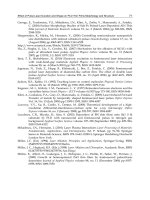
Figure 5.19
Periungual fibroma. (Courtesy of Akiro Kamumochi, Japan.)
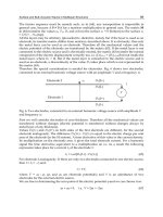
Figure 5.20
Koenen’s tumour associated with nail plate destruction.
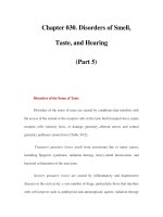
Figure 5.21
Periungual tissue disorders 133
Multiple Koenen’s tumours.
Koenen’s tumours are cured by simple excision. Usually no suture is necessary.
Tumours growing out from under the proximal nail fold are removed after reflecting the
p
roximal nail fold back by making lateral incisions down each margin in the axis of the
lateral nail grooves. Subungual fibromas are removed after avulsion of the corresponding
part of the nail plate.
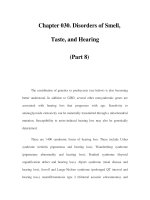
Acquired periungual fibrokeratoma
Acquired periungual fibrokeratomas are probably identical to acquired digital
fibrokeratomas and Steel’s ‘garlic clove’ fibroma. They are acquired, benign,
spontaneously developing, asymptomatic nodules with a hyperkeratotic tip and a narrow
base (Figures 5.22, 5.23). They most commonly occur in the periungual area or on othe
r
p
arts of the fingers. A case was described in which the lesion was located beneath the
nail, visible under the free margin of the great toe nail. Most periungual fibrokeratomas
emerge from the most proximal part of the nail sulcus growing on the nail and causing a
sharp longitudinal depression. Trauma is thought to be a major factor initiating acquired
periungual fibrokeratoma.
Microscopically, acquired periungual fibrokeratomas resemble hyperkeratotic ‘dermal
hernias’. The core consists of mature eosinophilic collagen fibres oriented along the main
vertical axis of the tumour. The fibroblastic cells are increased in number. Most fibromas
are highly vascular. The epidermis is thick and acanthotic. There is a marked
orthokeratotic horny layer, which may be parakeratotic and contains serum or blood at
the tip of the tumour. Elastic fibres are normal. Acid mucopolysaccharide levels are not
increased.
Figure 5.22
Acquired fibrokeratoma.
A text atlas of nail disorders 134
Figure 5.23
‘Garlic clove’ fibrokeratoma.
Surgical treatment is the same as for Koenen’s tumours and depends on the size and
location of the lesion.
The differential diagnosis of acquired periungual fibroma includes:
Subungual filamentous tumour
Subungual filamentous tumours are thread-like, horny, subungual lesions growing with
the nail plate and emerging from under the free edge of it. They may cause a longitudinal
rim. This entity is probably a narrow, extremely hyperkeratotic fibrokeratoma; it can be
pared down painlessly when the nail is cut.
• keloid
• Koenen’s tumour
• recurring digital fibrous tumours of childhood
• dermatofibrosarcoma
• fibrosarcoma
• acrochordon
• cutaneous horn
• eccrine poroma
• pyogenic granuloma
• verruca vulgaris
• exostosis.
Periungual tissue disorders 135
Recurring digital fibrous tumours of childhood (benign juvenile digital
fibromatosis)
Recurring digital fibrous tumours are round, smooth, firm tumours with a reddish or livid
red colour. They are located on the dorsal and axial surfaces of the fingers and toes,
characteristically sparing the thumbs and great toes (Figure 5.24). They may present a
t
b
irth or develop during infancy, although a single case of presentation in adulthood has
b
een described. There is no sex predominance. Fingers are more often affected than toes.
On reaching the nail unit the tumours may elevate the nail plate, leading to dystrophy but
not to
Figure 5.24
Benign juvenile digital fibromatosis (Courtesy of C.Moss, UK.)
destruction. Often the tumour is multicentric, occurring on several digits. Although an
infectious origin is probable, no virus has been isolated and viral particles have not been
demonstrated by electron microscopy. Up to 60% recur after excision. Spontaneous
regression was noted in 5 out of 61 cases; regression may be hastened by cryosurgery.
Radical surgical ablation of the area involved may rarely be necessary, including the nail
unit, leading to permanent loss of the nail. Firm plantar nodules may be associated with
these tumours.
Histological examination shows a diffuse, proliferative, cellular process in the dermis
with increased numbers of apparently normal fibroblasts with uniform, spindle-shaped
nuclei. Mitoses are absent or rare. Elastic tissue is decreased. In about 2% of the
fibroblasts, paranuclear inclusion bodies, 3–10µm in diameter, can be seen in adequately
fixed specimens with the use of stains such as iron haematoxylin, methyl green-
p
yronin
and phosphotungstic acid-haematoxylin. Electron microscopy shows that the inclusions
consist of fibrillar masses without a limiting membrane. On the basis of this evidence, it
has been suggested that the condition should be termed ‘elastodysplasia’.
A text atlas of nail disorders 136
Distal digital keratoacanthoma
Subungual and periungual keratoacanthomas may occur as solitary or multiple tumours.
They are rare, benign, rapidly growing, seemingly aggressive tumours usually situated
b
eneath the distal portion of the nail bed. The lesion starts as a small, painful keratotic
nodule visible beneath the free edge, growing rapidly to a 1 cm lesion within 4–8 weeks.
Its typical gross appearance, as a dome-shaped nodule with a central plug of horny
material filling the crater, is more obvious on an adequate histological specimen. Less
frequently the tumour grows out from under the proximal nail fold, which becomes
swollen and inflamed. In contrast to keratoacanthomas elsewhere, in distal digital tumors,
spontaneous regression is rare.
The tumour soon erodes the bone, but reconstitution of the defect can be achieved.
Glomus tumour
The glomus tumour was first described about 200 years ago as a painful, subcutaneous
‘tubercle’. Several cases were described as ‘malignant angiosarcomas’ or ‘colloid
sarcomas’. Seventy-five per cent of glomus tumours occur in the hand, especially in the
finger tips and particularly the subungual area. Between 1% and 2% of all hand tumours
are glomus tumours. The age at the time of diagnosis ranges from 30 years to 50 years.
Men are less frequently affected than women.
The tumour is characterized by intense, often pulsating pain that may be spontaneous
or provoked by the slightest trauma. Even changes in temperature, especially from warm
to cold, may trigger pain radiating up to the shoulder. Sometimes the pain is worse at
night: it may disappear when a tourniquet is applied.
The tumour is seen through the nail plate as a small, bluish to reddish-
b
lue spot several
millimetres in diameter, rarely exceeding 1 cm (Figure 5.25). Sometimes it causes a slight
rise in surface temperature which can be detected by thermography. Minor nail
deformities are caused by 50% of the tumours—ridging or a nail plate ‘gutter’
b
eing the
most common. A similar proportion cause a depression on the dorsal aspect of the distal
phalangeal bone, or even a cyst visible on X-ray. Probing, which elicits pain, an
d
transillumination may help to localize the tumour if it is not clearly visible through the
nail. If the tumour cannot be localized clinically or by X-ray, arteriography should be
performed; this will reveal a star-shaped telangiectatic zone. In selected cases, magnetic
resonance imaging (MRI) has been shown to help in the diagnosis of a glomus tumour o
f
the finger tip, revealing even very small lesions.
Many patients give a history of trauma. The most common misdiagnoses are neuroma,
causalgia, gout and arthritis. Histological examination shows a highly differentiated,
organoid tumour. It consists of an afferent arteriole, vascular channels lined with
endothelium and surrounded by irregularly arranged cuboidal cells with round, dark
nuclei and pale cytoplasm. Primary collecting veins drain into the cutaneous veins.
Myelinated and non-myelinated nerves are found and may account for the pain. The
tumour is surrounded by a fibrous capsule. Since all the elements of the normal glomus
are present, the glomus tumour may be considered as a hamartoma rather than a true
Periungual tissue disorders 137
tumour.
Figure 5.25
Glomus tumour.
Treatment is by surgical excision. Small tumours may be removed by punching a 6 mm
hole in the nail plate, incising the nail bed and enucleating the lesion. The small nail disc
is put back in its original position as a physiological dressing. Larger tumours may be
treated after removal of the proximal half of the nail plate; those in lateral positions are
removed by an L-shaped incision parallel to and 4–6 mm on the volar side of the lateral
nail fold. The nail bed is carefully dissected from the bone until the tumour is reached
and removed. This is usually curative, although the pain may take several weeks to
disappear. Recurrences occur in 10–20% of cases and may represent incomplete excision
or adjacent tumours overlooked at the initial operation, or genuine new growth. More
extensive surgery than is usual might achieve more first-time cures.
Subungual exostosis
Subungual exostoses are not true tumours but rather outgrowths of normal bone o
r
calcified cartilaginous remains (Figure 5.26). Whether or not subungual osteochondroma
is a different entity is not clear. Subungual exostoses are painful bony growths which
elevate the nail. They are particularly frequent in young people and are mostly located in
the great toe, although less commonly subungual exostoses also occur on the fingers.
They start as small elevations of the dorsal aspect of the distal phalanx and eventually
emerge from under the nail edge or destroy the nail plate. If the nail is shed, the surface
b
ecomes eroded and secondarily infected, sometimes mimicking ingrown toe nail.
Walking may be painful.
A text atlas of nail disorders 138
Trauma appears to be a major causative factor, although some authors claim that a
history of trauma only occurs in a minority. The triad of pain (the leading symptom), nail
deformation and radiographic features is usually diagnostic. The exostosis is a
trabeculated osseous growth with an expanded distal portion covered with radiolucent
fibrocartilage.
Osteochondroma, commonly presenting with the same symptoms, has a male
p
redominance. There is often a history of trauma. Its growth rate is slow. Radiographic
examination shows a well-defined, sessile, bony growth with a hyaline cartilage cap
which must be differentiated from primary subungual calcification (particularly in
Figure 5.26
(a) Exostosis; (b) X-ray of exostosis.
older women) and secondary subungual calcification due to trauma and psoriasis.
Treatment is by excision of the excess bone under full aseptic conditions. The nail
p
late is partially removed and a longitudinal incision made in the nail bed. The osseous
growth with its cartilaginous cap is carefully dissected, using fine skin hooks to avoid
damage to the fragile nail bed. The tumour is removed with a fine chisel, wheneve
r
possible through an L-shaped or ‘fish mouth’ incision, in order to avoid avulsion of the
nail plate.
Myxoid pseudocysts of the digits
The many synonyms for mixed pseudocyst of the digits reflect the controversial nature o
f
this lesion:
D
istorted nail shape may be due to a bone tumor
Periungual tissue disorders 139
Figure 5.27
Large periungual myxoid pseudocyst.
Whereas some authors regard it as a synovial cyst, most now believe it to be a
periarticular degenerative lesion.
Myxoid cysts occur more often in women. They are typically found in the proximal
nail fold of the fingers and rarely on toes (Figures 5.27–5.29). Usually asymptomatic,
these lesions vary from soft to firm, cystic to fluctuant, and may be dimpled, dome-
shaped or smooth-surfaced. Transillumination confirms their cystic nature. They are
always located to one side of the midline and rarely exceed 10–15 mm in diameter. The
skin over the lesion is thinned and may be verrucous or even ulcerated. Rarely, a
p
aronychial fistula may develop under the proximal nail fold, less commonly under the
nail plate. Longitudinal grooving of the nail results from pressure on the matrix.
Occasionally a series of irregular transverse grooves are seen, suggesting alternating
intermittent decompression and refilling of the cyst. Degenerative, ‘wear and tear’
osteoarthritis, frequently with Heberden’s nodes, is present in most cases.
• dorsal finger cyst
• synovial cyst
• recurring myxomatous cyst
• cutaneous myxoid cyst
• dorsal distal interphalangeal joint ganglion
• digital mucinous pseudocyst
• focal myxomatous degeneration
• mucoid cyst.
A text atlas of nail disorders 140
Figure 5.28
Nail plate gutter due to myxoid pseudocyst.
Figure 5.29
Subungual myxoid pseudocyst with nail plate disruption.
Histopathological investigation reveals the pseudocystic character. Cavities without
synovial lining are located in an ill-defined fibrous capsule. The structure is essentially
Periungual tissue disorders 141
myxomatous with interspersed fibroblasts. Areas of myxomatous degeneration may
merge to form a multilocular pseudocyst. In the cavities, a jelly-like substance is found
which stains positively for hyaluronic acid. In some cases a mesothelial-like lining is
found in the stalk connecting the pseudocyst with the distal interphalangeal joint. It has
b
een suggested that the lesion arises from the joint capsule or tendon sheath synovia, as
do ganglia in other areas.
A multitude of treatments have been recommended, including repeated incision and
drainage, simple excision, multiple needlings and expression of contents, X-irradiation (5
Gy, 50 kV, Al 1mm, three times at weekly intervals), electrocautery, chemical cautery
with nitric acid, trichloroacetic acid or phenol, massages or injection of proteolytic
substances, hyaluronidase, steroids (fluoran-drenolone tape, or injections) and sclerosing
solutions, cryosurgery, radical excision and even amputation.
The intralesional injection of corticosteroid crystal suspension has been recommended.
The cyst is first drained from a proximal point to avoid leakage of the steroid suspension
when the patient’s hand is lowered. Careful dissection and excision of the lesion gives the
highest cure rate. A tiny drop of methylene blue solution, diluted with a local anaesthetic
solution and mixed with fresh hydrogen peroxide, is injected into the distal
interphalangeal joint at the volar joint crease. The joint will accept only 0.1–0.2 ml o
f
dye. This clearly identifies the pedicle connecting the joint to the cyst, if one is present,
and also the cyst itself. This procedure sometimes reveals occult satellite cysts.
Alternatively, the methylene blue may be injected into the cyst to define the tract back
to its site of origin. The incision line is drawn on the finger, including a portion of the
skin directly over the cyst and continuing proximally in a gentle curve to end dorsally
over the joint. The lesion is meticulously dissected from the surrounding soft tissue and
the pedicle traced to its origins adjacent to the joint capsule and resected. Dumb-
b
ell
extension of cysts to each side of the extensor tendon is easily dissected by
hyperextending the joint. Osteophytic spurs adjacent to the joint must be removed with a
fine chisel or bone rongeur. Liquid nitrogen cryosurgery has been used with an 86% cure
rate. The field treated included the cyst and the adjacent proximal area to the transverse
skin creases overlying the terminal joint. Two freeze/thaw cycles were carried out, each
freeze time being 30 s after the ice field had formed, the intervening thaw time being at
least 4 min; if this method is adopted then longer freeze times must be avoided o
r
p
ermanent matrix damage may occur. If the cyst is first pricked and emptied of its
gelatinous contents, then equally good cure rates can be obtained with a single 20 s freeze
after initial ice formation. For distal posterior nail fold lesions, excision of the proximal
nail fold and associated cyst has been recommended.
Sclerosing agents may also be useful: after puncture and expression of cyst contents
0.20–0.30 ml of a 1 % solution of sodium tetradecyl sulphate is injected; a second or a
third injection may be required at monthly intervals.
M
yxoid pseudocysts rarely occur without ‘wear and tear’
osteoarthritis
A text atlas of nail disorders 142
Figure 5.30
Epidermoid carcinoma—verrucous periungual involvement.
Bowen’s disease (epidermoid carcinoma)
Bowen’s disease is a term for intra-epithelial squamous carcinoma (Figures 5.30–5.32). It
is not as rare as might be inferred from the medical literature.
Figure 5.31
Epidermoid carcinoma—subungual involvement.
Periungual tissue disorders 143
Figure 5.32
(a) Epidermal carcinoma; (b) epidermal carcinoma, with nail plate
trimmed back to show extension of invasion. (Courtesy of G. Cannata,
Italy.)
The clinical picture of Bowen’s disease of the nail unit is variable. It may show a
p
eriungual erythematous, squamous or eroded plaque. In the lateral nail wall and groove,
it usually presents as a recalcitrant hyperkeratotic or papillomatous, slowly enlarging
lesion. Distal involvement of the proximal nail fold results in the formation of a
characteristic whitish band. The fingers are far more frequently affected than the toes:
typically the thumbs, less often the index and middle fingers. The median age at
presentation is approximately 60 years, men predominating. Bowen’s disease evolves and
expands only slowly. Biopsies taken from the most indurated and warty area often reveal
invasive squamous cell carcinoma in contrast to the flat plaque. Many authorities
therefore no longer differentiate Bowen’s disease from squamous cell carcinoma,
preferring the term ‘epidermoid carcinoma’ for all cases.
Surgical removal of the affected area and a small margin of healthy tissue is the
treatment of choice. With some authorities, we prefer the Mohs fresh tissue removal
method. Despite the fact that cryosurgery is highly effective in treating Bowen’s disease
at other skin sites, it is only rarely effective for nail apparatus types.
I
ntra-epithelial squqmous carcinoma is not rare, the whole
tumor usually having local invasion at some point
A text atlas of nail disorders 144
Squamous cell carcinoma
Squamous cell carcinoma of the nail unit (also known as epidermoid carcinoma) is a low-
grade malignancy. Many cases have been reported, with a male predominance.
Trauma, chronic infection and chronic radiation exposure are possible aetiological
factors; human papillomavirus (HPV) has been incriminated in some cases. Two reported
cases had associated congenital ectodermal dysplasia. Most lesions occur on the fingers,
particularly the thumbs and index fingers (Figure 5.31). The presenting symptoms
include pain, swelling, inflammation, elevation of the nail, ulceration, a tumour ‘mass’,
ingrowing of the nail, ‘pyogenic granuloma’ and bleeding. Bone involvement is a rare,
very late sign. The duration of symptoms before diagnosis is greater than 12 months in
over half the cases. Only in one published case (with ectodermal dysplasia) has the
condition led to death, from rapid generalized metastases.
Subungual squamous cell carcinoma is slow-growing and may be mistaken for chronic
infection. This frequent misdiagnosis unduly prolongs the period between the onset of the
disease, diagnosis and therapy. Often it is not possible to determine whether the tumou
r
was present initially or developed later, secondary to trauma, warts or infection. As
mentioned above, invasive squamous cell carcinoma may develop from Bowen’s disease.
The possibility of a link with HPV strains 16, 34 and 35 sheds new light on the aetiology
of this type of cancer and suggests a logical cause for multiple digital Bowen’s disease.
Subungual melanotic lesions
The term ‘longitudinal melanonychia’ (LM) describes the presence of single or multiple
longitudinal pigmented streaks within the nail plate (Figures 5.33–5.35). A band of LM
may be due to one of four possible mechanisms:
Table 5.5 lists the causes of LM.
S
quamous cell carcinoma of the nail apparatus has a good
prognosis compared with other sites
• focal activation of the nail matrix melanocytes
• hyperplasia of the nail matrix melanocytes
• naevus of the nail matrix
• melanoma of the nail matrix.
Table 5.5
Causes of longitudinal melanonychia
Racial variation
Lau
g
ie
r
-Hunzike
r
-Baran s
y
ndrome (Fi
g
ure 5.34)
Inflammator
y
nail disorders
Dru
g
s
Periungual tissue disorders 145
Figure 5.33
Lateral band of longitudinal melanonychia.
Focal activation of the nail matrix melanocytes
This is the most common cause of LM, and is typified by the presence of melanocytes
with long dendrites located among nail matrix basal layers. There is no atypia or theque
formation.
Melanocyte activation occurs in 77% of African-Americans over 20 years of age and in
almost 100% of those over 50 years old. It is observed in 10–20% of Japanese individuals
as well as in people of Hispanic descent and other dark-skinned groups. It is uncommon
in white populations. Normal variant in black population is due to the number and size o
f
melanosomes produced. In white people melanosomes are small and aggregated in
complexes. In black people melanosomes are greater in length, larger in diameter and
distributed singly within keratinocytes.
Irradiation
Fun
g
al
Endocrine diseases
Trauma
N
eoplasms
AIDS
N
utritional
A text atlas of nail disorders 146
Figure 5.34
Laugier-Hunziker-Baran syndrome—nail and lip hyperpigmentation.
Melanocyte activation may be induced by repeated trauma to the nail matrix. Patients
who pick, break or chew the skin over the proximal nail fold frequently develop bands o
f
LM. This is usually associated with nail plate surface abnormalities due to repeated nail
matrix injury. Frictional LM is commonly observed in the toes of elderly individuals who
have foot deformities and/or unsatisfactory footwear. The melanonychia typically appears
at the site of friction with the tip of the shoes or under an overriding toe (see Chapter 9).
Inflammatory disorders of the nail may also produce nail pigmentation. Post-
inflammatory melanonychia has been described in lichen planus, Hallopeau’s
acrodermatitis and chronic radiodermatitis. Trichophyton rubrum and Scytalidium
dimidiatum (Hendersonula toruloidea) nail infection may also occasionally lead to LM.
Longitudinal melanonychia may be secondary to the inflammatory changes which induce
activation of nail matrix melanocytes, or due to direct melanin production by the fungi.
Activation of nail matrix melanocytes is occasionally seen in endocrine disorders such
as Addison’s disease, in pregnancy and in patients with human immunodeficiency virus
(HIV) infection, even in those not treated with zidovudine (azidothymine, AZT). Nail
matrix melanocytes may also be activated by drugs such as AZT, cance
r
chemotherapeutic agents and psoralens. Drug-induced melanonychia usually involves
several digits; it is reversible.
Melanocyte hyperplasia
Melanocyte hyperplasia is characterized by an increased number of melanocytes, which
are scattered between nail matrix keratinocytes without ‘nest’ formation. The
p
athogenesis of melanocyte hyperplasia is unknown. We have found this pathological
picture in patients with a single band of LM. Differential diagnosis from melanoma in
situ may be difficult, and these bands should be completely excised in order to perform
serial sections.
Periungual tissue disorders 147
Nail matrix naevus
Congenital and acquired melanocytic naevi may occur in the nail matrix and present as
LM. Nail matrix naevi are rare and only a few historically proven naevi of the nail matrix
have been reported. Naevi of the nail matrix are most commonly of the junctional type.
The architectural pattern of nail matrix naevi is similar to that of skin naevi. Naevus cells
are usually seen arranged in nests at the dermo-epidermal junction. Single naevus cells
can sometimes be found among nail matrix basal and suprabasal onychocytes. Dendritic
melanocytes are only occasionally present.
Immunostaining with HMB-45 of nail matrix naevi shows a positive reaction in the
cells of the epidermal and junctional component as usually seen in acquired skin naevi.
N
ail pigmentation due to congenital nail matrix melanocytic naevi may spontaneously
regress. However, fading of the pigmentation may only relate to decreased activity of the
naevus cells rather than regression of the naevus itself.
The frequency of progression from nail matrix naevi to nail matrix melanoma is not
known but a few cases have been well documented. Surgical excision of nail matrix naevi
is therefore a justified preventive measure.
Malignant melanoma
In the nail apparatus the most common initial sign of melanoma is acquired LM in white
Figure 5.35
Malignant melanoma.
individuals or broadening of an existing band in people of African or Asian descent. This
tumour and its practical differential diagnosis from other chromonychias and nail
dystrophies is described in this section; linear melanonychia from other causes is
A text atlas of nail disorders 148
considered in Chapter 7.
Melanoma of the nail region is now better understood since the identification and
analysis of acrolentiginous melanoma. It may be localized subungually or periungually
with pigmentation and/or dystrophy of the nail plate (Figure 5.35). Initial lesions may be
mistaken histologically for benign or atypical melanocytic hyperplasia, but serial sections
usually reveal the true nature of the disease.
Approximately 2–3% of melanomas in whites, and 15–20% in blacks are located in the
nail unit. However, malignant melanoma is rare in black people; thus the number of nail
melanomas does not significantly differ between these population groups. Most white
p
atients have a fair complexion, light hair, and blue or hazel eyes. There is no sex
p
redominance, although some reports show variable female or male predominance. The
mean age at onset is 55–60 years. Most tumours are found in the thumbs or great toes.
Melanoma of the nail region is often asymptomatic. Many patients only notice a
pigmented lesion after trauma to the area; only approximately two-thirds seek medical
advice because of the appearance of the lesion; pain or discomfort is rare, and nail
deformity, spontaneous ulceration, sudden change in colour, bleeding or tumour mass
b
reaking through the nail are even more infrequent. It is useful to remember that a
p
igmented subungual lesion is more likely to be malignant than benign. If the melanoma
is pigmented it may show one or more of the following characteristics:
Current experience has demonstrated that Hutchinson’s sign, while valuable, is not an
infallible predictor of melanoma, for the following reasons:
Total reliance on the (apparent) presence or absence of periungual pigmentation may lead
to over- or underdiagnosis of subungual melanoma. All relevant clinical and historical
A
cquired longitudinal melanonychia after puberty in a white-
skinned individual requires urgent biopsy
1
A spot appearing in the matrix, nail bed or plate. This may vary in colour from
brown to black; it may be homogeneous or irregular, and is seldom painful.
2
A longitudinal brown to black band of variable width running through the whole
visible nail.
3
Less frequently, Hutchinson’s sign—periungual extension of brown-black
pigmentation from LM onto the proximal and lateral nail folds—is an important
indicator of subungual melanoma (but note the reservations discussed below).
• Periungual pigmentation is present in a variety of benign disorders and, under these
circumstances, may lead to overdiagnosis of subungual melanoma.
• Periungual hyperpigmentation occurs in at least one non-melanoma skin cancer:
Bowen’s disease of the nail unit.
• Hyperpigmentation of the nail bed and matrix may reflect through the ‘transparent’
nail folds, simulating Hutchinson’s sign (‘pseudo-Hutchinson’s sign’). Each of the
above may incorrectly suggest a diagnosis of subungual melanoma. Table 5.6 lists
disorders in which pseudo-Hutchinson’s sign occurs.
Periungual tissue disorders 149
information, including the presence or absence of periungual pigmentation, must be
carefully evaluated in a patient suspected of having subungual melanoma. Ultimately, the
diagnosis of subungual melanoma is made histologically. Hutchinson’s sign is a single,
important clue to this diagnosis. The nail plate may also become thickened or fissured
and permanently shed.
A
p
proximately 25% of melanomas are amelanotic (pigmentation not an obvious o
r
prominent sign; Figure 5.36) and may mimic pyogenic granuloma, granulation tissue o
r
ingrowing nail. The risk of misdiagnosis is particularly high in these cases.
Malignant melanoma must be considered in the differential diagnosis (see Table 5.3) in
all cases of inexplicable chronic paronychia, whether painful or not, in torpid
granulomatous ulceration of the proximal nail fold and in pseudoverrucous keratotic
lesions of the nail bed and lateral nail groove. Subungual melanoma may also simulate
mycobacterial infections, mycotic onychodystrophy, recalcitrant paronychia and
ingrowing nail. Subungual haematoma is not rare and may present
Table 5.6
Disorders accompanied by pseudo-Hutchinson’s sign
D
isorde
r
Clinical
f
eatures
Beni
g
n
Illusory pigmentation Dark colour is visible because of the cuticle and thin
nail fold transparency and not because of melanin
localization within these tissues
Ethnic pigmentation Proximal nail fold of dark-skinned persons—lateral
nail folds not involved; LM may be present or
absent; often exa
gg
erated in thumbs
N
aevoid lenti
g
o Ma
y
recur after sur
g
ical removal
Laugier-Hunziker-Baran
s
y
ndrome
Macular pigmentation of lips, mouth and genitalia;
one or several fin
g
ers involved
Peutz-Jeghers syndrome Hyperpigmentation of fingers and toes, macular
p
igmentation of buccal mucosa and lips
Addison’s disease Diffuse tanning of both exposed and non-exposed
portions of the body; bluish-black discoloration of
the mucous membranes of the lips and mouth
X-ray therapy Treatment for finger dermatitis, psoriasis and chronic
p
aron
y
chia
Malnutrition Pol
y
dact
y
lous involvement
Minoc
y
cline Pol
y
dact
y
lous involvement
AIDS patients Polydactylous involvement; zidovudine produces
similar features
Trauma Due to friction, nail bitin
g
and pickin
g
, or boxin
g
Congenital or acquired
naevus after biopsy
Pigment recurrence after biopsy of LM in acquired
and congenital melanocytic naevi, often striking
c
y
tolo
g
ic at
y
pia
Regressing naevoid
melanosis in childhood
Monodactylous; initial increase in dyschromia
followed by subsequent pigment regression;
A text atlas of nail disorders 150
Figure 5.36
(a, b) Malignant melanoma—amelanotic.
without a history of severe trauma. It may follow repeated minor trauma which escapes
the patient’s attention, such as in ‘tennis toe’, or follow trauma from wearing hard ski
b
oots. Although haematoma following a single traumatic event usually grows out in one
p
iece, rather than as a longitudinal streak due to the continuous production of pigment,
repeated trauma may cause difficulties in differential diagnosis. It is recommended that
the lesion should be examined with a magnifying loupe after it has been covered with a
drop of oil. The pigmented nail should be clipped and tested with the argentaffin reaction
in order to rule out melanin pigmentation. Subungual haemoglobin is not degraded to
haemosiderin and is therefore negative to staining with Prussian blue. Scrapings or small
p
ieces of the nail boiled with water in a test tube give a positive benzidine reaction with
the conventional haemoglobin reagent strips. The difference between haemosiderinic and
p
erplexin
g
disorder
Subungual haematoma Exceptionally, blood spreads to nail folds and the
h
y
pon
y
chial area
Silver nitrate For treatment of granulation tissue; may produce a
b
lack halo
Mali
g
nant
Bowen’s disease Features clinicall
y
t
y
pical of subun
g
ual melanoma
(After Baran and Kichijian
(1996). LM, longitudinal
melanon
y
chia.
Periungual tissue disorders 151
melanotic pigment, sometimes difficult to discern by routine histological methods, is
easily seen by ultrastructural techniques: ferrous pigment is intercellular while melanin is
intracellular.
Because of its frequency, melanonychia striata in people with deeply pigmented skin is
considered a normal finding, but up to one-fifth of all melanomas in black patients are in
the subungual area, and these typically begin with a pigmented spot producing a
longitudinal streak. These spots are usually black rather than the normal brown. The
diagnosis may be aided by comparing them with the brown stripes in other nails or by the
occurrence of Hutchinson’s sign.
The following guidelines should be adhered to where possible to enable accurate tissue
diagnosis to be made and appropriate treatment carried out. As a first step, the anatomical
site of the matrix affected will be obtained from the level of the melanin pigment
identified with Fontana’s silver stain of a nail clipping obtained from the distal free edge.
The type of biopsy selected will then depend on the site of the matrix melanin production,
the width of the linear pigmentation, and the site of the band in the nail plate. If the
p
igment is located within the ventral portion of the nail plate, a decision has to be made
depending on the width of the band:
If the pigment involves the upper portion of the nail, it is obviously difficult to use the
two previous procedures to remove the source of melanin pigment, for anatomical
reasons and because of the risk of a secondary dystrophy, thus:
• A punch biopsy should be used when the width of the band is less than 3 mm. If the
b
ase of the nail plate is removed, the specimen may be released more easily, and the
integrity of the region distal to the biopsied matrix area may be checked.
• A transverse matrix biopsy should be used for a band wider than 3 mm.
• A rectangular block of tissue is excised using two parallel incisions down to the
bone. An L-shaped incision is carried back along the lateral nail wall, freeing this
flap. The lateral section may then be rotated medially and approximated to the
remaining nail segment.
• If the band is wider than 6 mm or if the whole thickness of the nail is involved by
the pigment, surgical removal of the nail apparatus seems the most logical method.
However, one (or even two) 3 mm punch biopsy is an alternative prior to more
radical treatment, especially in young women.
• When the band lies within the lateral third of the nail plate, lateral longitudinal
biopsy is more suitable.
• If LM is accompanied by periungual pigmentation (Hutchinson’s sign), removal of
the nail apparatus is required. Histological examination of acral lentiginous
melanoma requires great experience, and often serial sections are needed to classify
the lesion accurately. Grading according to Clark’s levels or Breslow’s maximum
tumour thickness is difficult and often inconclusive.
N
ail apparatus melanoma has a poorprognosis, with up to 50%
of patients dying within 5 years of the diagnosis
A text atlas of nail disorders 152
Subungual melanoma has a poor prognosis. The reported 5-year survival rates range from
35% to 50%. Most patients present with advanced subungual melanoma; however, even
early diagnosis is not a guarantee of a good prognosis. Women have a better prognosis
than men. Factors contributing to a poor prognosis are delay in diagnosis and, as a result
of this, inadequate treatment. The tumour may be mistaken for a traumatic dystrophy, and
valuable time may be lost before the diagnosis is made.
Treatment depends on the stage of the disease. Levels I and II melanomas may be
adequately treated by wide local excision, and repair of the defect with graft or flap.
Amputation is usually advised for melanoma at levels more advanced than II. When the
thumb has to be amputated, pollicization of a finger may provide a functional
replacement. There would appear to be no relationship between the prognosis and the
extent of the amputation, although metacarpo/metatarsophalangeal amputation is
considered to be inadequate because of local recurrences. The rationale for elective
lymph node dissection and/or isolated hyperthermic perfusion of the extremity with
cytotoxic drugs is still under discussion. Immune enhancement such as BCG (bacillus
Calmette-Guérin) therapy is used in some centres.
Table 5.7
Conditions in which nail pustulation may occur
Infective
(primar
y
cause)
Acute paron
y
chia (see p. 81)
Blisterin
g
distal dact
y
litis
Hand, foot and mouth disease
Herpes simplex (primar
y
and recurrent)
Gonorrhoea
Impeti
g
o
Veillonella infection (in newborns)
Non-infective
(secondar
y
infection ma
y
occur)
In
g
rowin
g
toe nail
Malali
g
nment in childhood
Common t
y
pe
Self-inflicted bullous lesions of newborn
Thumb suckin
g
(and paron
y
chia)
Dermatoses
Acrokeratosis paraneoplastica
Acropustulosis/psoriasis
Parakeratosis pustulosa
Reite
r
’s s
y
ndrome
Periungual tissue disorders 153
PUSTULES
The conditions in which nail apparatus pustulation may be a significant sign are listed in
Table 5.7.
Herpes simplex
Distal digital herpes simplex infection may affect the terminal phalanx as a primary
herpetic ‘whitlow’ or start as an acute, intensely painful paronychia (Figures 5.37, 5.38).
It is relatively common in dental staff, anaesthetists and those involved with the care o
f
the mouth and upper respiratory tract in unconscious patients. Recurrent forms are
generally less severe and have a milder clinical course than the initial infection.
After an incubation period of 3–7 days, during which local tenderness, erythema and
swelling may develop, a crop of vesicles appears at the site of origin in the skin. The
vesicles are typically distributed in the paronychia and on the volar digital skin,
resembling pyogenic infection of the finger tip. Close inspection, however, will reveal the
characteristic pale, raised vesicles surrounded by an erythematous border. An acutely
p
ainful whitlow may develop and extend under the distal free edge of the nail and into the
nail bed. A distinct predilection for the thumb, index and ring fingers on the dominant
hand has been noted, but any finger may be involved. Multiple lesions are rare. For 10
–
14 days the vesicles gradually increase in size, often coalescing into large, honeycombed
b
ullae. New crops of lesions may appear during this time. Vesicular fluid is clear early in
the disease but may become turbid, seropurulent or even haemorrhagic within days o
f
onset. At times, a pale yellow colour of the vesicles will suggest pyogenic infection, yet
frank pus is not usually obtained. Patients complain of tenderness and severe throbbing in
the affected digit. Coexisting primary herpetic infections of the mouth and finger nails
suggest auto-inoculation of the virus into the nail tissues as a result of nail biting or finge
r
sucking.
Radiating pain along the C7 spinal nerve distribution is sometimes noted before each
recurrence. Lymphangitis may start from the wrist and extend to the axilla with painful
lymphadenopathy. Numbness and hypo-aesthesia following the acute episode have been
observed.
The diagnosis of herpetic infection can be made by examining the base of the vesicles
for the characteristic multinucleated ‘balloon’ giant cells, in stained smears. The presence
of intranuclear inclusions is also significant. Viral cultivation, usually positive within 24
hours of onset, is confirmatory; the active viral phase lasts up to 4–5 days in primary
attacks but only 2–3 days in recurrent episodes.
Differential diagnosis
It is important to exclude primary or recurrent herpes simplex infection in the differential
diagnosis of every vesiculopustular finger infection. The typical appearance of the lesions
with disproportionately severe pain, the absence of pus in the confluent, multiloculated,
A text atlas of nail disorders 154
vesiculopustular lesions and the lack of increased tension in the finger pulp aid in
differentiating this slow-healing infection from a bacterial foreign body or paronychia.
Figure 5.37
Primary herpes simplex—herpetic ‘whitlow’.
Figure 5.38
Subungual recurrent herpes simplex.
Herpes zoster infections, which may affect the proximal nail fold like herpes simplex,
also involve the entire sensory dermatome. The pustules of primary cutaneous Neisseria
Periungual tissue disorders 155
g
onorrhoeae infection may resemble herpes simplex on the rare occasion when it occurs
on the finger. The diagnosis is established by positive Gram staining and bacteriological
culture.
Treatment
Treatment is aimed primarily at symptomatic relief and the avoidance of secondary
infection. Topical acyclovir may shorten the course of any one attack; given orally the
drug may prevent recurrences while it is being taken. On cessation of the treatment
relapses are unfortunately common. This is a preventable infection. Gloves should always
b
e worn on both hands for procedures such as intubation, removal of dentures o
r
providing oral care, despite the additional costs involved.
Veillonella infection in the newborn
Many epidemics of subungual infection have been described among infants in postnatal
wards and special care baby units. The number of fingers affected per patient ranged
from one to ten; the thumbs are less frequently involved than other fingers; toe nails are
not affected. Three stages occur: first, a small amount of clear fluid appears under the
centre of the nail, along with mild inflammation at the distal end of the finger. This initial
vesicle lasts approximately 24 hours; it sometimes enlarges but never extends to the edge
of the nail. Some small lesions bypass the second, pustular stage, going directly to the
third stage. As a rule the fluid becomes yellow after 24 hours, the pus remaining for 24
–
48 hours, before gradually turning brown and being absorbed. The colour fades
progressively over a period of 2–6 weeks, leaving the nail and nail bed completely
normal.
Subungual pus obtained by aseptic puncture of the nails shows tiny, Gram-negative
cocci about 0.4 µm in diameter. These organisms resemble Veillonella, a group o
f
anaerobes of dubious pathogenicity found as commensals in the saliva, vagina and
respiratory tract. Systemic antibiotics do not change the clinical course of the nail lesions,
which do not differ from those observed in other untreated and affected newborn
children.
Impetigo
The dorsal aspect of the distal phalanx may be involved by impetigo (Figure 5.39). I
t
presents in two forms:
The latter is characterized by the appearance of large, localized, intra-epidermal bullae
that persist for longer periods than the transient vesicles of streptococcal impetigo which
subsequently rupture spontaneously to form very thin crusts. The lesions of bullous
impetigo may mimic the non-infectious bullous diseases (such as drug-induced types o
r
1
Vesiculopustular, with its familiar honey-crusted lesions, usually due to beta-
haemolytic streptococci.
2
Bullous, usually due to phage type 71 staphylococci.
A text atlas of nail disorders 156
pemphigoid). Oral therapy of bullous impetigo with a penicillinase-resistant penicillin
should be instituted and continued until the lesions resolve. Cephalexin and erythromycin
are acceptable alternatives. The lesions should be cleansed several times daily and topical
aureomycin (3%) applied to all the affected areas.
Figure 5.39
Impetigo of the nail apparatus.
Blistering distal dactylitis
Blistering distal dactylitis is a variant of streptococcal skin infection. It presents as a
superficial, tender, blistering beta-haemolytic streptococcal infection over the anterior fat
pad of the distal phalanx of the finger (Figure 5.40). The lesion may or may not have a
p
aronychial extension. This blister, containing thin, white pus, has a predilection for the
tip of the digit and extends to the subungual area of the free edge of the nail plate. The
area may provide a nidus for the beta-haemolytic streptococcus and act as a focus o
f
chronic infection similar to the nasopharynx. The age range of affected patients is 2–16
years. For local care incision, drainage and antiseptic soaking are indicated, giving a
more rapid response than systemic antibiotic therapy alone: effective regimens include
benzylpenicillin (penicillin G) in a single intramuscular dose, a 10-day course of oral
p
henoxymethylpenicillin or eryhromycin ethyl succinate. This type of treatment
decreases the reservoir of streptococci by preventing spread to family contacts. This
infection has been described as a complication of ingrowing toe nail. The differential
diagnosis includes blisters resulting from friction, thermal and chemical burns, infectious
states such as herpetic whitlow, staphylococcal bullous impetigo and the Weber-
Cockayne variant of epidermolysis bullosa simplex.
Periungual tissue disorders 157