Báo cáo y học: " Perforated gastric corpus in a strangulated paraesophageal hernia: a case report" ppt
Bạn đang xem bản rút gọn của tài liệu. Xem và tải ngay bản đầy đủ của tài liệu tại đây (268.51 KB, 3 trang )
Case report
Open Access
Perforated gastric corpus in a strangulated paraesophageal hernia:
a case report
Alexis E Shafii
1
, Steven C Agle
2
and Emmanuel E Zervos
2
*
Address:
1
Department of Thoracic and Cardiovascular Surgery, The Cleveland Clinic, 9500 Eucid Avenue, Cleveland, Ohio 44195, United States
and
2
Department of Surgery, East Carolina University, 600 Moye Boulevard, Greenville, North Carolina 27834, United States
Email: AES - ; SCA - ; EEZ* -
* Corresponding author
Published: 7 May 2009 Received: 8 December 2008
Accepted: 9 February 2009
Journal of Medical Case Reports 2009, 3:6507 doi: 10.1186/1752-1947-3-6507
This article is available from: />© 2009 Shafii et al; licensee Cases Network Ltd.
This is an Open Access article distributed under the terms of the Creative Commons Attribution License (
/>which permits unrestricted use, distribution, and reproduction in any medium, provided the original work is properly cited.
Abstract
Introduction: Patients with paraesophageal hernias often present secondary to chr onic
symptomatology. Infrequently, acute intestinal ischemia and perforation can occur as a consequence
of paraesophageal hernias with potentially dire consequences.
Case presentation: An 86-year-old obtunded male presented to the emergency department with
hypotension and severe back and abdominal pain. An emergency abdominal CT scan was ordered
with a presumptive diagnosis of ruptured abdominal aortic aneurysm. CT topograms revealed
extensive free intra-abdominal air and herniated abdominal viscera into the right hemithorax. Prior to
completion of the CT study, the patient sustained a cardiopulmonary arrest. Surgery was consulted,
but the patient was unable to be revived. Post-mortem examination revealed gross contamination
within the abdomen and a giant, incarcerated, hiatal hernia with organoaxial volvulus and ischemic
perforation.
Conclusion: Current recommendations call for prompt repair of giant hiatal hernias before they
become symptomatic due to the increased risk of strangulation. Torsion of the stomach in large hiatal
hernias frequently leads to a fatal complication such as this warranting elective repair as soon as
possible.
Introduction
Paraesophageal hernias occur when intra-abdominal
contents herniate through the esophageal hiatus into the
mediastinum. There are four types: type I occurs when the
stomach slides into the mediastinum thus displacing the
gastroesophageal junction into the thorax (sliding hiatal
hernia), type II and III paraesophageal hernias result from
herniation of the stomach through the esophageal hiatus
and subsequent o rganoaxial and mesoaxial rotation
respectively, and type IV hernias involve organs other
than the stomach herniating through the hiatus into the
thorax. Current recommendations are for prompt repair
secondary to the possibility of complications including
hemorrhage, ischemia, and perforation [1].
Page 1 of 3
(page number not for citation purposes)
Case Presentation
An 86-year-old white, American, male presented to the
emergency department hypotensive and obtunded with
severe abdominal and back pain of unknown duration. A
ruptured abdominal aortic aneurysm was initially sus-
pected and the patient was taken for an abdominal CT
scan at the request of the emergency room physicians.
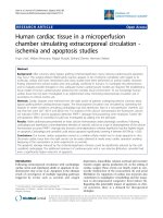
After completion of a thoracoabdominal topogram, a large
quantity of free intra-abdominal air was seen on the lateral
view and herniated abdominal viscera were identified in
the right chest on the supine view (Figure 1). Prior to
completion of the scan, the patient succumbed to a
cardiopulmonary arrest. A postmortem examination of the
abdomen and chest was performed. Upon entering the
abdominal cavity there was gross contamination as well as
a giant incarcerated hiatal hernia. The entire stomach was
herniated up and into the posterior mediastinum with
only the pylorus visible at the hiatus (Figure 2). Remnants
of a failed hiatal hernia repair were found along the
diaphragmatic extension of the left crus. Once the stomach
was freed from adhesions and reduced into the abdominal
cavity the site of an ischemic perforation of the gastric
fundus was identified (Figure 3).
The patient's past medical history was uncovered post-
mortem and was significant for a prior coronary artery
bypass, congestive heart failure, and previous hiatal hernia
repair. The initial discovery of a giant hiatal hernia was
made thirteen years prior by esophagogastroduodeno-
scopy during an evaluation for coffee ground emesis and
chronic anemia. Repair was performed at that time and
consisted of primary crural re-approximation and gastro-
pexy. Late recurrence of the giant hiatal hernia was also
documented but re-operation was not undertaken due to
his poor cardiac reserve.
Conclusion
Torsion of the stomach in these very large hiatal hernias
can lead to fatal complications with considerable fre-
quency, and as a result, elective repair is warranted upon
discovery except in the moribund patient [2]. Emergent
surgical intervention in the case of a complete gastric
volvulus involves reduction of the volvulus and hiatal
repair [3]. Patients with this condition often present with a
classic triad composed of retching, epigastric pain, and
failure to place a nasogastric tube. Partial gastrectomy may
also be required in cas es of infarcted stomach or
perforation. Optimal elective repair involves reduction of
the hernia, excision of the hernia sac, and repair of the
hiatal defect, which if excessively large, may require
prosthetic mesh reinforcement [4]. Collis-Nissen fundo-
plication may be added to the repair to accommodate
relative esophageal shortening but not without risk of
dysmotility of the distal esophagus [5]. While traditionally
these repairs were approached via celiotomy or thoracot-
omy, the majority of cases are now amenable to laparo-
scopic approaches with excellent outcomes [6]. Indeed, in
the referenced study, 200 consecutive patients underwent
Figure 1. Intraabdominal free air seen on lateral abdominal
topogram and herniated abdominal viscera on supine view.
Figure 2. Pointer on pylorus at esophageal hiatus.
Figure 3. Perforation of gastric fundus.
Page 2 of 3
(page number not for citation purposes)
Journal of Medical Case Reports 2009, 3:6507 />laparoscopic repair of paraesophageal hernias with only
one death, low morbidity, and a 2.5% recurrence rate.
It is evident that this patie nt’s pathology was the
consequence of a chronically incarcerated giant hiatal
hernia left untreated, which ultimately led to his demise.
While it remains unclear as to what his true surgical risks
were, we currently recommend that most patients can be
repaired with low morbidity and nearly zero mortality.
List of abbreviations
CT, Computerized tomography.
Consent
Written informed consent has been attained from the
deceased patient’s family to publish information related to
the case as well as images associated with the case.
Competing interests
The authors declare that they have no competing interests’.
Authors’ contribution
AS and EZ were both involved in the conception and data
gathering for the case report. SA was involved in drafting
and revising the manuscript. AS, EZ and SA were involved
in the literature review and obtaining the critical intellec-
tual content used in this case report. These three authors
have also given final approval for publication.
References
1. Krähenbühl L, Schäfer M, Farhadi J, Renzulli P, Seiler CA, Büchler MW:
Laparoscopic treatment of large paraesophageal hernia
with totally intrathoracic stomach. J Am Coll Surg. Sep 1998,
187(3):231-237.
2. Maruyama T, Fukue M, Imamura F, Nozue M: Incarcerated
paraesophageal hernia associated with perforation of the
fundus of the stomach: report of a case. Surg Today 2001,
31:454-457.
3. Hill, L D: Incarcerated Paraesophageal Hernia. A surgical
emergency. Am J Surg 1973, 126:286-291.
4. Oelschlager BK, Pellegrini CA, Hunter J, Soper N, Brunt M, Sheppard
B, Jobe B, Polissar N, Mitsumori L, Nelson J, Swanstrom L: Biologic
prosthesis reduces recurrence after laparoscopic paraeso-
phageal hernia repair: a multicenter, prospective, rando-
mized trial. Annals of Surgery 2006, 244(4):481-490.
5. Jobe BA, Horvath KD, Swanstrom LL: Postoperative function
following laparoscopic collis gastroplasty for shortened
esophagus. Arch Surg. 1998 Aug, 133(8):867-74.
6. Pierre A, Luketich J, Fernando H, Christie N, Buenaventura P, Litle V,
Schauer P: Results of laparoscopic repair of giant paraesopha-
geal hernias: 200 consecutive patients. Ann Thorac Surg 2002,
74:1909-1915.
Page 3 of 3
(page number not for citation purposes)
Journal of Medical Case Reports 2009, 3:6507 />Do you have a case to share?
Submit your case report today
• Rapid peer review
• Fast publication
• PubMed indexing
• Inclusion in Cases Database
Any patient, any case, can teach us
something
www.casesnetwork.com