Báo cáo y học: " Late presentation of arrhythmogenic right ventricular cardiomyopathy: a case report" potx
Bạn đang xem bản rút gọn của tài liệu. Xem và tải ngay bản đầy đủ của tài liệu tại đây (948.92 KB, 4 trang )
Case report
Open Access
Late presentation of arrhythmogenic right ventricular
cardiomyopathy: a case report
Georgios I Papaioannou
1
*, Theodoros Apostolopoulos
2
, Sotiria Stambola
2
,
Antonios Zilidis
1
and John Gialafos
2
Addresses:
1
Cardiac Catheterization Laboratory, Athens Medical Center, Athens, Greece
2
Department of Cardiology, Division of Electrophysiology, Athens Medical Center, Athens, Greece
Email: GIP* - ; TA - ; SS - ; AZ - ; JG -
* Corresponding author
Received: 11 March 2008 Accepted: 14 April 2009 Published: 4 August 2009
Journal of Medical Case Reports 2009, 3:7235 doi: 10.4076/1752-1947-3-7235
This article is available from: />© 2009 Papaioannou et al.; licensee Cases Network Ltd.
This is an Open Access article distributed under the terms of the Creative Commons Attribution License (
/>which permits unrestricted use, distribution, and reproduction in any medium, provided the original work is properly cited.
Abstract
Introduction: Arrhythmogenic right ventricular cardiomyopathy is an inherited myocardial disease
affecting predominantly young people and manifests as sustained ventricular tachycardia with left
bundle branch block morphology, sudden death or isolated right or biventricular heart failure.
However, its first manifestation as sustained ventricular tachycardia in older patients without preceding
symptoms of heart failure is infrequent. To our knowledge, our patient is among the oldest reported in
the literature presenting with ventricular tachycardia because of arrhythmogenic right ventricular
cardiomyopathy without preceding symptoms of heart failure.
Case presentation: We present an unusual case of a very late presentation of a right ventricular
cardiomyopathy in a 72-year-old white Caucasian man. The patient was admitted with symptoms of
weakness, dizziness and chest discomfort for several hours. His electrocardiogram showed a wide-
complex tachycardia with left bundle branch block morphology and left axis deviation. Because of
continuing hemodynamic instability, the patient was cardioverted to sinus rhythm with a single 300 J
shock. His post-cardioversion electrocardiogram, cardiac echocardiogram, coronary angiogram,
magnetic resonance imaging and electrophysiological study confirmed the diagnosis of arrhythmo-
genic right ventricular cardiomyopathy. The patient was treated with an implantable cardioverter
defibrillator and discharged on sotalol.
Conclusion: This case report demonstrates that arrhythmogenic right ventricular cardiomyopathy may
have a very late presentation and this diagnosis should be considered as a potential cause of sustained
ventricular tachycardia of right ventricular origin among the elderly and should be treated accordingly.
Introduction
Arrhythmogenic right ventricular cardiomyopathy (ARVC)
is an inherited m yocardial disease affecting predomi-
nantly young people. It manifests as sustained ventricular
tachycardia (VT) with left bundle branch block (LBBB)
morphology, sudden death or isolated right or biventri-
cular heart failure with the majority of cases been diagnosed
before the age of 40, while heart failure symptoms and
Page 1 of 4
(page number not for citation purposes)
signs typically appear later in life. Its first presentation
as sustained VT in o lder patients without preced ing
symptoms of heart failure is infrequent.
Case presentation
A 72-year-old white Caucasian man, without prior history
of heart disease was admitted with symptoms of weakness,
dizziness and chest discomfort for several hours. Physical
examination revealed low blood pressure (85/50 mmHg)
and a weak, regular and rapid pulse. The electrocardio-
gram (ECG) showed a wide-complex tachycardia with
LBBB morphology and left axis deviation. Because of con-
tinuing hemodynamic instability, the patient was cardio-
verted to sinus rhythm with a single 300 J shock.
The ECG during the episode of the tachycardia was
consistent with sustained VT of right ventricular origin.
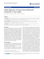
Post-cardioversion ECG showed negative T-waves at the
inferior and precordial leads with a QRS duration of
110 ms and the p resence o f an e psilon wave in V
1
(Figure 1 ).
Chemistries were normal with the exception of troponin
I which was positive. The patient was subsequently started
on metoprolol. Continuous ECG monitoring revealed
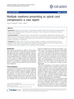
multiple episodes of non-sustained VT. Cardiac ultra-
sound examination showed a normal left ventricle and a
slightly enlarged right ventricle with local dyskinesia and
diastolic bulging of the free wall (Figure 2A).
As a diagnosis of ARVC was most likely, the patient
underwent simultaneous cardiac catheterization and
electrophysiological study. His coronary arteries and left
ventricle were normal. Right ventriculography in the right
anterior oblique view confirmed the local hypokinesia of
the free wall with diastolic bulging. During the electro-
physiological study, a sustained VT was provoked with
identical morphology to the one on admission. The VT
responded to antitachycardia pacing and the defibrillation
threshold was 17 J.
A magnetic resonance imaging (MRI) scan that followed
revealed diffuse areas of fat tissue at the right ventricular
wall especially localized at the free and lateral
segments, while multiple small aneurysms were also
present (Figure 2B-D). Right ventricular ejection fraction
was 30%. Left ventricular free wall and intraventricular
septum were free of disease. Finally, a signal-averaged ECG
was positive for late potentials. Based on the clinical
presentation and subsequent work-up, a definite diagnosis
of ARVC was made. The patient was treated with an
implantable cardioverter defibrillator (ICD) and dis-
charged on sotalol.
Discussion
ARVC is an inherited myocardial disease primarily affect-
ing the right ventricle and is characterized by the gradual
replacement of myocytes by adipose and fibrous tissue.
It affects young people and may cause sudden death,
especially during athletic activity [1]. Diagnostic criteria for
ARVC were proposed in 1996 and include major and
minor criteria [2]. Our patient had two major (epsilon
wave in lead V1 and MRI findings of multiple localized
aneurysms of the right ventricle) and five minor criteria
(LBBB type ventricular tachycardia, inverted T waves in
precordial leads, regional right ventricular hypokinesia,
reduction of right ventricular ejection fraction and late
potentials on single-averaged ECG), making the diagnosis
of ARVC definite. Despite the extensive T wave changes
recorded in the inferior and precordial leads, our patient
only had evidence of fibrofatty tissue at the right ventricle,
while there was no indication of left ventricular involve-
ment from MRI at the time of presentation.
ARVC manifests as sustained VT with LBBB morphology,
sudden death or isolated right or biventricular heart
failure. At least 80% of cases are diagnosed before the
age of 40, while heart failure symptoms and signs typically
appear in the fourth and fifth decades of life. To our
knowledge, our patient is among the oldest reported in the
literature presenting with VT because of ARVC without
symptoms of heart failure.
Although the inheritance pattern is typically autosomal
dominant, autosomal recessive ARVC (Naxos disease)
with a characteristic phenotype and mutation of plako-
globin, a protein that forms cell-to-cell junctions, is more
prevalent in Greece [3]. However, even this recessive form
of ARVC almost always manifests before the age of 40. Our
patient had never experienced any symptoms that could be
related to ARVC and he lacked any specific phenotype or
family history that could raise the suspicion of ARVC.
The management of patients with ARVC is targeted toward
prevention of sudden cardiac death and treatment of
symptoms of heart failure in the case of biventricular
Figure 1. Patient’s electrocardiogram after successful
cardioversion. The QRS duration in leads V
1
to V
3
is greater
than 110 ms and there is an evident epsilon wave in V
1
(arrow).
Page 2 of 4
(page number not for citation purposes)
Journal of Medical Case Reports 2009, 3:7235 />involvement. While antiarrhythmic medications including
beta-adrenergic blocking agents sotalol and amiodarone
can be used to prevent recurrent cardiac arrhythmia, treat-
ment with ICD should be considered in individuals at
high risk [4,5]. Radiofrequency ablation [6,7] can be at-
tempted in patients who are unresponsive or intolerant to
antiarrhythmic drugs but is frequently unsuccessful and
may require multiple attempts because of the patchy
nature of the disease. Individuals with ARVC should be
prohibited from vigorous exercise and after ARVC is
diagnosed, all first-degree relatives should be screened.
Conclusion
Our case demonstrates that arrhythmogenic right ventri-
cular cardiomyopathy may have a very late presentation
and this diagnosis should be considered as a potential
cause of sustained ventricular tachycardia of right ven-
tricular origin among the elderly.
Consent
Written informed consent was obtained from the patient
for publication of this case report and any accompanying
images. A copy of the written consent is available for
review by the Editor-in-Chief of this journal.
Competing interests
The authors declare that they have no competing interests.
Authors’ contributions
All authors participated in the management of this patient.
The first author prepared and revised the manuscript and
all authors approved the final draft.
B
A
C
D
Figure 2. Panel (A) Cardiac echocardiogram (4 chamber view) shows slightly enlarged right ventricle with local dyskinesia
and diastolic bulging of the free wall (arrow). Panel (B) Axial T1 weighted spin echo image shows diffuse hyperintense signal
(arrows) in the free wall of the right ventricle. Panel (C) T2 weighted cine image with demonstration of multiple small
aneurysms at the free wall of the right ventricle (arrows). Panel (D) Using an axial T1 weighted spin echo image with fat
suppression, the hyperintense signal in the right ventricle wall was suppressed and identified as fatty tissue (arrows).
Page 3 of 4
(page number not for citation purposes)
Journal of Medical Case Reports 2009, 3:7235 />References
1. Furlanello F, Bertoldi A, Dallago M, Furlanello C, Fernando F, Inama G,
Pappone C, Chierchia S: Cardiac arrest and sudden death in
competitive athletes with arrhythmogenic right ventricular
dysplasia. Pacing Clin Electrophysiol 1998, 21:331-335.
2. McKenna WJ, Thiene G, Nava A, Fontaliran F, Blomstrom-
Lundqvist C, Fontaine G, Camerini F: Diagnosis of arrhythmo-
genic right ventricular dysplasia/cardiomyopathy: task force
of the working group myocardial and pericardial disease of
the European Society of Cardiology and of the Scientific
Council of Cardiomyopathies of the International Society
and Federation of Cardiology. Br Heart J 1994, 71:215-218.
3. Antoniades L, Tsatsopoulou A, Anastasakis A, Syrris P, Asimaki A,
Panagiotakos D, Zambartas C, Stefanadis C, McKenna WJ,
Protonotarios N: Arrhythmogenic right ventricular cardiomyo-
pathy caused by deletions in plakophilin-2 and plakoglobin
(Naxos disease) in families from Greece and Cyprus:
genotype-phenotype relations, diagnostic features and prog-
nosis. Eur Heart J 2006, 27:2208-2216.
4. Buja G, Estes NA 3rd, Wichter T, Corrado D, Marcus F, Thiene G:
Arrhythmogenic right ventricular cardiomyopathy/dysplasia:
risk stratification and therapy. Prog Cardiovasc Dis 2008, 50:
282-293.
5. Zipes DP, Camm AJ, Borggrefe M, Buxton AE, Chaitman B, Fromer M,
Gregoratos G et al.: ACC/AHA/ESC 20 06 guidelines fo r
management of patients with ventricular arrhythmias and
the prevention of sudden cardiac death. Europace 2006, 8:
746-837.
6. Fontaine G, Tonet J, Gallais Y, Lascault G, Hidden-Lucet F, Aouate P,
Halimi F, Poulain F, Johnson N, Charfeddine H, Frank R: Ventricular
tachycardia catheter ablation in arrhythmogenic right ven-
tricular dysplasia: a 16-year experience. Curr Cardiol Rep 2000,
2:498-506.
7. Kottkamp H, Hindricks G: Catheter ablation of ventricular
tachycardia in ARVC: is curative treatment at the horizon?
J Cardiovasc Electrophysiol 2006, 17:477-479.
Page 4 of 4
(page number not for citation purposes)
Journal of Medical Case Reports 2009, 3:7235 />Do you have a case to share?
Submit your case report today
• Rapid peer review
• Fast publication
• PubMed indexing
• Inclusion in Cases Database
Any patient, any case, can teach us
something
www.casesnetwork.com