Báo cáo y học: " Rare case of autonomic instability of the lower limb presenting as painless Complex Regional Pain Syndrome type I following hip surgery: two cas" pps
Bạn đang xem bản rút gọn của tài liệu. Xem và tải ngay bản đầy đủ của tài liệu tại đây (744.21 KB, 3 trang )
Case report
Open Access
Rare case of autonomic instability of the lower limb presenting
as painless Complex Regional Pain Syndrome type I following
hip surgery: two case reports
AJ Shyam Kumar
1
*, SKS Wong
2
and JG Andrew
2
Address:
1
All Wales Higher Specialist Training Scheme, Rhos Gwyn, Abergele Road, Colwyn Bay LL29 9AE, UK and
2
Department of Trauma &
Orthopaedics, Ysbyty Gwynedd, Penrhosgarnedd, Bangor LL57 2PW, UK
Email: AJSK* - ; SKSW - ; JGA -
* Corresponding author
Published: 29 May 2009 Received: 9 May 2008
Accepted: 22 January 2009
Journal of Medical Case Reports 2009, 3:7271 doi: 10.1186/1752-1947-3-7271
This article is available from: />© 2009 Kumar et al; licensee Cases Network Ltd.
This is an Open Access article distributed under the terms of the Creative Commons Attribution License (
/>which permits unrestricted use, distribution, and reproduction in any medium, provided the original work is properly cited.
Abstract
Introduction: According to the International Association for the Study of Pain criteria of 1994, pain
is a diagnostic requirement for Complex Regional Pain Syndrome type I. However, other authors
have suggested that patients can rarely present with the sensory and vascular symptoms of Complex
Regional Pain Syndrome without pain. This entity has not been reported following hip surgery in the
English medical literature.
Case presentation: We present two cases of Complex Regional Pain Syndrome-like symptoms
following hip surgery and with the total absence of pain. The first case was a 29-year-old Caucasian
woman who had a reattachment of the g reater trochanter fo llowing non-union of an
intertrochanteric osteotomy of the hip. Five weeks later, the patient presented with features of
Complex Regional Pain Syndrome but with the absence of pain. The second patient was a 20-year-old
Caucasian woman who had undergone an open debridement and repair of a torn acetabular labrum.
Ten days later, the patient presented with features suggestive of Complex Regional Pain Syndrome
which was again painless. Both patients were non-weight bearing at presentation and the symptoms
resolved following recommencement of weight bearing.
Conclusions: The authors believe these symptoms are manifestations of vascular changes to the
lower limb as a result of non-weight bearing status. Painless Complex Regional Pain Syndrome-like
symptoms may occur in patients who are kept non-weight bearing following hip surgery. However,
vascular insufficiency and deep venous thrombosis must be excluded before this diagnosis is made. If
the clinical situation permits, early weight bearing may relieve symptoms. Orthopaedic and vascular
surgeons should be aware of this entity when a postoperative patient presents to them with the
above clinical picture. This is also relevant to general practitioners who are likely to see the patients
in the postoperative period.
Page 1 of 3
(page number not for citation purposes)
Introduction
Pain out of proportion to the injury is an essential criterion
for the diagnosis of Complex Regional Pain Syndrome
(CRPS) type I [1-3]. We present two cases of CRPS like
symptoms following hip surgery but with the complete
absence of pain.
Case presentation
Patient 1
A 29-year-old Caucasian woman had a varus intertrochan-
teric osteotomy with trochanteric advancement for an old
malunited femoral neck fracture. The femoral neck fracture
was sustained in a childhood injury and was treated
conservatively. The patient underwent reattachment of the
greater trochanter for a failed trochanteric fixation
approximately 4 weeks after her initial operation. She
was discharged 2 days after the second procedure.
Approximately 5 weeks later, the general practitioner
referred her to the vascular surgeons with painless
discolouration of the right leg. Clinical examination
revealed a discoloured right foot associated with mild
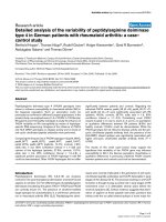
swelling and several small blisters on her toes. The most
prominent feature was that the right foot exhibited bluish
discolouration which evidently subsided to near-normal
colour on elevation (Figure 1). The femoral, popliteal,
dorsalis pedis and posterior tibialis pulses were present
and normal.
Handheld Doppler examination demonstrated a relative
decrease in the audible quality of the biphasic pulse when
compared to the normal side. Duplex scan of the right
lower limb excluded the possibility of any superficial or
deep venous thrombosis. The patient was commenced on
50% weight bearing status and was discharged home. A
recent follow-up demonstrated significant improvement
in symptoms since commencement of weight bearing
status.
Patient 2
A 20-year-old Caucasian woman who presented with
symptoms of right hip impingement underwent open
debridement and repair of a torn acetabular labrum. The
patient was discharged on the 5th postoperative day
following an uneventful recovery period. She was to
remain non-weight bearing for 6 weeks. Ten days
following discharge, she presented to the clinic complain-
ing of her right foot going blue and cold and with the
complete absence of pain. The discolouration occurred
when the limb was in a dependent position. Clinically, all
peripheral pulses were present and normal. The patient
was reviewed by a vascular surgeon who excluded the
possibility of arterial insufficiency and deep venous
thrombosis.
The patient was discharged with the advice to commence
on weight bearing of the operated hip. In the follow-up
clinic 5 months later, she was found to have complete
resolution of her vascular symptoms.
Discussion
According to the International Association for the Study of
Pain (IASP) criteria of 1994, pain, evidence of change in
blood flow or abnormal sudomotor activity and the
absence of conditions that would otherwise account for
symptoms are essential diagnostic entities of Complex
Regional Pain Syndrome type I. Eisenberg and Melamed
[4] reported a series of five patients with various foot
pathologies who had presented with all of the criteria of
Complex Regional Pain Syndrome except pain. The
authors are not aware of any English medical literature
with reports of painless Complex Regional Pain Syndrome
following hip surgery.
Veldman et al. reported a series of 829 patients with reflex
sympathetic dystrophy (RSD) and among them, 7% of the
patients did not have pain as a symptom [5]. Although is
not clear from the article if any of the Complex Regional
Pain Syndrome-like symptoms developed following hip
surgery, this does substantiate the possibility of Complex
Regional Pain Syndro me-like symptoms in the total
absence of pain.
The authors of this article postulate that in the hip, CRPS-
like symptoms develop following a period of non-weight
bearing. In both of our patients, the symptoms occurred
during non-weight bearing and subsided after weight
bearing was commenced. Unilateral lower limb
Figure 1. Right foot shows bluish discolouration compared to
the left when it is in the dependent position. Note the
generalized swelling of the right foot with loss of wrinkles
compared to the left.
Page 2 of 3
(page number not for citation purposes)
Journal of Medical Case Reports 2009, 3:7271 />suspension experiments in normal patients have shown an
increase in flow mediated dilatation of arteries of the
lower limb along with a decrease in venous capacitance
[6]. This mechanism may explain the vascular changes
which were more intense with the foot in the dependent
positions. In such a clinical setting, vascular insufficiency
and deep venous thrombosis should be excluded. Unless
surgically contraindicated, weight bearing should be
commenced at the earliest time possible.
Conclusion
Painless Complex Regional Pain Syndrome-like symptoms
may occur in patients who are kept non-weight bearing
following hip surgery. However, vascular insufficiency and
deep venous thrombosis must be excluded before this
diagnosis is made. If the clinical situation permits, early
weight bearing may relieve symptoms.
Consent
Written informed consent was obtained from the patients
for publication of this case report and any accompanying
images. A copy of the written consent is available for
review by the Editor-in-Chief of this journal.
Competing interests
The authors declare that they have no competing interests.
Authors’ contributions
JGA postulated the concept and designed the study,
interpreted the data and revised the final manuscript.
AJSK identified and re-examined the cases, acquired the
data and analysed it, reviewed the literature and wrote the
article. SKSW also re-examined the cases for the study,
assisted in data acquisition, analysed the data and assisted
in writing up the article.
References
1. Reinders MF, Geertzen JHB, Dijkstra PU: Complex regional pain
syndrome type 1: Use of the International Association for the
Study of Pain diagnostic criteria defined in 1994. Clin J Pain
2002, 18:207-215.
2. Boas RA. Complex regional pain syndrome: Symptoms, signs
and differential diagnosis.InReflex Sympathetic Dystrophy: A
ReappraisalJaning W, Stanton-Hicks M. Seattle, IASP Piers; 1996:79-92.
3. Stanton-Hicks M, Janig W, Harsenlusch S, Haddox JD, Boas R,
Wilson P: Reflex sympathetic dystrophy: Changing concepts
and taxonomy. Pain 1995, 63:127-133.
4. Eisenberg E, Melamed E: Can complex regional pain syndrome
be painless? Pain 2003, 106:263-267.
5. Veldman PHJM, Reyne n HM, Arnt z IE, Goris RJ: Signs and
symptoms of reflex sympath etic dystrophy: prospective
study of 829 patients. Lancet 1993, 342:1012-1016.
6. Bleekers MWP, De Groot PCE, Poelkens F, Rongen GA, Smits P,
Hopman MT: Vascular adaptation to 4 weeks of deconditioning
by unilateral lower limb suspension. Am J Physiol Heart Circ Physiol
2005, 288:1747-1755.
Page 3 of 3
(page number not for citation purposes)
Journal of Medical Case Reports 2009, 3:7271 />Do you have a case to share?
Submit your case report today
• Rapid peer review
• Fast publication
• PubMed indexing
• Inclusion in Cases Database
Any patient, any case, can teach us
something
www.casesnetwork.com