Kỹ thuật mới trong quản lý vết thương docx
Bạn đang xem bản rút gọn của tài liệu. Xem và tải ngay bản đầy đủ của tài liệu tại đây (653.67 KB, 9 trang )
Vol 10, No 5, September/October 2002
303
Vacuum-assisted wound closure
(VAC) was introduced by Argenta
and Morykwas
1
based on the effects
of pulling on the tissue of a wound
cavity by means of suction. VAC
exposes the wound bed to mechani-
cally induced negative pressure,
thereby removing fluid from the
extravascular space, improving cir-
culation, and enhancing the prolifer-
ation of reparative granulation tis-
sue. Essentially, an evacuation tube
is embedded in reticulated polyure-
thane foam dressing that is placed
into the wound. The open-cell na-
ture of the dressing ensures equal
distribution of the negative pressure.
When the negative pressure is ap-
plied, effluent from the wound is
drawn through the evacuation tube
into a reservoir. This open-cell vacu-
um technique has been used by a
variety of surgical disciplines,
2-5
although its use with orthopaedic
patients has been limited and pre-
liminary.
6-10
In Europe, Fleischmann, one of
the early proponents of vacuum
therapy, began using a comparable
technique independently of Argenta
and Morykwas.
6,10
The Fleischmann
technique uses a polyvinyl alcohol
sponge with a smaller pore size than
that of the polyurethane dressing
of Argenta and Morykwas. The
wound is closed over evacuation
tubes, whose fenestrated ends are
placed in contact with the sponge,
and negative pressure is applied
through the tubes to the sponge.
Fleischmann et al
6,10
have reported
encouraging results with this tech-
nique for open fractures and infec-
tion.
Animal Studies
Several animal models have validat-
ed the efficacy of VAC.
11
In a group
of 20-kg pigs, paired wounds were
created equidistant from the dorsal
midline. Laser Doppler needle
probes were inserted adjacent to the
wounds to allow continuous record-
ing of perfusion units. Subatmos-
pheric pressure was applied to the
wounds in increments of 25 mm Hg
(range, 0 to 400 mm Hg) for 15-
minute intervals. Intermittent ap-
plications of negative pressure (on
for 1 to 10 minutes, off for 1 to 5
minutes) as well as continuous set-
tings were studied. Peak increases
in blood flow (four times baseline)
Dr. Webb is Professor and Chief, Orthopedic
Trauma, Department of Orthopedic Surgery
and Rehabilitation, Wake Forest University
School of Medicine, Winston-Salem, NC.
Reprint requests: Dr. Webb, Medical Center
Boulevard, Winston-Salem, NC 27157-1070.
Copyright 2002 by the American Academy of
Orthopaedic Surgeons.
Abstract
Vacuum-assisted wound closure (VAC) is a wound management technique that
exposes the wound bed to negative pressure by way of a closed system. Edema
fluid is removed from the extravascular space, thus eliminating an extrinsic
cause of microcirculatory embarrassment and improving blood supply during
this phase of inflammation. In addition, the mechanical tension from the vacu-
um may directly stimulate cellular proliferation of reparative granulation tis-
sue. Orthopaedic indications for VAC include traumatic wounds after débride-
ment, infection after débridement, and fasciotomy wounds for compartment
syndrome. VAC also can be used as a dressing for anchoring an applied split-
thickness skin graft. The technique is contraindicated in patients with thin, eas-
ily bruised or abraded skin; those with neoplasm as part of the wound floor; and
those with allergic reactions to any of the components that contact the skin.
Clinical experience with the technique has resulted in a low incidence of minor,
reversible irritation to surrounding skin and no major complications. Further
experience is required, as well as clinical and basic research, to define optimal
indications and benefits compared with traditional methods of wound manage-
ment.
J Am Acad Orthop Surg 2002;10:303-311
New Techniques in Wound Management:
Vacuum-Assisted Wound Closure
Lawrence X. Webb, MD
Perspectives on Modern Orthopaedics
were noted at 125 mm Hg below
ambient pressure in the intermittent
mode. The optimum intermittent
cycle was 5 minutes on and 2 min-
utes off.
Five animals were used to study
granulation tissue formation by ini-
tially assessing wound volume.
Subatmospheric pressure (−125 mm
Hg) was applied to one of the
wounds of each animal, and the
control wound was managed with
sterile saline-moistened dressings.
Volume of the wounds was mea-
sured every 48 hours. The mean
increase in the rate of granulation
tissue formation for saline dress-
ing–treated wounds was 63.3% ±
26.1%. In wounds treated with
intermittent subatmospheric pres-
sure (5-minute-on, 2-minute-off
cycle), the granulation response was
103.4% ± 35.3%.
Bacterial clearance studies were
conducted by infecting wounds with
Staphylococcus aureus and S epider-
midis. Subatmospheric pressure was
used for one wound and a moist-
ened saline dressing for the paired
control. Punch biopsies of wound
tissue were obtained from the base of
each wound at 24-hour intervals for
2 weeks. Bacterial levels remained
below 10
5
organisms/g of tissue for
all treated wounds. Bacterial levels
in control wounds remained above
10
5
organisms/g of tissue until day
11; levels were highest at day 5.
Flap survival also was evaluated
using dorsally based flaps assigned
to one of four treatment groups: (1)
preoperative and postoperative
exposure to negative pressure, (2)
only preoperative exposure to nega-
tive pressure, (3) only postoperative
exposure to negative pressure, and
(4) no exposure to negative pressure
(controls). Groups 1 and 2 were ex-
posed to subatmospheric pressure of
−125 mm Hg continuously for 4
days before surgery. Groups 1 and 3
had continuously applied subatmos-
pheric pressure for 72 hours after
surgery. A percent flap survival
was calculated, with the viable sur-
face areas of each flap expressed as a
percentage of the entire flap surface
area. The flaps treated both before
and after surgery (group 1) had the
greatest survival (72.2%), followed
by the flaps treated only postopera-
tively (group 3 [67.4%]). The flaps
with only preoperative exposure
(group 2) had 64.8% survival, and
the control flaps (group 4) had the
lowest flap survival (51.2%). The dif-
ference between groups 1 and 4 was
statistically significant (P < 0.01).
11
Fabian et al
12
compared four
treatment groups using a hypoxic
full-thickness wound model in New
Zealand white rabbits: (1) VAC
dressing alone (n = 21), (2) VAC
dressing plus hyperbaric oxygen
alone (n = 20), (3) VAC dressing to
suction alone (n = 21), and (4) VAC
dressing to suction and hyperbaric
oxygen (n = 20). Parameters mea-
sured to assess healing rate included
peak granulation tissue, granulation
tissue gap, and epithelialization tis-
sue gap. A statistically significant
(P < 0.05) difference was found
between vacuum treatment with or
without hyperbaric oxygen versus
dressings alone. The authors con-
cluded that vacuum treatment in-
creases the rate of healing in a rabbit
ischemic wound model compared
with controls, with or without hy-
perbaric oxygen, and that hyperbar-
ic oxygen did not significantly alter
the rate of healing.
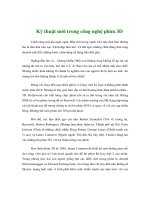
Application of the VAC
System
A VAC system consists of several
essential elements (Fig. 1, A). A ster-
ile reticulated polyurethane sponge
is cut to conform with the surface of
the wound and then is placed into
the wound to make contact with the
entire wound surface. A plastic
egress tube runs from the sponge to
another tube, which is connected to
a reservoir and programmable vacu-
um pump. An adherent plastic
sheet with adhesive on one side is
placed over the sponge around the
tubing. The sheet passes onto and
into the sponge and adheres to the
surrounding skin to seal it and
thereby form a closed system for the
wound. The settings for the vacuum
pump are adjustable for levels of
negative pressure from −50 mm Hg
to −200 mm Hg. The pump settings
can be adjusted for either continu-
ous or intermittent operation.
Before application of the VAC
system, basic wound care principles
are followed, with removal of all
devitalized and contaminated mate-
rial. This material is a focus for bac-
terial growth, which impedes the
wound-healing process. Also, thor-
ough débridement is critical before
application of VAC.
The reticulated polyurethane
sponge, available in three sizes,
comes in sterile packaging with two
transparent plastic self-adhesive
sheets. The sponge can be cut to
match the shape of the wound. It
should be placed so that it has direct
contact with the entire wound sur-
face, particularly at its depth (Fig. 1,
B). If this is not done, the wound
tissue can proliferate above the
deepest part of the wound and pos-
sibly wall off a “dead space,” which
could cause abscess formation and
ultimately prolong time to healing.
The sponge should be loose and
expanded, not tightly packed.
The plastic tube is fenestrated at
the end opposite the reservoir and
is either inserted into the sponge
through a hole cut with scissors or
placed on its surface (Fig. 1, C). The
sponge and tube are then sealed to
each other and anchored to the skin
with the clear flexible plastic sheet
cut to an appropriate size, prefer-
ably with a mesentery between the
tube (where it lifts from the sponge)
and the corresponding skin. When
the components are properly placed,
a closed system is created consisting
of the wound, the sponge, the
Vacuum-Assisted Wound Closure
Journal of the American Academy of Orthopaedic Surgeons
304
lumen of the tube, and the collecting
reservoir. The reservoir is then
placed into the receiving slot on the
vacuum pump, and negative pres-
sure is generated (Fig. 1, D). A con-
tinuous pressure of 125 mm Hg
below ambient pressure is the most
commonly used setting. The inter-
mittent setting was originally de-
signed into the system because of
the animal experiments that showed
beneficial effects on blood flow,
granulation tissue formation, and
random flap survival with an inter-
mittent 3-minutes-on, 5-minutes-off
cycle.
11
However, when the ambi-
ent pressure cycles back to 0 mm
Hg, the sponge re-expands. This
causes some motion at the wound
surface, which creates pain. There-
fore, the intermittent setting is sel-
dom used. For a weeping wound, a
lower negative pressure setting (−50
mm Hg) is used to minimize the
irritation of the intact skin at the
wound margin.
Obtaining an airtight seal is diffi-
cult in some situations, for example,
when the adjacent tissue is moist
Lawrence X. Webb, MD
Vol 10, No 5, September/October 2002
305
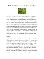
from a burn or avulsion or is close
to an external fixation pin or a tube.
Sterile hydrocolloid gel helps pro-
vide a secure seal when applied cir-
cumferentially to these areas as well
as around the pin about an inch
above the skin (Fig. 2).
The sponge is usually changed at
48-hour intervals as a bedside pro-
cedure if this schedule is tolerated
by the patient and the wound size is
limited. Local anesthetic (topical 1%
lidocaine placed in the sponge after
the vacuum has been turned off) has
successfully alleviated pain during
sponge changes. Lidocaine dosage
must be monitored carefully be-
cause the wound surface can serve
as an entry portal to the systemic
circulation. The sponge change is
Figure 1 A, Vacuum-assisted wound closure system.
B, The sponge is packed into the wound gently and should
not overlap at the skin margin. C, The tip of the drainage
tube is placed in a hole cut in the sponge. The sponge and
surrounding skin are then covered with an adhesive plastic
dressing. D, Once the vacuum is established, the sponge
collapses.
Adherent plastic sheet
Egress tube
Reservoir inserts into side
wall of programmable pump
Programmable pump
Sponge on wound
A
B C D
Figure 2 External fixator stabilizing an open tibia fracture. The leg was degloved (includ-
ing the peroneal nerve). Sterile hydrocolloid gel was placed around the pins (arrows) to
create a seal in the area of the wound draped with the flexible plastic sheet.
performed as a clean (but not ster-
ile) procedure, with normal blood
and body fluid precautions. In some
patients with extensive, semiacute
wounds, general anesthesia in an
operating room is required.
The volume of fluid produced in
the first 24 to 48 hours can be sub-
stantial—as much as 500 to 1,000
mL—but this depends on the size,
location, and nature of the wound
as well as the general condition of
the patient. Wounds in areas that
are edematous (whether because of
congestive heart failure, low protein
level, or other disorders) produce
more fluid as the vacuum pulls this
third-space fluid from the wound.
For patients with extensive wounds
with large surface areas in locations
characterized by regional edema or
in patients with systemic edema,
careful monitoring of fluid volume,
hemodynamics, and electrolyte bal-
ance may best be conducted in an
intensive care unit, an intermediate
care unit, or a burn unit. Antico-
agulated patients should be moni-
tored carefully. The VAC technique
has been used on debilitated pa-
tients without adverse impact on
electrolyte balance, kidney or liver
function, or other vital systems. In
most cases, there is no need for spe-
cialized monitoring.
Indications by Wound
Classification
Wounds treated with the VAC tech-
nique can be grouped into nine
descriptive categories:
13
(1) wounds
to which a split-thickness skin graft
is applied, (2) infected wounds
(after débridement), (3) open frac-
ture wounds, (4) acute soft-tissue
wounds (with exposed tendon,
hardware, bone, and/or joint), (5)
fasciotomy wounds after compart-
ment syndrome, (6) chronic wounds
(>3 months’ duration), (7) surgical
wounds that are difficult to close
because of tension, (8) wounds with
external fixation pins or tubes or
catheter sites with irritation and
drainage, and (9) surgical wounds
that weep serous fluid after the sec-
ond postoperative day.
For wound types 1 through 7, the
pressure setting is −125 mm Hg and
the sponge is changed at 48-hour
intervals. For wound types 2, 3, 4,
and 6, the VAC technique is appro-
priate only after complete débride-
ment. In infected wounds or severely
contaminated acute wounds, it may
be appropriate to wait for a “second
look” débridement to be confident
that all of the devitalized tissue has
been removed. For wound type 8
(external fixation pin irritation), the
pressure setting is −50 mm Hg and
the sponge at the base of the pin can
be changed about once per week.
Sterile hydrocolloid gel is useful cir-
cumferentially around the pin a
short distance (1 in) above the skin
to aid in sealing the sponge with the
adhesive sheet. When the VAC
technique is applied for wound type
9 (weeping surgical wounds), the
sponge is also applied with the pres-
sure setting lowered to −50 mm Hg
so as not to irritate the skin.
When used to bolster a split-
thickness skin graft (wound type 1),
the sponge is applied directly over
the split graft, which covers the
entire wound surface. The pressure
is set at −125 mm Hg and the
sponge left in place for 4 days. (If a
portion of the graft lifts from the
recipient bed at the time of sponge
removal, the graft and sponge are
reapplied to the bed and sealed, and
the negative pressure is reestab-
lished for 48 hours.) Some surgeons
prefer to use petrolatum-impregnat-
ed gauze as an intermediary be-
tween the graft and the sponge.
1,14
Early Results
The success of VAC depends on
the indication for which it is used.
For traumatic wounds, a successful
transition to wound closure or sta-
ble wound coverage is an adequate
end point. In general, the method
appears to be useful in accelerating
wound healing by promoting
wound granulation. The granula-
tion is often exuberant and will
cover small areas of exposed hard-
ware, bone, fascia, and tendon, pro-
vided these structures are clean. In
a number of such cases, use of VAC
has either circumvented the need
for or enhanced the success of flap
coverage of a wound
15
(Figs. 3 and
4). The VAC method is not a substi-
tute for débridement, however, and
with infected wounds, contaminated
and devitalized tissues and/or
retained implants ordinarily will
require removal. Small-surface-area
exposures of bone or hardware in
well-vascularized tissue, on the
other hand, are quite amenable to
VAC treatment, which encourages
the overgrowth of healthy granula-
tion tissue. This overgrowth allows
for secondary epithelialization or
simple wound closure or split-skin
coverage of the area. Larger areas
of exposed or infected hardware
may be amenable to VAC treatment
with the techniques described by
Fleischmann et al,
6
whose results
are encouraging but preliminary
and await longer follow-up.
Split-thickness skin grafts have
been observed to heal more pre-
dictably with this technique.
16
This
may be because of the evacuation of
the serous fluid that forms on the
surface of the wound. This fluid
might otherwise get between the
undersurface of the graft and the
recipient tissue bed and thereby act
as a barrier to oxygen diffusion.
When the VAC system is used as
a dressing for fasciotomy wounds
after compartment syndrome,
edema can be minimized and viable
muscle preserved. A retrospective
analysis comparing this technique
to simple saline dressings for fas-
ciotomy wounds to treat compart-
ment syndrome of the leg demon-
Vacuum-Assisted Wound Closure
Journal of the American Academy of Orthopaedic Surgeons
306
strated several advantages of the
VAC technique. These include
more rapid resolution of edema
fluid from the tissue, allowing earli-
er definitive closure/coverage com-
pared to a control group. In addi-
tion, a greater proportion of VAC-
treated wounds underwent primary
closure rather than skin grafting for
wound coverage.
17
For weeping wounds with con-
firmed drainage beyond the second
postoperative day, a successful tran-
sition to a clean, dry wound free of
infection is the desired end point.
About 10% of patients undergoing
elective surgery of the hip or knee
have wound drainage to an extent
that requires continued surgical
dressings at or beyond the second
postoperative day. These wounds
have been shown to heal better
when the seroma is drained.
18
Application of the VAC system at
low negative pressure (−50 mm Hg)
for this indication resulted in suc-
cessful transition to a dry wound
that healed uneventfully in 54
patients with one 24-hour applica-
tion. The two remaining wounds
required two or more applications.
This experience has prompted the
use of the sponge as a dressing on
elective surgical wounds when the
wound is prone to weep (eg, after a
prolonged surgery with a large inci-
sion in obese patients or those with
localized or generalized edema.)
This method is also used when, in
the judgment of the surgeon, the
wound needs to be isolated (eg, for
an anticipated prolonged stay in the
intensive care unit where antibiotic-
resistant nosocomial organisms are
known risks).
A recent randomized prospective
trial demonstrated the superiority of
VAC over wet-to-moist saline dress-
ings applied to chronic nonhealing
wounds.
19
In this study, the differ-
ence was most pronounced for re-
duction of the measured wound
depth (66% for VAC versus 20% for
saline). Histologic evaluation of
these wounds demonstrated a con-
sistent difference between those
treated with VAC (characterized by
reparative granulation tissue forma-
tion) and those managed with
dressing changes (characterized by
inflammation and fibrosis). This
would indicate an enhancement in
the quality of the healing tissue with
vacuum treatment, in addition to
the improved rate of healing.
The ability to directly inspect the
vacuum tube and reservoir allows
the surgeon to assess the character
and volume of the drainage fluid.
This feature may provide an advan-
tage over dressing changes, which
often allow assessment of the drain-
age only by examining the removed
dressing. Examination of dressing
requires wound manipulation and
the removal and reapplication of ad-
hesive tape, and furthermore does
not provide accurate assessment of
Lawrence X. Webb, MD
Vol 10, No 5, September/October 2002
307
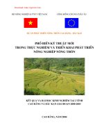
A B C
D E F
Figure 3 A, Anteroposterior radiograph of a 20-year-old woman who sustained an ankle
fracture 3 years previously. Because of ankle pain, plate removal was attempted in the
office, but failed and was complicated by an infection 10 days later. B, During débride-
ment, the hardware was removed and the wound left open with exposed bone at its base.
C, A VAC system was applied to the wound, with the sponge changed at 48-hour inter-
vals. D, By the third sponge change, there was a healthy lawn of granulation tissue over
the bone. A split-thickness skin graft was applied and secured to the wound bed, with a
vacuum sponge used as a bolster. Continuous pressure of −125 mm Hg pressure was
applied for 4 days. E, With removal of the sponge, there was a complete take of the graft.
Note the rash from the overlap of the sponge on the skin just over the lower margin of the
wound. The rash completely resolved in 36 hours. F, One year later, the grafted area is
healed and stable with no signs of indolent infection.
the amount or turbidity of drainage.
Maintenance of a closed system, with
its preclusion of repetitive dressing
changes and lower likelihood of con-
tamination of the wound, may be an
added advantage.
20
In addition, the
VAC system allows wound fluid to
be collected for future analysis. A
randomized prospective trial com-
paring the VAC system to standard
dressing changes for weeping surgi-
cal wounds is underway.
Complications and
Contraindications
Complications with the use of the
VAC technique have been few. The
most common is a rash on the skin
resulting from contact with the suc-
tion sponge (Fig. 3, E), which usually
resolves in 24 hours. This occurred
in 6 of 270 patients (2.2%) in one
study and resolved within 48 hours
in each case.
13
The rash was not
associated with any itching or pain.
To minimize this complication, care
should be taken to confine the
sponge to the wound and avoid any
overlap onto the normal skin. If
overlap of the skin is unavoidable,
such as with application over a sur-
gical wound, a setting of −50 mm
Hg can be used.
If a patient has thin skin, as with
elderly patients on long-term sys-
temic steroids, shearing avulsion
may occur during sponge exchange
when lifting the adhesive plastic
from the skin. The technique is
therefore contraindicated in individ-
uals who are intolerant (for either
allergic or mechanical reasons) of
adhesives on the skin.
If the sponge is left deep in a
wound for more than 48 hours, it can
be difficult to extract because of the
overgrowth of exuberant granula-
tions. Once the sponge is extracted,
minor bleeding may occur, which is
typical of this hypervascular tissue;
this is easily controlled with pres-
sure.
Vacuum-Assisted Wound Closure
Journal of the American Academy of Orthopaedic Surgeons
308
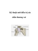
A B
C D
E F
Figure 4 A, A 22-year-old male polytrauma victim sustained a type III open fracture of
the tibia with a traumatic below-knee amputation of the contralateral leg. The wound was
débrided and the fracture stabilized with an external fixator. B, The skin was closed over
the exposed bone, leaving only a small area exposed (arrow). C, Two VAC sponges were
applied to the open wounds, and changed at 48-hour intervals. D, At day 6, with good
granulation tissue, a split-thickness graft was applied. The sponge system was reapplied
for 4 days, with good graft incorporation. Medial (E) and anterior (F) views of the leg at 2
years with a stable soft-tissue coverage and no infection.
The effects of the VAC technique
on neoplasm are unknown; accord-
ingly, VAC should not be used when
neoplasm is part of the wound.
Careful monitoring is important in
anticoagulated patients or those
with a bleeding disorder, or when
wounds are extensive and a large
amount of fluid evacuation is antici-
pated (eg, large-surface-area wounds
or burn wounds).
Treatment Costs
Philbeck et al
21
demonstrated the
cost effectiveness of VAC in an out-
patient setting. They reviewed the
records of 1,262 Medicare patients
with advanced-stage pressure ulcers
of the trunk or trochanter who had
failed to respond to previous inter-
vention and subsequently under-
went VAC treatment. The wounds
were categorized, and wound-heal-
ing rates were calculated for each
type and compared with rates in
other published reports.
22
Costs
based on wound-closure rate, days
to heal, material cost per day, and
daily nursing visit cost were used to
arrive at overall estimates.
Discussion
Wounds heal by progression through
phases. After the injury or wound-
ing mechanism, there is an initial
inflammatory phase characterized
by an array of vascular, cellular,
and humoral events. These events
include release of vasoactive sub-
stances and triggers for local white
cell migration with an outpouring
of tissue fluid. Over time, this
phase gives way to the reparative
phase, which is characterized by
angiogenesis, tissue granulation,
heightened collagen production,
and re-epithelialization.
The VAC technique acts to pull
off the fluid from the tissue space,
thereby lowering capillary afterload
in the zone of stasis. Because the
embarrassment to microcirculation
is removed, delivery of oxygen and
nutrients is enhanced while removal
of inhibitory factors and toxins is
facilitated. In addition, laser Dopp-
ler flow studies have documented a
notably increased blood flow adja-
cent to the wound during the course
of treatment.
1,11
These factors may
account for the successful preven-
tion of the progression of partial-
thickness burns in the animal model
reported by Morykwas et al.
23
They
may also account for the prevention
of ulcers after injection of doxoru-
bicin in a swine model.
24
Because bacterial colonization
hampers wound healing,
25-27
the ef-
fect of lowering the wound bacterial
count
11
may give vacuum treatment
an advantage over other methods
for management of open contami-
nated wounds or wounds with a
history of infection. Despite this ad-
vantage, VAC is best regarded as an
adjunct to wound management, not
a substitute for appropriate surgical
débridement. Larger areas of ex-
posed/infected hardware are best
handled with traditional techniques;
most necessitate removal of the inert
material as part of the management
of the infection.
28,29
Contraction of the sponge under
the influence of the vacuum creates
tension on the cells that comprise
the surface of the wound. This
mechanical tensile stress may stimu-
late angiogenesis and a proliferation
of primitive mesenchymal cells and
fibroblasts.
30-32
This may account
for the increased rate of skin graft
donor site re-epithelialization ob-
served by Genecov et al,
33
as well as
the success of the technique in man-
aging wounds with exposed tendon
and bone in the lower extremity.
15
This relationship between the ten-
sile forces on cells and angiogenesis
and tissue growth was first postu-
lated nearly a century ago.
34
More
recent studies
35-37
have documented
the effects on tissue regeneration
and proliferation in the setting of
bone distraction as well as tissue
expansion.
Animal experiments have shown
the beneficial effect of VAC on evac-
uation of wound edema, bacterial
clearance from the wound, im-
proved local blood flow, and stimu-
lation of the formation of healing
granulation tissue.
11
Others, work-
ing with a rabbit model, demon-
strated that VAC accelerates wound
healing more than do simple dress-
ings at normal pressure or with
hyperbaric oxygen.
12
In the clinical evaluation, the end
points for wound healing are some-
what subjective. This makes it diffi-
cult to produce solid clinical re-
search that clearly demonstrates the
superiority of VAC or other tech-
niques compared with alternatives.
Although further animal research
may help to circumvent some of
these difficulties, creatively designed
and well-controlled comparative
clinical studies are needed to docu-
ment or refute the advantages of
wound management by VAC in the
clinical setting.
Summary
VAC appears to offer some distinct
advantages over traditional wound-
closure methods. These include
evacuation of wound edema, hasten-
ing and promoting the formation of
hypervascular wound granulation,
and rapid and complete incorpora-
tion of meshed split-thickness skin
grafts. The system is closed, which
lowers the likelihood of wound con-
tamination by resistant hospital
organisms. Preliminary cost analy-
ses in outpatient settings have
demonstrated cost advantages of
vacuum treatment over conventional
management with dressing changes.
Because wound fluid is collected on
an ongoing basis, VAC may prove to
be a valuable research tool, helping
the clinician assess the character and
Lawrence X. Webb, MD
Vol 10, No 5, September/October 2002
309
volume of wound drainage. Mor-
bidity has been minor, with no major
complications. There is a low inci-
dence of minor, reversible rash that
occurs when the sponge is posi-
tioned with any overlap on the nor-
mal skin. The VAC technique is not
a substitute for wound débridement;
rather, it is an adjunct to wound
management. The clinical benefits
need to be further scrutinized with
well-controlled prospective studies.
Vacuum-Assisted Wound Closure
Journal of the American Academy of Orthopaedic Surgeons
310
References
1. Argenta LC, Morykwas MJ: Vacuum-
assisted closure: A new method for
wound control and treatment: Clinical
experience. Ann Plast Surg 1997;38:
563-577.
2. Argenta PA, Rahaman J, Gretz HF III,
Nezhat F, Cohen CJ: Vacuum-assisted
closure in the treatment of complex
gynecologic wound failures. Obstet
Gynecol 2002;99:497-501.
3. Garner GB, Ware DN, Cocanour CS, et
al: Vacuum-assisted wound closure
provides early fascial reapproximation
in trauma patients with open abdo-
mens. Am J Surg 2001;182:630-638.
4. Harlan JW: Treatment of open sternal
wounds with the vacuum-assisted clo-
sure system: A safe, reliable method.
Plast Reconstr Surg 2002;109:710-712.
5. Chang KP, Tsai CC, Lin TM, Lai CS, Lin
SD: An alternative dressing for skin
graft immobilization: Negative pressure
dressing. Burns 2001;27:839-842.
6. Fleischmann W, Strecker W, Bombelli
M, Kinzl L: Vacuum sealing as treat-
ment of soft tissue damage in open
fractures [German]. Unfallchirurg
1993;96:488-492.
7. Webb LX, Argenta LC, Lange R, et al:
Abstract: Vacuum assisted wound clo-
sure (VAC therapy): Experience with
its use in 150 patients. Orthopaedic
Trauma Association Final Program, 14th
Annual Meeting. Rosemont, IL, Ortho-
paedic Trauma Association, 1996, pp
340-341.
8. Greer S, Kasabian A, Thorne C, Borud
L, Sims CD, Hsu M: Letter: The use of
a subatmospheric pressure dressing to
salvage a Gustilo grade IIIB open tibial
fracture with concomitant osteomye-
litis to avert a free flap. Ann Plast Surg
1998;41:687.
9. Mullner T, Mrkonjic L, Kwasny O,
Vecsei V: The use of negative pressure
to promote the healing of tissue
defects: A clinical trial using the vacu-
um sealing technique. Br J Plast Surg
1997;50:194-199.
10. Fleischmann W, Lang E, Russ M:
Treatment of infection by vacuum
sealing [German]. Unfallchirurg 1997;
100:301-304.
11. Morykwas MJ, Argenta LC, Shelton-
Brown EI, McGuirt W: Vacuum-assist-
ed closure: A new method for wound
control and treatment. Animal studies
and basic foundation. Ann Plast Surg
1997;38:553-562.
12. Fabian TS, Kaufman HJ, Lett ED, et al:
The evaluation of subatmospheric
pressure and hyperbaric oxygen in
ischemic full-thickness wound healing.
Am Surg 2000;66:1136-1143.
13. Webb LX, Schmidt U: Wound manage-
ment with vacuum therapy [German].
Unfallchirug 2001;104:918-926.
14. Schneider AM, Morykwas MJ, Argenta
LC: A new and reliable method of
securing skin grafts to the difficult
recipient bed. Plast Reconstr Surg 1998;
102:1195-1198.
15. DeFranzo AJ, Argenta LC, Marks MW,
et al: The use of vacuum-assisted clo-
sure therapy for treatment of lower
extremity wounds with exposed bone.
Plast Reconstr Surg 2001;108:1184-1191.
16. Blackburn JH II, Boemi L, Hall WW, et
al: Negative-pressure dressings as a
bolster for skin grafts. Ann Plast Surg
1998;40:453-457.
17. Chang D, Castle J, Webb LX: Abstract:
Vacuum-assisted closure for fascioto-
my wounds after compartment syn-
drome of the leg. Orthopaedic Trauma
Association Final Program, 17th Annual
Meeting. Rosemont, IL, Orthopaedic
Trauma Association, 2001, p 57.
18. Varley GW, Milner SA: Wound drains
in proximal femoral fracture surgery:
A randomized prospective trial of 177
patients. J R Soc Med 1995;88:42P-44P.
19. Joseph E, Hamori CA, Bergman S, Roaf
E, Swann NF, Anastasi GW: A pro-
spective randomized trial of vacuum-
assisted closure versus standard therapy
of chronic nonhealing wounds. Wounds
2000;12:60-67.
20. Tscherne H: The management of open
fractures, in Tscherne H, Gotzen L (eds):
Fractures With Soft-Tissue Injuries. New
York, NY: Springer-Verlag, 1984, pp 1-18.
21. Philbeck TE Jr, Whittington KT,
Millsap MH, Briones RB, Wight DG,
Schroeder WJ: The clinical and cost
effectiveness of externally applied neg-
ative pressure wound therapy in the
treatment of wounds in home health-
care Medicare patients. Ostomy Wound
Manage 1999;45:41-50.
22. Ferrell BA, Osterweil D, Christenson
PA: A randomized trial of low-air-loss
beds for treatment of pressure ulcers.
JAMA 1993;269:494-497.
23. Morykwas MJ, David LR, Schneider
AM, et al: Use of subatmospheric pres-
sure to prevent progression of partial-
thickness burns in a swine model. J
Burn Care Rehabil 1999;20(1 Pt 1):15-21.
24. Morykwas MJ, Kennedy A, Argenta
JP, Argenta LC: Use of subatmospher-
ic pressure to prevent doxorubicin
extravasation ulcers in a swine model.
J Surg Oncol 1999;72:14-17.
25. Hunt TK: The physiology of wound heal-
ing. Ann Emerg Med 1988;17:1265-1273.
26. Seiler WO, Stahelin HB, Sonnabend W:
Effect of aerobic and anaerobic germs
on the healing of decubitus ulcers
[German]. Schweiz Med Wochenschr
1979;109:1594-1599.
27. Daltrey DC, Rhodes B, Chattwood JG:
Investigation into the microbial flora
of healing and non-healing decubitus
ulcers. J Clin Pathol 1981;34:701-705.
28. Gristina AG, Costerton JW: Bacterial
adherence to biomaterials and tissue:
The significance of its role in clinical
sepsis. J Bone Joint Surg Am 1985;67:
264-273.
29. Gristina AG, Barth E, Webb LX:
Microbial adhesion and the pathogene-
sis of biomaterial-centered infections, in
Gustilo RB, Gruninger RP, Tsukayama
DT (eds): Orthopaedic Infection: Diagno-
sis and Treatment. Philadelphia, PA: WB
Saunders, 1989.
30. Aronson J: The biology of distraction
osteogenesis, in Bianchi-Maiocchi A,
Aronson J (eds): Operative Principles of
Ilizarov: Fracture Treatment, Nonunion,
Osteomyelitis, Lengthening, Deformity
Correction. Baltimore, MD: Williams
and Wilkins, 1991, pp 42-52.
31. Aronson J: Experimental assessment
of bone regenerate quality during dis-
traction osteogenesis, in Brighton CT,
Friedlaender GE, Lane JM: Bone
Formation and Repair. Rosemont, IL:
American Academy of Orthopaedic
Surgeons, 1994, pp 441-463.
32. Aronson J, Harrison BH, Stewart CL,
Harp JH Jr: The histology of distraction
osteogenesis using different external
fixators. Clin Orthop 1989;241:106-116.
33. Genecov DG, Schneider AM, Morykwas
MJ, Parker D, White WL, Argenta LC: A
controlled subatmospheric pressure
dressing increases the rate of skin graft
donor site reepithelialization. Ann Plast
Surg 1998;40:219-225.
34. Thoma R: Über die histomechanik des
gefaßsystems und die pathogenese der
angiosklerose. Virchows Arch F, Path
Anat 1911;204:1-74.
35. Ilizarov GA: The tension-stress effect
on the genesis and growth of tissues:
I. The influence of stability of fixation
and soft-tissue preservation. Clin
Orthop 1989;238:249-281.
36. Ilizarov GA: The tension-stress effect
on the genesis and growth of tissues:
II. The influence of the rate and fre-
quency of distraction. Clin Orthop
1989;239:263-285.
37. Argenta LC, Marks MW: Tissue ex-
pansion, in Georgiade GS, Georgiade
NG, Riefkohl R, Barwick WJ (eds):
Textbook of Plastic, Maxillofacial and
Reconstructive Surgery, ed 2, vol 1.
Baltimore, MD: Williams and Wilkins,
1992, pp 103-113.
Lawrence X. Webb, MD
Vol 10, No 5, September/October 2002
311