Báo cáo y học: " Avascular necrosis of humeral head in an elderly patient with tuberculosis: a case report" pps
Bạn đang xem bản rút gọn của tài liệu. Xem và tải ngay bản đầy đủ của tài liệu tại đây (248.75 KB, 3 trang )
BioMed Central
Page 1 of 3
(page number not for citation purposes)
Journal of Medical Case Reports
Open Access
Case report
Avascular necrosis of humeral head in an elderly patient with
tuberculosis: a case report
Renu Agarwal
1
, Ruchika Gupta
2
, Sompal Singh*
1
, Kusum Gupta
1
and
Madhur Kudesia
1
Address:
1
Department of Pathology, Hindu Rao Hospital, Malka Ganj, Delhi-110007, India and
2
Department of Pathology, All India Institute of
Medical Sciences, Ansari Nagar, New Delhi-110029, India
Email: Renu Agarwal - ; Ruchika Gupta - ; Sompal Singh* - ;
Kusum Gupta - ; Madhur Kudesia -
* Corresponding author
Abstract
Introduction: Osteonecrosis (avascular necrosis) is known to be caused by high-dose
corticosteroid therapy, alcoholism and rarely by infections. However, a tubercular etiology of this
condition is very rare. A review of the literature yielded only a few cases of polyarticular
tuberculosis with osteonecrosis in immunosuppressed individuals. No case of monoarticular
tubercular osteonecrosis diagnosed by aspiration cytology was found. Since tuberculosis is a
curable disease, an early and accurate diagnosis is essential.
Case presentation: A 60-year-old Indian man presented with diffuse swelling and pain in the left
shoulder for the previous 6 months. A computed tomography scan of the left shoulder revealed
crescentic lucency in the humeral head, suggestive of osteonecrosis. Fine needle aspiration cytology
smears from the swelling showed features of an acute suppurative lesion. Stain for acid-fast bacillus
was positive and thus, a final clinico-pathological diagnosis of osteonecrosis of humeral head with
tubercular etiology was rendered. The patient was initiated on anti-tuberculous therapy with
symptomatic improvement in his condition.
Conclusion: Osteonecrosis, a debilitating disease, may rarely occur due to tuberculosis, especially
in endemic areas. Fine needle aspiration cytology is an effective and inexpensive modality for an
early diagnosis of the tubercular etiology of osteonecrosis.
Introduction
Osteonecrosis, also known as avascular necrosis (AVN),
occurs in people with risk factors such as high-dose corti-
costeroid therapy, excessive alcohol intake, injury, malig-
nancy, systemic lupus erythematosus, and hematologic
disorders such as sickle cell disease [1]. Among infectious
causes, Human Immunodeficiency Virus (HIV) and
meningococcemia have been reported to cause AVN [2,3].
However, AVN in association with tuberculosis has been
reported in only a few cases [4,5]. In one case, described
by Cheung et al. in 1995, polyarticular tuberculosis with
AVN was identified in a HIV positive patient [4]. No case
of monoarticular tuberculosis associated with AVN has
been reported in the available literature.
Published: 4 December 2008
Journal of Medical Case Reports 2008, 2:361 doi:10.1186/1752-1947-2-361
Received: 6 February 2008
Accepted: 4 December 2008
This article is available from: />© 2008 Agarwal et al; licensee BioMed Central Ltd.
This is an Open Access article distributed under the terms of the Creative Commons Attribution License ( />),
which permits unrestricted use, distribution, and reproduction in any medium, provided the original work is properly cited.
Journal of Medical Case Reports 2008, 2:361 />Page 2 of 3
(page number not for citation purposes)
Our case depicts a rare association of monoarticular tuber-
culosis with AVN in an immunocompetent patient.
Case presentation
A 60-year-old man, Indian by origin, presented with
swelling and pain in the left shoulder of 6 months dura-
tion. There was associated anorexia and loss of weight.
However, there was no history of preceding trauma, corti-
costeroid therapy or significant medical or surgical treat-
ment. He was a non-alcoholic and non-smoker.
On local examination, a diffuse, soft and tender swelling,
4 × 4 cm, was seen at the left shoulder. There was mild
restriction of movement of the left shoulder. The overly-
ing skin was warm and erythematous. Hematological
investigations revealed peripheral blood lymphocytosis
and increased erythrocyte sedimentation rate (ESR) (35
mm in the first hour). Mantoux test using 5 tuberculin
units (TU) of purified protein derivative (PPD) showed
significant induration after 72 hours (13 mm). Serological
tests for HIV, rheumatoid factor and anti-nuclear antibod-
ies (ANA) were negative. Chest X-ray did not show any
evidence of active/healed pulmonary tuberculosis. Radio-
graphs of the left shoulder joint did not reveal any bony
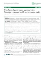
abnormality. Computed tomography (CT) scan of the left
shoulder showed a crescentic lucency in the humeral head
with associated soft tissue swelling, consistent with a diag-
nosis of osteonecrosis (Figure 1). The patient was referred
for fine needle aspiration (FNA) cytology of the soft tissue
swelling to assist in etiological diagnosis.
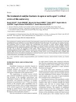
FNA yielded a purulent aspirate, smears which showed an
acute suppurative lesion with intact and degenerated neu-
trophils in a proteinaceous background along with a few
lymphocytes and histiocytes (Figure 2). No epithelioid
cell granulomas were noted. Ziehl Neelsen staining
showed occasional acid-fast bacilli (Figure 2, inset). A
diagnosis of tubercular etiology of osteonecrosis was ren-
dered. The patient was put on antitubercular therapy, after
which the pain and swelling reduced markedly.
Discussion
Osteonecrosis, also known as avascular necrosis (AVN),
aseptic necrosis or ischemic necrosis, results from tempo-
rary or permanent loss of blood supply to a part of bone.
As a result of the loss of blood supply, the bone may ulti-
mately collapse [1].
Numerous risk factors have been associated with AVN
including corticosteroid therapy, alcohol intake and bony
injury. Other associations include systemic malignancy,
lupus erythematosus, sickle cell disease, Gaucher's dis-
ease, Caissons disease, gout, vasculitis, osteoarthritis,
osteoporosis, radiation therapy, chemotherapy and organ
transplantation, particularly renal transplant [1]. A rare
causal association with infections such as HIV and menin-
gococcemia (with disseminated intravascular coagula-
tion) has been reported [2,3]. However, a large number of
cases do not have any obvious etiologic factor and are
reported as idiopathic [1].
Various imaging techniques have been used for diagnosis
of AVN. Plain X-ray has a low sensitivity and shows bone
damage only in later stages of disease. CT scan is better
than X-rays, however, its sensitivity compared to magnetic
resonance imaging (MRI) is still low, especially in early
Computed tomography scan at the level of the upper humerus showing crescentic lucency as evidence of osteonecrosisFigure 1
Computed tomography scan at the level of the upper
humerus showing crescentic lucency as evidence of
osteonecrosis.
Fine needle aspiration smear showing many viable and degen-erating neutrophils in a thin necrotic backgroundFigure 2
Fine needle aspiration smear showing many viable
and degenerating neutrophils in a thin necrotic back-
ground. Inset shows an acid-fast bacillus (Giemsa Stain
×200, Inset: Ziehl Neelsen Stain ×400).
Journal of Medical Case Reports 2008, 2:361 />Page 3 of 3
(page number not for citation purposes)
stages of the disease [1]. MRI is currently the accepted
standard for noninvasive diagnosis. The classical MRI
appearance of AVN is that of a segmental area of low sig-
nal density in the subchondral bone on T1-weighted
images [1]. In our patient, a plain radiograph did not
reveal AVN, which could be picked up on CT scan. MRI
could not be performed due to lack of facilities.
The goal of treatment of AVN is to improve the use of the
affected joint, stop further damage to the bone and ensure
bone and joint survival. The underlying cause of AVN has
to be ascertained and eliminated if possible. Surgical
intervention, including arthroscopic debridement, core
decompression, vascularized bone grafting and bone
reconstruction, is advocated when symptoms are persist-
ent and signs of collapse are evident [1].
Tuberculosis, caused by Mycobacterium tuberculosis, is a
common infectious disease in the developing countries
and is re-emerging in developed nations due to the
human immunodeficiency virus (HIV) pandemic [6].
Osteoarticular tuberculosis results from hematogenous
dissemination of Mycobacterium tuberculosis from a pri-
mary infected visceral focus to the skeletal system [7]. Our
present case adds to the myriad of radiological presenta-
tions of osteoarticular tuberculosis, i.e. avascular necrosis.
AVN with tuberculosis as an etiological cause of
osteonecrosis has only been mentioned in rare case
reports. Two cases of AVN of femoral capital epiphysis fol-
lowing intertrochanteric tubercular osteomyelitis have
been reported [5]. There is a recent report describing AVN
in a HIV positive patient with polyarticular tuberculosis
[4]. To the best of our knowledge, the present case is the
first report documenting an association of monoarticular
tuberculosis with AVN in an immunocompetent patient,
where the etiology of osteonecrosis was confirmed on
aspiration cytology. In this patient, FNA showed an acute
suppurative lesion with a few acid-fast bacilli. This case
underlines the utility of Ziehl-Neelsen stain to diagnose
tubercular etiology in cases with radiological diagnosis of
AVN, especially when the patient is a resident of an
endemic zone.
Conclusion
This case report emphasizes that tuberculosis should be
retained as one of the important differential diagnoses in
cases of osteonecrosis, especially in endemic areas, after
other more common etiological disorders have been
excluded. Aspiration cytology offers a rapid, yet inexpen-
sive method for diagnosis leading to appropriate therapy
being initiated.
Abbreviations
ANA: anti-nuclear antibody; AVN: avascular necrosis; CT:
computed tomography; ESR: erythrocyte sedimentation
rate; FNA: fine needle aspiration; HIV: Human Immuno-
deficiency Virus; MRI: magnetic resonance imaging; PPD:
purified protein derivative; TU: tuberculin units.
Consent
Written informed consent was obtained from the patient
for publication of this case report and any accompanying
images. A copy of the written consent is available for
review by the Editor-in-Chief of this journal.
Competing interests
The authors declare that they have no competing interests.
Authors' contributions
RA performed the fine needle aspiration cytology and
wrote the case outline. RG was a major contributor in
writing the manuscript and revising it. SS assisted in
reviewing the slides, provded images and helped in final
drafting of the manuscript. KG assisted in the literature
review and writing of the manuscript. MK interpreted the
fine needle aspiration cytology and critically reviewed the
manuscript for its intellectual content. All authors have
read and approved the final manuscript to be published.
All authors have participated sufficiently to take public
responsibility of the content of the manuscript.
References
1. Assouline-Dayan Y, Chang C, Greenspan A, Shoenfeld Y, Gershwin
ME: Pathogenesis and natural history of osteonecrosis. Semin
Arthritis Rheum 2002, 32:94-124.
2. Qaqish RB, Sims KA: Bone disorders associated with the
human immunodeficiency virus: pathogenesis and manage-
ment. Pharmacotherapy 2004, 24:1331-1346.
3. Campbell WN, Joshi M, Sileo D: Osteonecrosis following menin-
gococcemia and disseminated intravascular coagulation in
an adult: case report and review. Clin Infect Dis 1997,
24:452-455.
4. Cheung NT, Saklatvala J, Dawes PT: Multiple joint avascular
necrosis: beware of tuberculosis and human immunodefi-
ciency virus – a rare but important cause. Br J Rheumatol 1995,
34:387-391.
5. Kemp HBS, Lloyd-Roberts GC: Avascular necrosis of the capital
epiphysis following osteomyelitis of the proximal femoral
metaphysis. J Bone Joint Surg Br 1974, 56:688-697.
6. Raviglione MC, Snider DE Jr, Kochi A: Global epidemiology of
tuberculosis: morbidity and mortality of a worldwide epi-
demic. JAMA 1995, 273:220-226.
7. Kumar R: Spinal tuberculosis: with reference to the children
of northern India. Child's Nerv Syst 2005, 21:19-26.