Báo cáo y học: "A varicocoele mimicking a hydrocoele in a man with portal hypertension: a case report" pot
Bạn đang xem bản rút gọn của tài liệu. Xem và tải ngay bản đầy đủ của tài liệu tại đây (216.09 KB, 3 trang )
BioMed Central
Page 1 of 3
(page number not for citation purposes)
Journal of Medical Case Reports
Open Access
Case report
A varicocoele mimicking a hydrocoele in a man with portal
hypertension: a case report
George Yardy*
1
, Akkib Rafique
2
, Iain Sellers
3
, Lawrence Berman
3
and
Nigel Bullock
1
Address:
1
Department of Urology, Addenbrooke's Hospital, Cambridge, UK,
2
Department of Radiology, Ealing Hospital, London, UK and
3
Department of Radiology, Addenbrooke's Hospital, Cambridge, UK
Email: George Yardy* - ; Akkib Rafique - ; Iain Sellers - ;
Lawrence Berman - ; Nigel Bullock -
* Corresponding author
Abstract
Introduction: Hydrocoele is a condition frequently encountered in adult urological practice. It is
usually of benign aetiology and often diagnosed on clinical grounds. Surgical repair, if indicated, is
generally straightforward.
Case presentation: We report a 53-year-old man with liver cirrhosis and clinical features of a
hydrocoele, in whom flow was demonstrated using Doppler ultrasonography in the fluid around
the testis, which communicated via varices with the left renal vein.
Conclusion: In this patient with misleading clinical signs, diagnosis was established radiologically.
Had surgery proceeded without this investigation, significant intra-operative bleeding would have
been likely.
Introduction
A hydrocoele causes fluctuant non-tender unilateral scrotal
swelling which is irreducible and may be tense or lax. It is
caused by an abnormal quantity of fluid within the tunica
vaginalis. In adults, it is usually idiopathic, but may be sec-
ondary to trauma, infection, neoplasia or lymphatic
obstruction. Paediatric hydrocoele is usually associated
with a patent processus vaginalis. A careful history and clin-
ical examination usually establishes the diagnosis and fur-
ther investigations are not always required. Treatment is
often not offered unless the condition troubles the patient
particularly. Hydrocoele repair is, however, a frequently
performed relatively minor procedure. We present a patient
with a hydrocoele on clinical grounds, in whom further
radiological investigation demonstrated that the fluid sur-
rounding the testis was blood within a scrotal varix which
had developed as a result of a portal-systemic anastomosis
involving the splenic and left renal veins.
Case presentation
A 53-year-old man was referred for assessment of a left
scrotal swelling which was slightly uncomfortable and
had been particularly noticeable for 3 months. He had a
history of non-insulin-dependent diabetes mellitus and
hepatic cirrhosis, which was probably secondary to non-
alcoholic steatohepatitis. He had oesophageal varices
which were asymptomatic and were being monitored. He
was examined in the outpatients department and the clin-
ical notes recorded that, "what he appears to have is a lax
left hydrocoele which is absolutely typical and of textbook
nature."
Published: 4 December 2008
Journal of Medical Case Reports 2008, 2:363 doi:10.1186/1752-1947-2-363
Received: 4 January 2008
Accepted: 4 December 2008
This article is available from: />© 2008 Yardy et al; licensee BioMed Central Ltd.
This is an Open Access article distributed under the terms of the Creative Commons Attribution License ( />),
which permits unrestricted use, distribution, and reproduction in any medium, provided the original work is properly cited.
Journal of Medical Case Reports 2008, 2:363 />Page 2 of 3
(page number not for citation purposes)
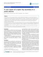
As the testis was difficult to feel within the fluid collection,
a scrotal ultrasound (US) examination was arranged to
exclude a testicular tumour as the source of the hydro-
coele. This showed no focal lesion within either testis (Fig-
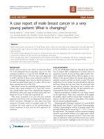
ure 1). However, Doppler ultrasonography revealed flow
within the fluid surrounding the left testis (Figure 2),
communicating via a large scrotal varicocoele with large
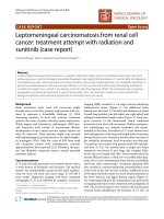
varices. Figure 3 shows flow demonstrated within these
dilated vessels which could be followed through the left
inguinal canal (Figure 3) to the region of the left renal
vein. Extensive spleno-renal varices were recorded.
Conservative treatment was advised as a result of this
investigation.
Discussion
Unusual portal-systemic shunts in portal hypertension
have been recorded including communication between a
coronary vein varicocoele and patent umbilical vein,
superior mesenteric vein and inferior vena cava, splenic
vein and abdominal wall, spleno-retroperitoneal and
omphalo-ilio-caval anastomosis [1]. Large scrotal varico-
coeles secondary to portal hypertension have been
described [2,3].
Ultrasound image – left testis focally normalFigure 1
Ultrasound image – left testis focally normal.
Colour Doppler ultrasound image – flow demonstrated in fluid surrounding left testisFigure 2
Colour Doppler ultrasound image – flow demon-
strated in fluid surrounding left testis.
Colour Doppler ultrasound image – variceal gonadal vein in left inguinal canalFigure 3
Colour Doppler ultrasound image – variceal gonadal
vein in left inguinal canal.
Publish with BioMed Central and every
scientist can read your work free of charge
"BioMed Central will be the most significant development for
disseminating the results of biomedical research in our lifetime."
Sir Paul Nurse, Cancer Research UK
Your research papers will be:
available free of charge to the entire biomedical community
peer reviewed and published immediately upon acceptance
cited in PubMed and archived on PubMed Central
yours — you keep the copyright
Submit your manuscript here:
/>BioMedcentral
Journal of Medical Case Reports 2008, 2:363 />Page 3 of 3
(page number not for citation purposes)
Our patient had cirrhosis and a unilateral scrotal swelling
which appeared on clinical grounds to be a hydrocoele
but US examination established that it was a varicocoele.
It is a noteworthy case because the varicocoele did not
have the typical "bag of worms" appearance of multiple
varicosities within the hemiscrotum: there was actually a
solitary shunt vessel enveloping the testis. This vein could
be traced along the inguinal canal and into the abdomen,
communicating with abnormal dilated vessels arising
from the spleen. We report an unusual form of spleno-
renal shunt.
Conclusion
Attempted hydrocoele repair in this patient would have
resulted in marked unanticipated blood loss and sudden
ligation of the scrotal varicocoele may have precipitated
rupture of other portal-systemic bypasses. We conse-
quently advocate cautious assessment of possible hydro-
coele in patients with portal hypertension.
Consent
Written informed consent was obtained from the patient
for publication of this case report and any accompanying
images. A copy of the written consent is available for
review by the Editor-in-Chief of this journal.
Competing interests
The authors declare that they have no competing interests.
Authors' contributions
GY prepared the manuscript. AR and LB undertook the
ultrasound examinations. NB instigated the report and
critiqued the manuscript.
Acknowledgements
Source of funding: UK National Health Service.
References
1. Di Candio G, Campatelli A, Mosca F, Santi V, Casanova P, Bolondi L:
Ultrasound detection of unusual spontaneous portosystemic
shunts associated with uncomplicated portal hypertension. J
Ultrasound Med 1985, 4(6):297-305.
2. Pinggera GM, Herwig R, Pallwein L, Frauscher F, Judmaier W, Mitter-
berger M, Bartsch G, Mallouhi A: Isolated right-sided varicocele
as a salvage pathway for portal hypertension. Int J Clin Pract
2005, 59(6):740-742.
3. Schulte-Baukloh H, Kammer J, Felfe R, Sturzebecher B, Knispel HH:
Surgery is inadvisable: massive varicocele due to portal
hypertension. Int J Urol 2005, 12(9):852-854.