Báo cáo Y học: Oxidation of propionate to pyruvate in Escherichia coli Involvement of methylcitrate dehydratase and aconitase pot
Bạn đang xem bản rút gọn của tài liệu. Xem và tải ngay bản đầy đủ của tài liệu tại đây (337.88 KB, 11 trang )
Oxidation of propionate to pyruvate in
Escherichia coli
Involvement of methylcitrate dehydratase and aconitase
Matthias Brock
1,
*, Claudia Maerker
1,
*, Alexandra Schu¨tz
1
,UweVo¨ lker
1,2,†
and Wolfgang Buckel
1
1
Laboratorium fu
¨
r Mikrobiologie, Fachbereich Biologie, Philipps-Universita
¨
t, Marburg, Germany;
2
Abteilung Biochemie,
Max-Planck-Institut fu
¨
r terrestrische Mikrobiologie, Marburg, Germany
The pathway of the oxidation of propionate to pyruvate in
Escherichia coli involves five enzymes, only two of which,
methylcitrate synthase and 2-methylisocitrate lyase, have
been thoroughly characterized. Here we report that the
isomerization of (2S,3S)-methylcitrate to (2R,3S)-2-methyl-
isocitrate requires a novel enzyme, methylcitrate dehydratase
(PrpD), and the well-known enzyme, aconitase (AcnB), of
the tricarboxylic acid cycle. AcnB was purified as 2-methyl-
aconitate hydratase from E. coli cells grown on propionate
and identified by its N-terminus. The enzyme has an
apparent K
m
of 210 l
M
for (2R,3S)-2-methylisocitrate but
shows no activity with (2S,3S)-methylcitrate. On the other
hand, PrpD is specific for (2S,3S)-methylcitrate
(K
m
¼ 440 l
M
) and catalyses in addition only the hydration
of cis-aconitate at a rate that is five times lower. The product
of the dehydration of enzymatically synthesized (2S,3S)-
methylcitrate was designated cis-2-methylaconitate because
of its ability to form a cyclic anhydride at low pH. Hence,
PrpD catalyses an unusual syn elimination, whereas the
addition of water to cis-2-methylaconitate occurs in the
usual anti manner. The different stereochemistries of
the elimination and addition of water may be the reason for
the requirement for the novel methylcitrate dehydratase
(PrpD), the sequence of which seems not to be related to any
other enzyme of known function. Northern-blot experi-
ments showed expression of acnB under all conditions tested,
whereas the RNA of enzymes of the prp operon (PrpE, a
propionyl-CoA synthetase, and PrpD) was exclusively pre-
sent during growth on propionate. 2D gel electrophoresis
showed the production of all proteins encoded by the prp
operon during growth on propionate as sole carbon and
energy source, except PrpE, which seems to be replaced by
acetyl-CoA synthetase. This is in good agreement with
investigations on Salmonella enterica LT2, in which disrup-
tion of the prpE gene showed no visible phenotype.
Keywords: 2-methylisocitrate; aconitase; methylcitrate dehy-
dratase; propionate metabolism; prp operon.
Several bacteria and fungi are able to oxidize propionate via
methylcitrate to pyruvate. Initially propionyl-CoA conden-
ses with oxaloacetate to (2S,3S)-methylcitrate, which iso-
merizes to (2R,3S)-2-methylisocitrate. Cleavage leads to
pyruvate and succinate. The consecutive oxidative regener-
ation of oxaloacetate from succinate completes the methyl-
citrate cycle. Initially this cycle was discovered by growing a
mutant strain of the yeast Candida lipolytica on odd-chain
fatty acids. The accumulation of a tricarboxylic acid was
observed during growth and identified as methylcitrate [1].
Further investigations revealed other enzymes necessary for
a functional methylcitrate cycle. The enzymes, however,
were only partially characterized and no genomic sequences
were identified [2–6]. More recently it was discovered that
propionate oxidation in aerobically growing Gram-negative
bacteria, especially Escherichia coli [7] and Salmonella
enterica serovar Thyphimurium LT2 [8], also proceeds via
methylcitrate. The purification of one of the key enzymes of
the methylcitrate cycle, methylcitrate synthase, led to the
identification of an operon necessary for propionate degra-
dation. In E. coli and S. enterica this prp operon is
composed of the genes prpB, prpC, prpD and prpE.PrpB
and PrpC were identified as 2-methylisocitrate lyase [9] and
methylcitrate synthase [7], respectively. PrpE was shown to
catalyse the activation of propionyl-CoA [10]. It remained
unclear, however, by which mechanism the dehydration and
rehydration of (2S,3S)-methylcitrate is performed to yield
(2R,3S)-2-methylisocitrate. In S. enterica it was reported
that the first reaction, the dehydration of methylcitrate, is
Correspondence to W. Buckel, Laboratorium fu
¨
r Mikrobiologie,
Fachbereich Biologie, Philipps-Universita
¨
t, D-35032 Marburg,
Germany. Fax: + 49 6421 2828979, Tel.: + 49 6421 2821527,
E-mail:
Abbreviations: Acs, acetyl-CoA synthetase; AcnB, aconitase B
(2-methylisocitrate dehydratase); PrpB, 2-methylisocitrate lyase;
PrpC, methylcitrate synthase; PrpD, methylcitrate dehydratase; PrpE,
propionyl-CoA synthetase.
Enzymes: acetyl-CoA synthetase (Acs, EC 6.2.1.1); aconitase B [AcnB,
2-methylisocitrate dehydratase (2S,3R)-3-hydroxybutane-1,2,3-tri-
carboxylate hydro-lyase, EC 4.2.1.3, also 4.2.1.99]; citrate synthase
(EC 4.1.3.7); fumarase (EC 4.2.1.2); isocitrate lyase (EC 4.1.3.1);
malate dehydrogenase (EC 1.1.1.37); malate synthase (EC 4.1.3.2);
methylcitrate dehydratase [(2S,3S)-2-hydroxybutane-1,2,3-tricarboxy-
late hydro-lyase, PrpD, EC 4.2.1.79]; methylcitrate synthase
(EC 4.1.3.31); 2-methylisocitrate lyase (EC 4.1.3.30); phosphoglycerate
mutase (EC 5.4.2.1); propanol-preferring alcohol dehydrogenase
(EC 1.1.1.1); propionyl-CoA synthetase (EC 6.2.1.17); pyruvate kinase
(EC 2.1.4.70); succinate dehydrogenase (EC 1.3.5.1).
*Present address: Institut fu
¨
r Mikrobiologie der Universita
¨
t,
Herrenha
¨
user Str. 2, D-30167 Hannover, Germany. These two authors
contributed equally to this work.
Present address: Funktionelle Genomforschung, Medizinische
Fakulta
¨
t, Ernst-Moritz-Arndt-Universita
¨
t, Walther-Rathenau-Str.
49A, D-17489 Greifswald, Germany.
(Received 28 July 2002, revised 24 October 2002,
accepted 28 October 2002)
Eur. J. Biochem. 269, 6184–6194 (2002) Ó FEBS 2002 doi:10.1046/j.1432-1033.2002.03336.x
catalysed by the PrpD protein [11]. However, the product of
this reaction was not further analysed. It was suggested that
2-methyl-cis-aconitate was formed. Interestingly, this reac-
tion would involve the unusual syn elimination of water,
whereas in all other analysed derivatives of malate this
b-elimination occurs in an anti manner; for a review see [12].
Aconitase from bovine heart follows this rule by dehydra-
ting both substrates, citrate and (2R,3S)-isocitrate, in an anti
manner. Furthermore the enzyme is able to hydrate
2-methyl-cis-aconitate to threo-2-methylisocitrate in an anti
manner, but cannot use methylcitrate as substrate [13].
Surprisingly, investigations of the PrpD protein showed that
this enzyme is not able to catalyse the hydration of
2-methyl-cis-aconitate to 2-methylisocitrate. There is genetic
evidence that an aconitase-like protein or even one of the
aconitases (AcnA or AcnB) from S. enterica catalyse this
hydration [11]. Other studies on the PrpD protein of E. coli
revealed the existence of an iron-sulfur cluster essential for
catalytic activity [14]. However, this is in disagreement with
results on the S. enterica PrpD protein, in which such a
cluster was not found [11]. Therefore, the biochemical
characterization of the E. coli PrpD protein will also focus
on the activity of this enzyme in the presence of chelating
agents such as EDTA and o-phenanthroline.
In this paper we report the in vitro reconstitution of the
oxidation of propionyl-CoA to pyruvate by the use of
purified PrpC, PrpD, AcnB and PrpB from E. coli.PrpD
and AcnB involved in the conversion of methylcitrate into
2-methylisocitrate were biochemically characterized. Fur-
thermore, expression of the genes involved in propionate
metabolism was studied in 2D protein gel electrophoresis
and Northern-blot experiments.
EXPERIMENTAL PROCEDURES
Bacteria and culture conditions
For purification of wild-type enzymes and for expression
studies, the E. coli K12 derivative W3350 (F
–
gal r
+
m
+
k
sensitive) was used [15]. For overexpression of the genes prpD
and prpB, E. coli TOP10 cells (Invitrogen) were used,
containing plasmids with the corresponding genes and an
N-terminal cloned histidine tag. For purification of wild-type
enzymes and expression studies, cells were grown aerobically
at 37 °C in minimal medium containing 60 m
M
K
2
HPO
4
,
33 m
M
KH
2
PO
4
,76m
M
(NH
4
)
2
SO
4
,2m
M
trisodium
citrate, 0.1% (v/v) trace element solution without chelating
agent [16], 1 m
M
MgSO
4
,and50m
M
sodium propionate,
sodium acetate or glucose. For overproduction of proteins,
cells were grown in Standard I medium (peptone, 15.6 gÆL
)1
;
yeast extract, 2.8 gÆL
)1
;100m
M
NaCl; 5 m
M
glucose;
Merck, Darmstadt, Germany) and induced with isopropyl
thio-b-
D
-galactoside. Cells were harvested by centrifugation
at 10 000 g and used directly or stored at )80 °C.
Purification of 2-methylisocitrate dehydratase (AcnB)
from
E. coli
W3350
For a standard purification, 18 g (wet weight) propionate-
grown cells was used. All purification steps were carried out
in an anaerobic chamber (95% N
2
,5%H
2
). Cells were
thawed on ice and suspended in 20 mL anaerobic buffer I
(20 m
M
potassium phosphate, pH 7.5, 1 m
M
trisodium
citrate and 1 m
M
dithiothreitol). Cells were broken by
sonication (Branson sonifier; 3 · 5 min at 60% pulse and
80% of full power). Cell debris was removed by ultracen-
trifugation at 96 000 g for 45 min. This crude extract was
filtered (0.45 lm pore size; Sarsted, Nu
¨
mbrecht, Germany)
and loaded on to a hydroxyapatite column (20 mL bed
volume) equilibrated with buffer I. Unless otherwise indi-
cated, the FPLC system and columns from Amersham
Biosciences were used. The hydroxyapatite column was
washed with buffer I. The flow through was concentrated
with an Amicon chamber over a PM 30 size-exclusion filter
(Millipore) and diluted in buffer II (20 m
M
Tris/HCl,
pH 7.5, with 1 m
M
trisodium citrate and 1 m
M
dithiothrei-
tol). The enzyme was loaded on to a Q-Sepharose column
(30 mL bed volume), previously equilibrated with buffer II.
The enzyme was eluted with buffer III (20 m
M
Tris/HCl,
pH 7.5, with 1 m
M
trisodium citrate, 1 m
M
dithiothreitol
and 1
M
NaCl) with a linear NaCl gradient of
150 )200 m
M
. Active fractions were pooled, and solid
(NH
4
)
2
SO
4
was added to a final concentration of 1
M
,
filtered and loaded on to a phenyl-Sepharose column (bed
volume 30 mL), previously equilibrated with buffer IV
(20 m
M
Tris/citrate, pH 8.0, with 1 m
M
dithiothreitol and
1
M
(NH
4
)
2
SO
4
). The enzyme was eluted with a linear
(NH
4
)
2
SO
4
gradient of 1.0–0
M
in buffer V (20 m
M
Tris/
citrate, pH 8.0, with 1 m
M
dithiothreitol) between 0.2 and
0
M
(NH
4
)
2
SO
4
and was concentrated as described above by
changing to buffer II. The enzyme was loaded on to a UnoQ
column (Bio-Rad; bed volume 6 mL) equilibrated with
buffer II and eluted with buffer III. The purity of the eluted
fractions was checked by electrophoresis on a 15% poly-
acrylamide gel in the presence of SDS.
Overproduction and purification of PrpB and PrpD
with N-terminal histidine tags
The source of PrpB protein, the 2-methylisocitrate lyase,
was described elsewhere [9]. The prpD ORF from the prp
operon of wild-type E. coli W3350 was amplified with Taq
polymerase. Primers were constructed with the complete
restriction sites of BamHI (primer: 5¢-CGGGATCCT
CAGCTCAAATCAACAACATCCGC-3¢)andPstI
(5¢-AACTGCAGTTAAATGACGTACAGGTCGAGAT
AC-3¢), respectively. After restriction of the PCR product
with both enzymes, the product was cloned into the
previously restricted pQE30 vector (Qiagen) for overexpres-
sion with an N-terminal His tag. Chemically competent
E. coli cells (TOP10) were transformed with the plasmid.
Overproduction of the PrpD protein was performed by
growing the cells in Standard I medium until D
578
¼ 0.8
and induction with 1 m
M
of isopropyl thio-b-
D
-galactoside
followed by incubation overnight. Overproduction of
methylcitrate dehydratase in four different clones was
confirmed by SDS/PAGE. All clones exhibted an induced
protein at 54 kDa (data not shown).
Cells from a 1.2-L culture (D
578
3) were induced for
10 h and harvested by centrifugation. Cells were washed
with 50 m
M
potassium phosphate, pH 7.0, centrifuged, and
suspended in the same buffer. Cells were broken by
sonication and centrifuged at 96 000 g. The resulting cell-
free extract was loaded on to a gravity flow Ni/nitrilotri-
acetic acid/agarose column with a bed volume of 5 mL.
The column was washed with 20 mL 50 m
M
potassium
Ó FEBS 2002 Isomerization of methylcitrate to 2-methylisocitrate (Eur. J. Biochem. 269) 6185
phosphate, pH 7.0, containing 20 m
M
histidine to remove
unspecifically bound proteins. PrpD was eluted with 50 m
M
potassium phosphate buffer, pH 7.0, containing 200 m
M
histidine. Active fractions were concentrated and desalted
over a PM 30 size-exclusion filter. After addition of glycerol
to a final concentration of 50% (v/v), the protein could be
stored at )20 °C without loss of activity.
Enzymatic synthesis of (2
S
,3
S
)-methylcitrate
Methylcitrate was produced with the methylcitrate syn-
thases PrpC from E. coli [7] or McsA from the filamentous
fungus Aspergillus nidulans [17]. The reaction was carried
out at room temperature for 20 h. Propionyl phosphate was
synthesized chemically by a modified synthesis described by
Stadtman [18], in which acetic acid anhydride was replaced
by propionic acid anhydride. Propionyl phosphate was
converted into propionyl-CoA with the help of phospho-
transacetylase from Bacillus stearothermophilus (Sigma,
Taufkirchen, Germany). A typical reaction for the synthesis
of methylcitrate was carried out in a final volume of 60 mL
and contained 50 m
M
propionyl phosphate, 100 m
M
oxaloacetic acid (neutralized with KHCO
3
), 0.2 m
M
CoASH, 500 U phosphotransacetylase and 50 U methyl-
citrate synthase. The reaction was buffered at pH 7.5 in
20 m
M
potassium phosphate. After incubation, the enzymes
were denatured by heat treatment for 20 min at 80 °Cand
centrifuged at 10 000 g for 10 min. The supernatant was
concentrated to a final volume of 10 mL in a rotary
evaporator. Precipitated salts were removed by centrifuga-
tion as described above, and the supernatant was loaded on
to a Dowex 1x8 column (Cl
–
form, bed volume 10 mL).
Methylcitrate was eluted with 1
M
HCl. The methylcitrate-
containing fractions, as tested enzymatically with the PrpD
protein, were concentrated by evaporation. The residual
brownish oil was checked for purity by
1
H-NMR
(500 MHz, CDCl
3
): d ¼ 1.19 (3H, d,
3
J ¼ 6.9 Hz CH
3
),
2.90 (1H, q,
3
J ¼ 6.9 Hz, CH), 2.90 (1H, d,
2
J ¼ 16.6 Hz,
CHH), 3.17 (1H, d,
2
J ¼ 16.6 Hz, CHH). Both, the E. coli
and the A. nidulans enzyme produced the same enantiomeric
pure (2S,3S)-methylcitrate (99.9%) as checked by enantio-
selective multidimensional capillary gas chromatography
(kindly performed by Professor A. Mosandl, Universita
¨
t
Frankfurt/Main, Germany).
Enzyme assays
2-Methylisocitrate lyase (PrpB) was assayed with the
coupled NADH-dependent assay as described previously
[9]. Methylcitrate dehydratase (PrpD) activity was measured
at 240 nm with a Kontron, model Uvikon 943 double-beam
UV/visible spectrophotometer, and the formation of the
double bond during dehydration of methylcitrate was
monitored. The absorption coefficient, e
240
,wastakenas
4.5 m
M
)1
Æcm
)1
[4]. The composition of the assay mixture
was 50 m
M
potassium phosphate, pH 7.5, and 1.3 m
M
methylcitrate in a final volume of 1 mL.
The racemic mixture of chemically synthesized threo-2-
methylisocitrate [9] was used to follow the dehydration and
the formation of the double bond in 2-methyl-cis-aconitate
at 240 nm; e
240
¼ 4.5 m
M
)1
Æcm
)1
[4]. The composition of
the assay was 50 m
M
potassium phosphate, pH 7.5, and
0.3 m
M
threo-2-methylisocitrate in a final volume of 1 mL.
To measure 2-methylisocitrate dehydratase (AcnB), a
coupled assay was performed in the reverse direction. The
reaction was followed at 340 nm under anaerobic condi-
tions with e
340
¼ 6.2 m
M
)1
Æcm
)1
. The composition of the
assay mixture was 50 m
M
potassium phosphate buffer,
pH 7.5, 2 m
M
MgCl
2
,0.2m
M
NADH, 0.64 m
M
methyl-
citrate, 0.2 U PrpD, 0.2 U PrpB, 0.3 m
M
dithiothreitol, 3 U
lactate dehydrogenase from rabbit muscle (Roche) and a
sample of purified AcnB in a final volume of 1 mL.
Gel electrophoresis and blotting of proteins
The protein fractions obtained from the purification of
2-methylisocitrate dehydratase were analysed by SDS/
PAGE. The apparent molecular mass of the 2-methylisoci-
trate dehydratase subunit was determined by measuring the
mobility by SDS/PAGE (15% acrylamide) [19] with stand-
ard proteins as molecular mass markers. Purified 2-methyl-
isocitrate dehydratase was blotted from the gel (10%
acrylamide) on a poly(vinylidene difluoride) membrane
(Millipore) with the transblot SD semidry transfer cell (Bio-
Rad), as described in the manufacturer’s protocol, and was
then N-terminally sequenced by Edman degradation (kindly
performed by D. Linder, Universita
¨
t Gießen, Germany).
Re-activation and inactivation of AcnB
AcnB was inactivated by exposure to air and by addition
of either EDTA or o-phenanthroline (both 2.5 m
M
final concentration). For reactivation, 98.3 mg FeSO
4
·
(NH
4
)
2
SO
4
· 6H
2
O (final concentration 5 m
M
)and136mg
cysteine hydrochloride (monohydrate) (15 m
M
)weredis-
solved under anaerobic conditions in 45 mL water, and the
pH was adjusted to 7.5 by dropwise addition of 1
M
NaOH.
Water was added to a final volume of 50 mL. One part of
enzyme solution was mixed with one part of re-activation
mixture and incubated for 60 min at room temperature
under anaerobic conditions.
Iron–sulfur cluster and metal cofactors
Purified PrpD protein was concentrated to 4 mg pro-
teinÆmL
)1
in 20 m
M
Hepes buffer, pH 7.5, and the activity
was measured with methylcitrate as substrate. An aliquot
was diluted and a spectrum was determined in the range
220–900 nm. A second 0.5-mL aliquot was taken and
incubated for 60 min at room temperature under anaerobic
conditions in re-activation mixture (0.5 mL) as described
above for the re-activation of the 2-methylisocitrate dehy-
dratase. PrpD was separated from the re-activation mixture
by the use of a Sephadex-NAP column (Pharmacia Biotech)
andelutedin20m
M
Hepes buffer, pH 7.5. Activity was
tested and a spectrum was determined as described above. A
third and fourth aliquot were taken and incubated with a
5m
M
final concentration of o-phenanthroline or 10 m
M
EDTA, respectively, and incubated for 20 min at room
temperature. PrpD was desalted, and activity and a
spectrum were determined as described above.
Synthesis of digoxygenin-labelled RNA probes
For the detection of mRNAs of the genes acs, acnB, prpD
and prpE, specific RNA probes labelled with digoxygenin
6186 M. Brock et al.(Eur. J. Biochem. 269) Ó FEBS 2002
were produced using the T7 polymerase. Oligonucleotides
were designed that contained the sequence of the T7
promoter in the reverse primer (the full sequences of all
primers are shown in Table 1). A PCR was performed with
Taq polymerase, and genomic DNA of E. coli W3350 was
used as a template. PCR products were separated by
electrophoresis in a 1% agarose gel and purified by the
Geneclean Kit II (BIO 101) as described in the manufac-
turer’s protocol. For in vitro transcription, 0.5–1.0 lgPCR
product was mixed with 2 lL NTP labelling mixture
containing UTP (Dig RNA Labelling Kit T7; Roche),
2 lL reaction buffer (Ambion), 1 lL RNase inhibitor (Dig
RNA Labelling Kit T7; Roche), 2 lL T7 polymerase
(20 UÆlL
)1
; Ambion) and diethyl pyrocarbonate-treated
water to a final volume of 20 lL. Transcription was carried
out at 37 °C for 1 h. RNase-free DNase I was added, and
the mixture was incubated at 37 °C for a further 15 min.
RNA was precipitated by the addition of 2.5 lL1
M
LiCl
and 90 lL 100% ethanol and incubated for 1 h at )80 °C.
After centrifugation (12 000 g,4°C), RNA pellets were
dried and dissolved in 100 lL nuclease-free water. The
intensity of the digoxygenin label of the probes was checked
by cross-linking the specific probes on a nylon membrane
and detection of the label by standard methods.
2D gel electrophoresis
After harvesting of the bacteria by centrifugation, cells were
washed in 10 m
M
Tris/HCl (pH 7.5)/1 m
M
EDTA, and the
cell pellet was suspended in the same buffer. Cells were
disrupted by several passages through a French pressure
cell, and debris was removed by centrifugation at 4 °Cand
20 000 g for 30 min. The protein concentration of the
supernatant fraction was assayed by the method of Brad-
ford [20]. For 2D gel electrophoresis, 400 lg crude protein
extract was solubilized in a hydration solution containing
8
M
urea, 2
M
thiourea, 2% (w/v) 3-[(3-chloramidopro-
pyl)dimethylammonio]propane-1-sulfonate (Chaps), 28 m
M
dithiothreitol, 1.3% (v/v) Pharmalytes, pH 3–10, and
bromophenol blue. After hydration in the protein-contain-
ing solution for 24 h under low-viscosity paraffin oil,
Immobiline DryStrips (IPG-strips; Amersham Biosciences)
covering the pH range 4–7 or 3–10 were subjected to
isoelectric focusing. The following voltage/time profile was
used: a linear increase from 0 to 500 V for 1000 Vh, 500 V
for 2000 Vh, a linear increase from 500 to 3500 V for 10 000
Vh and a final phase of 3500 V for 35 000 Vh (pH 4–7) or
for 21 000 Vh (pH 3–10). IPG-strips were consecutively
incubated for 15 min each in equilibration solution A and
B. Solution A contained 50 m
M
Tris/HCl, pH 6.8, 6
M
urea,
30% glycerol, 4% SDS and dithiothreitol (3.5 mgÆmL
)1
).
Solution B contained iodoacetamide (45 mgÆmL
)1
)instead
of dithiothreitol. In the second dimension, proteins were
separated on SDS/12.5% polyacrylamide gels with the
Investigator
TM
System (Perkin–Elmer Life Sciences,
Cambridge, UK) at 2 W per gel. Gels were stained with
PhastGel BlueR according to the manufacturer’s (Amer-
sham Biosciences) instructions. After scanning, the 2D
PAGE images were analysed with the Melanie3Ò software
package (Bio-Rad Laboratories GmbH). Three separate
gels of each condition and two independent cultivations
were analysed, and only spots displaying the same pattern in
all parallels were selected for further characterization.
Protein identification by peptide mass fingerprinting
Protein spots were excised from PhastGel BlueR-stained 2D
gels, destained, and digested with trypsin (Promega);
peptides were then extracted [21]. Peptide mixtures were
purified with C18-tips according to the manufacturer’s
(Millipore) instructions and directly eluted on to a sample
template of a MALDI-TOF mass spectrometer with an
eluent containing 50% (v/v) acetonitrile, 0.1% (v/v) tri-
fluoroacetic acid, saturating amounts of a-cyano-3-
hydroxycinnamic acid and calibration peptides. Peptide
masses were determined in the positive ion reflector mode in
a Voyager DE RP mass spectrometer (Applied Biosystems)
with internal calibration. Mass accuracy was better
than 50 p.p.m. Peptide mass fingerprints were compared
with databases using the
MASCOT
program (http://www.
matrixscience.com/cgi/index.pl?page= /home.html). The
searches considered oxidation of methionine, pyroglutamic
acid formation at the N-terminal glutamine, and modifica-
tion of cysteine by carbamidomethylation as well as partial
cleavage leaving a maximum of one internal site uncleaved.
RNA isolation and Northern blot
E. coli W3350 cells were grown on propionate, acetate or
glucose minimal medium to an D
578
of 0.8 under vigorous
shaking at 37 °C. The cultures (20 mL) were mixed with
20 mL frozen Ôkilling bufferÕ (20 m
M
Tris/HCl, pH 7.5,
5m
M
MgCl
2
,20m
M
NaN
3
; diethyl pyrocarbonate treated)
and centrifuged for 10 min at 4000 g.Cellpelletswere
suspended in 200 lL killing buffer, and frozen in
liquid nitrogen. Cells were broken in a frozen state in a
Table 1. Oligonucleotides used for the generation of RNA probes. The reverse primer contains the promoter region for the T7 polymerase at the 5¢
end. An asterisk denotes the end of the promoter region.
Probe Reverse primer Forward primer
Acs 5¢-
TAATACGACTCACTATAGGGA*5¢-AACACACCATTCCTGCCAAC-3¢
CCACCACAGGTCGCGCC-3¢
AcnB 5¢-
TAATACGACTCACTATAGGGA*5¢-CTCACACGCTGCTGATGTTC-3¢
CGTGGTTACGCACTTCACC-3¢
PrpD 5¢-
TAATACGACTCACTATAGGGA*5¢-AACATCGGCGCGATGATCC-3¢
TCGCTGCTTCAACTGCCG-3
PrpE 5¢-
TAATACGACTCACTATAGGGA*5¢-ACCGGAGCAGTTCTGGGC-3¢
GATTCCAGCCACGCCACC-3¢
Ó FEBS 2002 Isomerization of methylcitrate to 2-methylisocitrate (Eur. J. Biochem. 269) 6187
Micro-Dismembrator (Braun Biotech International) at
2600 r.p.m. for 2 min. Cell extracts were mixed with 4 mL
lysis solution containing 4
M
guanidine thiocyanate, 25 m
M
sodium acetate, pH 5.2, and 0.5% N-lauroylsarcosine (w/v)
at 37 °C. One part of this solution was mixed with one part of
acidic phenol/chloroform/3-methylbutan-1-ol (50 : 48 : 2,
by vol.), shaken at room temperature for 5 min and
centrifuged at 12 500 g for 5 min. The upper layer was
mixed with 1 mL acidic phenol/chloroform/
3-methylbutan-1-ol, shaken again for 5 min, and spun down
as described above. The upper layer was mixed with 800 lL
chloroform/3-methylbutan-1-ol (24 : 1, v/v), and centri-
fuged as described above. The aqueous phase was collected,
and 80 lL3
M
sodium acetate (pH 5.2) and 1.1 mL propan-
2-ol were added. RNA was precipitated by incubation at
)80 °C for 1 h. The RNA was centrifuged (23 000 g,4°C,
15 min) and the pellet was washed with 70% ice-cold
ethanol. The RNA was dried at room temperature and
dissolved in 30 lL diethyl pyrocarbonate-treated water.
Quality and quantity of isolated RNA was checked with the
Agilent 2100 Bioanalyzer (Agilent Technologies, Bo
¨
blingen,
Germany) as described in the manufacturer’s protocol.
RNA (8 lgin4.5lL) was mixed with 10.5 lLdenatur-
ation solution [90 lL formamide, 18 lL formaldehyde,
18 lL10· Mops (200 m
M
Mops, 50 m
M
sodium acetate,
10 m
M
EDTA, dissolved in diethyl pyrocarbonate-treated
water and adjusted to pH 7.0)] and loaded on to a 1.4%
(w/v) agarose gel containing 1.8
M
formaldehyde. RNA was
separatedat70 Vfor3handtransferredtoanylontransfer
membrane (Schleicher and Schuell) [22]. The RNA blot was
saturated with blocking reagent and hybridized with the
digoxygenin-labelled antisense RNA probes overnight.
After a wash, specific hybridization signals were detected
by incubation with alkaline phosphatase-conjugated anti-
digoxygenin Ig (Roche) and monitoring the conversion of
the ECF-vistra substrate with a STORM 860 fluorimager
(Amersham Biosciences).
Determination of 2-methyl-
cis
-aconitate by anhydride
formation
Enzymatically synthesized methylcitrate (0.8 m
M
)wasdis-
solved in a final volume of 5 mL 20 m
M
Hepes, pH 7.5, and
incubated with 0.5 U PrpD. A 1-mL aliquot was taken, and
the reaction was monitored at 240 nm until the equilibrium
of the reaction was reached. PrpD was inactivated by
heating the whole sample for 15 min at 80 °C. Denatured
protein was removed by centrifugation. Of this solution,
900 lL was mixed with 100 lL water, and a UV/visible
spectrum in the range 220–400 nm was recorded. A second
sample was prepared by using another 900 lLofthe
solution and addition of 100 lL8
M
HCl. The anhydride
formation was followed at 259 nm until no further change
in absorbance was observed. A second spectrum in the
range 220–400 nm was recorded, and the difference spec-
trum between the neutral and the acidified sample was
calculated using the Microsoft Excel worksheet. As a
control, a methylcitrate solution without addition of PrpD
was treated as described above. No change in absorbance
was detectable during acidification.
RESULTS
Biochemical analysis of 2-methylisocitrate dehydratase
E. coli cells were grown to D
578
¼ 1.2 in the presence of
50 m
M
propionate in the minimal medium. 2-Methylisoci-
trate dehydratase was identified in extracts of these cells by
monitoring the decrease in A
340
with enzymatically prepared
(2S,3S)-methylcitrate as substrate and with PrpD, PrpB and
lactate dehydrogenase as auxiliary enzymes (Fig. 1). Start-
ing from 18 g wet cells, the protein was purified from a
specific activity in crude extracts of 0.16 UÆmg
)1
to
1.9 UÆmg
)1
with a yield of 3.6%. Purification was per-
formed by chromatography on hydroxyapatite, Q-Seph-
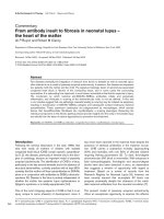
arose, phenyl-Sepharose and UnoQ (Table 2). A major
band was revealed in the resulting protein fractions by
SDS/PAGE (Fig. 2, lanes 6 and 7) with an apparent
molecular mass of 94 kDa and a turnover number of 3 s
)1
.
Comparison of the forward reaction (hydration of
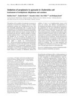
Fig. 1. Pathway of propionate oxidation to pyruvate. The enzymes are
indicatedinitalics.
Table 2. Purification protocol for AcnB. A unit is defined as the oxidation of 1 lmol NADHÆmin
)1
in the coupled assay.
Purification step
Activity
(U)
Protein
(mg)
Specific activity
(UÆmg
)1
)
Yield
(%)
Purification
factor
Cell-free extract 217 1360 0.16 100 1.0
Hydroxyapatite 142 720 0.20 66 1.2
Q-Sepharose 91.4 158 0.58 42 3.6
Phenyl-Sepharose 24.0 30.8 0.78 11 4.9
UnoQ (Fr. 48) 3.7 2.2 1.7 1.7 10.6
UnoQ (Fr. 49) 1.9 1.0 1.9 0.9 11.9
6188 M. Brock et al.(Eur. J. Biochem. 269) Ó FEBS 2002
2-methyl-cis-aconitate) in the coupled assay with the activity
of the back reaction measured by the dehydration of
chemically synthesized threo-2-methylisocitrate yielded a
ratio of 1 : 0.7. The K
m
of the purified enzyme with threo-2-
methylisocitrate as substrate was determined as 210 l
M
,
which is somewhat higher than K
m
¼ 51 l
M
for (2R,3S)-
isocitrate determined with the aconitase, AcnB [23]. The
enzyme showed no detectable activity with (2S,3S)-methyl-
citrate as substrate.
N-Terminal sequence determination of the purified
protein by Edman degradation revealed the peptide
sequence, MLEEYXKXVAEXAAE, where X denotes
unclear amino acids. Comparison of this sequence with
the databases showed 100% identity with the N-terminal
sequence of the E. coli citrate cycle aconitase, AcnB
(
SWISSPROT
P36683), with the sequence, MLEEYRKH
VAERAAE. The calculated molecular mass of AcnB from
its genomic sequence is 93 498 Da, which is in good
agreement with the apparent molecular mass of 94 kDa
derived from the SDS/PAGE analysis.
The enzyme was rapidly inactivated by exposure to air,
which is already known for aconitases [24], as well as during
the purification procedure, especially during chromatogra-
phy on the phenyl-Sepharose column. Activity was partially
restored by incubation in re-activation mixture under
anaerobic conditions as described in Experimental proce-
dures. Addition of EDTA or o-phenanthroline totally
inactivated enzymatic activity. This is in good agreement
with the requirement for a functional [4Fe)4S] cluster for
aconitase activity.
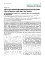
Cloning and characterization of PrpD
The prpD gene was cloned and overexpressed as described
in Experimental Procedures. The overproduced protein was
purified to a specific activity of 11.4 UÆ(mg protein)
)1
by
chromatography on a Ni/nitrilotriacetate/agarose column
(Fig. 3). PrpD showed maximum activity with enzymati-
cally produced (2S,3S)-methylcitrate as substrate (K
m
¼
440 l
M
). Another substrate was cis-aconitate, whereas
citrate and (2R,3S)-isocitrate (natural occurring stereoisom-
er) showed no significant activity. Other related compounds
such as trans-aconitate, threo-2-methylisocitrate and eryth-
ro-2-methylisocitrate, and (S)-malate and (R)-malate gave
no activity at all (Table 3). Unfortunately, authentic
2-methyl-cis-aconitate was not available. The enzyme does
not require any metal cofactors for full enzymatic activity.
In UV/visible spectra, no extra band beside that at 280 nm
could be seen. Neither the spectra nor the activity changed
after incubation of PrpD with o-phenanthroline or with the
re-activation mixture as described for aconitase.
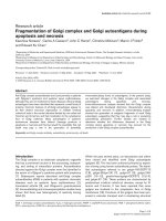
The most likely product of the dehydration reaction of
(2S,3S)-methylcitrate by PrpD is postulated to be 2-methyl-
cis-aconitate. Acidification of the reaction mixture with HCl
led to an increased A
259
as shown in Fig. 4. This can be
explained by the formation of a planar five-membered cyclic
anhydride from 2-methyl-cis-aconitate under acidic
conditions. This condensation would be less likely with
2-methyl-trans-aconitate, which would lead to a nonplanar
six-membered cyclic anhydride. A comparable example of a
five-membered cyclic anhydride formation between two
carboxylic acid groups orientated in a cis conformation is
observed in 2,3-dimethylmaleate, which is formed by a
d-isomerase reaction from (R)-3-methylitaconate (2-methy-
lene-3-methylsuccinate) during the nicotinate fermentation
Fig. 3. Analysis of purified PrpD by SDS/PAGE. The protein was
overproduced with an N-terminal His tag and purified by chroma-
tography on a Ni/nitrilotriacetate/agarose column. Lane 1, sample of
purified PrpD; lane M, molecular mass standard.
Fig. 2. Analysis of the purification of AcnB by SDS/PAGE. Lane 1,
crude extract (21 lg); lane 2, hydroxyapatite (30 lg); lane 3, Q-Seph-
arose (14 lg); lane 4, phenyl-Sepharose (5 lg); lane M, molecular mass
standard; lane 5, UnoQ column fraction 40 (2 lg); lane 6, UnoQ
column fraction 48 (2 lg); lane 7, UnoQ column fraction 49 (1 lg).
Lanes 6 and 7 show the purified AcnB protein at 94 kDa as determined
by Edman degradation.
Table 3. Substrate specificity of PrpD. No activity (< 0.01 UÆmg
)1
)
was found with threo-2-methylisocitrate and erythro-2-methylisoci-
trate, trans-aconitate,
D
-malate and
L
-malate, fumarate, maleate,
D
-tartrate and meso-tartrate,
D
-citramalate and
L
-citramalate,
mesaconate, citraconate, itaconate, and (R,S)-3-methylitaconate.
Substrate
Concentration
(m
M
)
Activity
UÆmg
)1
%
(2S,3S)-Methylcitrate 1.0 11.4 100
cis-Aconitate 0.5 2.3 20
Citrate 50–100 0.12 1.1
(2R,3S)-Isocitrate 5–75 0.09 0.8
Ó FEBS 2002 Isomerization of methylcitrate to 2-methylisocitrate (Eur. J. Biochem. 269) 6189
by Eubacterium barkeri. Under acidic conditions, dimethyl
maleate spontaneously forms the anhydride with a maximal
absorbance at 256 nm [25]. The formation of 2-methyl-
cis-aconitate from (2S,3S)-methylcitrate is further suppor-
ted by the substrate specificity of PrpD. cis-Aconitate is a
moderate substrate, whereas no activity is detectable with
trans-aconitate.
In vitro
reconstitution of the methylcitrate cycle
For in vitro reconstitution, purified enzymes and enzy-
matically produced (2S,3S)-methylcitrate were used.
(2S,3S)-Methylcitrate (1.3 m
M
) was converted into
2-methyl-cis-aconitate by the PrpD protein. 2-Methyl-
cis-aconitate acted as substrate for AcnB and was hydrated
to (2R,3S)-2-methylisocitrate. This product was cleaved by
PrpB into succinate and pyruvate (Fig. 1). To monitor the
reaction and to pull the equilibrium to the side of pyruvate
formation, lactate dehydrogenase and NADH as cosub-
strate were used. This coupled assay was also used to
monitor the purification of the aconitase AcnB as described
above. Absence of the aconitase or any other enzyme
resulted in a loss of pyruvate formation. This result clearly
demonstrates that both proteins, PrpD and AcnB, are
essential for the conversion of methylcitrate into 2-methyl-
isocitrate.
Northern-blot analysis of
prpD
,
prpE
,
acnB
and
acs
transcripts
The four genes were selected for the following reasons.
Transcription levels of acs, the gene coding for acetyl-CoA
synthetase (Acs), were used for comparison of the specificity
of transcription during growth on acetate and propionate,
respectively. Furthermore, this gene was of interest because
of the ability of the Acs to activate propionate to the
corresponding CoA ester. In S. enterica it was shown earlier
thatastraincarryingadeletionoftheacs gene was still able
to grow on propionate but not on acetate. A propionyl-
CoA synthetase mutant was able to grow on propionate as
well as on acetate. A double mutant with deletion of both
genes did not grow on acetate or propionate [10]. Therefore
we postulated that transcripts of acs may be visible under
both growth conditions, whereas the transcripts for propio-
nyl-CoA synthetase, prpE, and methylcitrate dehydratase,
prpD, should be exclusively formed during growth on
propionate. In contrast, transcription of acnB coding for
AcnB is expected to achieve similar levels under all
conditions tested. AcnB is an essential enzyme of the citrate
cycle, as well as of the glyoxylate cycle. During growth on
propionate, pyruvate is formed, which is oxidized to acetyl-
CoA, a substrate for the citrate and the glyoxylate cycle [7].
Furthermore, AcnB acts as 2-methylisocitrate dehydratase
Fig. 4. UV spectra and difference spectrum of 2-methyl-cis-aconitate
and 2-methyl-cis-aconitate anhydride. Methylcitrate was incubated with
methylcitrate dehydratase until the equilibrium of the reaction was
reached. A UV spectrum was recorded (bold line). Another sample was
acidified with HCl and incubated until no further change in A
259
was
recorded. A second spectrum was recorded (thin line). The difference
spectrum (inset) was calculated by the use of the Microsoft Exel
worksheet.
Fig. 5. Northern-blot analysis of transcripts of acnB, acs, prpE and
prpD under different growth conditions. Lane 1, glucose-grown cells;
lane 2, propionate-grown cells; lane 3, acetate-grown cells. Equal
amounts of total RNA were added in each lane. Specific transcripts
were detected with digoxygenin-labelled antisense RNA probes (see
also Table 4 and Fig. 7). An arrow denotes expected transcript sizes;
the additional asterisk indicates alternative transcript sizes. The sizes
(kb) are: 2.0 and 5.7* for acs;4.6and5.9*forprpE and prpD.Further
explanations are given in the Results section. The small box on the
right shows the region of the rRNA, to show that the same amount of
RNA was applied to each lane.
Fig. 6. Scheme of the structure of the E. coli and S. enterica prp oper-
ons. All genes are located in the same orientation, and the encoded
proteins show sequence identities of 76–96%. The E. coli operon
contains an additional repetitive extragenic palindromic element
(REP-element) between the prpB and prpC coding sequence. The
DNA sequence of the intergenic region containing the REP-element is
shown in the upper part of the figure. Bold and italic letters highlight
thesinglerepetitiveelements,respectively.
6190 M. Brock et al.(Eur. J. Biochem. 269) Ó FEBS 2002
and therefore comprises a twofold function during growth
on propionate.
For the transcription experiments, RNA was purified
from E. coli W3350 cells grown on glucose, acetate or
propionate as sole carbon and energy source. Cells were
harvested in the early exponential growth phase
(D
578
¼ 0.7–1.2) and broken as described in Experimental
Procedures. Quality and quantity of the RNA used in each
experiment was confirmed by the use of the Agilent 2100
Bioanalyzer. For each probe, the same quantity of RNA
from cells grown on glucose, acetate or propionate was used
(Fig. 5). Arrows denote main transcripts. Those with an
additional asterisk denote larger transcripts, which may be
formed by a read-through and can be observed from high
gene expression or because of an alternative starting point of
transcription. The first possibility may be correct for the acs
gene (2.0 kb), which is not located in an operon, but may be
transcribed together with the consecutive genes yjcH, yjcG
and yjcF (5.6 kb). The second possibility may be correct for
prpE and prpD, because the prp operon of E. coli,in
contrast with that of S. enterica, is interrupted by a so-called
repetitive extragenic palindromic element. This element is
located between prpB and prpC (Fig. 6) and may be
responsible for the two transcript sizes (4.6 and 5.9 kb),
because these elements are suspected to be involved in
transcriptional regulation [26]. As expected, acnB is
expressed under all conditions tested and shows a single
transcript (Fig. 5). Transcripts of prpD and prpE are
exclusively formed during growth on propionate. It can be
concluded that acetate is not able to induce transcription of
the specific genes involved in propionate catabolism. In
contrast, a strong signal for the transcript of acs was
observed on acetate as well as on propionate. This coincides
with the investigations in S. enterica described above [10].
The acs gene is able to replace prpE but not vice versa.
Furthermore, propionate may be able to induce all genes of
a functional glyoxylate cycle, because activity measurements
for malate synthase of E. coli grown on propionate medium
as compared with acetate showed specific activities of
0.50 UÆmg
)1
and 0.48 UÆmg
)1
, respectively [7]. The weak
acs signal detected on glucose is in agreement with the
observation of acetate excretion and consumption during
growth on glucose medium [27].
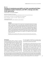
2D gel electrophoresis
2D gel electrophoresis was carried out to monitor differ-
ences in the protein pattern of cells grown on acetate or
propionate. E. coli W3350 cells were grown on propionate
or acetate minimal medium and crude extracts were
prepared from exponentially growing cells as described in
Experimental procedures. Figure 7 exemplarily displays the
protein profile of E. coli W3350 grown with either acetate or
propionate as carbon source. Protein spots, which displayed
significantly different intensities under the two growth
conditions, were isolated from the gels and identified by
peptide mass fingerprinting (Table 4). Proteins induced in
Fig. 7. Comparison of the protein profile of E. coli grown in minimal
medium with acetate (A) or propionate (B) as carbon sources. Crude
protein extracts were prepared and separated by 2D gel electrophor-
esis. After staining with PhastGel BlueR, the gels were scanned with an
imaging system and analysed with the Melanie 3.0 software package.
Protein spots induced or repressed by propionate are marked with
arrowheads or boxes, respectively. Proteins identified by peptide mass
fingerprinting are labelled with their gene names. The acnB gene
product was identified by MS analysis of coseparated purified AcnB
and a comparison with previous 2DE data [33]. (C) Alkaline sections
of gels covering the pH range 3–10 and containing PrpC are displayed.
Ó FEBS 2002 Isomerization of methylcitrate to 2-methylisocitrate (Eur. J. Biochem. 269) 6191
the presence of propionate at a higher or lower level than in
the presence of acetate are labelled with arrowheads and
boxes, respectively (Fig. 7). PrpB, PrpC and PrpD encoded
by the prp operon were exclusively produced during growth
on propionate. PrpE, the propionyl-CoA synthetase, was
detected on neither acetate nor propionate minimal
medium. However, Acs seems to be present in high
amounts, suggesting that it can also serve as a propionate-
activating enzyme. Furthermore, increased levels of malate
synthase (AceB) were found to be present during growth on
propionate. Therefore, the main anaplerotic source of
oxaloacetate appears to be the glyoxylate cycle rather than
carboxylation of pyruvate or phosphoenolpyruvate as
proposed previously [7]. Six proteins, including phospho-
glycerate mutase 1 (GpmA), a propanol-preferring alcohol
dehydrogenase (AdhP), and pyruvate kinase (PykF) seemed
to be present in reduced amounts in propionate-grown cells
compared with cultures grown in the presence of acetate.
DISCUSSION
AcnB purified from E. coli W3350 cells grown on propion-
ate as sole carbon and energy source was the only enzyme
that displayed activity as a 2-methylisocitrate dehydratase.
Similar results were obtained from S. enterica.AcnAand
AcnB from this organism were overproduced, and enzy-
matic activity for the dehydration of 2-methylisocitrate was
studied [11]. This was in agreement with earlier investiga-
tions performed on horse and bovine heart aconitases,
which both catalyse the reversible hydration of 2-methyl-cis-
aconitate to 2-methylisocitrate, but not to methylcitrate
[13,28]. The aconitase from E. coli (AcnB) completes the
methylcitrate cycle. AcnB possesses a twofold function; it
acts as 2-methylisocitrate dehydratase and a citrate/iso-
citrate isomerase in the citrate cycle. The latter is also active
during growth on propionate, because a-oxidation of
propionate via methylcitrate yields pyruvate, which is
converted into acetyl-CoA and funnelled into the citrate
cycle [7]. The observation that AcnB was purified instead of
AcnA is in agreement with the different expression of the
two genes. AcnB was identified as the major citrate cycle
enzyme, whereas AcnA is an anaerobic stationary-phase
enzyme which is specifically induced by iron and redox
stress [29].
Interestingly, two enzymes are involved in the conversion
of methylcitrate into 2-methylisocitrate. PrpD is involved in
the dehydration of (2S,3S)-methylcitrate to 2-methyl-cis-
aconitate. The elimination of water from (2S,3S)-methyl-
citrate to 2-methyl-cis-aconitate is an unusual reaction,
because it displays a syn elimination, which has not
previously been found in any other dehydration of a
derivative of malate. This may explain why PrpD shows no
significant identities with other proteins with known func-
tion except deduced proteins from prp operons of many
proteobacteria, e.g. S. enterica (Fig. 6). In addition, PrpD
shows sequence identities with deduced proteins from the
Gram-positive Bacillus subtilis (61%, Mmge, accession no.
P45859), the eukaroytes Saccharomyces cerevisiae (57%,
Pdh1p, accession no. NP-015326) and Mus musculus (14%,
immune responsive protein 1, accession no. XP-127883), as
well as the archaeon Sulfolobus tokodaii (23%, long
hypothetical Mmge protein, accession no. BAB66901).
The PrpD protein from E. coli possesses high substrate
specificity. The best substrate was stereochemically pure
(2S,3S)-methylcitrate produced by methylcitrate synthases
from E. coli or A. nidulans. Partial activity was also
observed with cis-aconitate. As the activity with citrate
was very low and that with (2R,3S)-isocitrate was almost
absent, it would be of interest to identify the product of the
syn hydration of cis-aconitate, perhaps one enantiomer of
erythro-isocitrate. No significant activity was detected with
many other hydroxy or unsaturated dicarboxylic and
tricarboxylic acids such as trans-aconitate, threo-2-methyl-
isocitrate and erythro-2-methylisocitrate,
D
-malate and
L
-malate, and (R)-citramalate and (S)-citramalate (Table 3).
In E. coli the dehydration of methylcitrate is independent of
any metal cofactors, which was also shown for the PrpD
protein from S. enterica [11], but is in disagreement with
another investigation [14], in which the specific activity of
the purified PrpD from a genetically amplified source
Table 4. Summary of propionate-induced proteins identified by peptide mass fingerprint matching (see also Figs 5 and 7). The theoretical isoelectric
point and molecular mass were calculated with the
COMPUTE
pI/mw tool of the proteomics tools collection at the ExPASy Molecular Biology Server
( />Protein pI
Molecular
mass (kDa) Function
SWISSPROT
acc. no.
Sequence
coverage (%)
1. Proteins induced at a higher level as compared with growth on acetate:
AceB 5.39 60.3 Malate synthase P08997 49
AcnB 5.24 93.5 Aconitase B P36683 10
Acs 5.50 72.1 Acetyl-CoA synthetase P27550 27
PrpB 5.44 32.1 2-Methylisocitrate lyase
(carboxyphosphoenolpyruvate phosphonomutase)
P77541 52
PrpC 6.66 43.1 Methylcitrate synthase P31660 22
PrpD 5.68 54.0 Methylcitrate dehydratase P77243 49
MglB 5.68 35.7 Galactose-binding protein P02927 38
MalE 5.22 40.7 Maltose-binding protein P02928 71
2. Proteins induced at a lower level as compared with growth on acetate:
AdhP 5.94 35.4 Propanol-preferring alcohol dehydrogenase P39451 54
GpmA 5.86 28.4 Phosphoglycerate mutase 1 P31217 39
PykF 5.77 50.7 Pyruvate kinase P14178 50
6192 M. Brock et al.(Eur. J. Biochem. 269) Ó FEBS 2002
(1.65 UÆmg
)1
protein) was significantly underestimated. The
substrate had been produced with the commercially avail-
able citrate synthase from pig heart, which yielded all four
possible stereoisomers rather than enantiomeric pure
(2S,3S)-methylcitrate as obtained with methylcitrate syn-
thases. Furthermore, the only active stereoisomer is pro-
duced in the lowest amount [13,30]. Our own observations
on the maximum activity of the PrpD protein with a
racemic mixture of all four stereoisomers of chemically
synthesized methylcitrate revealed a 10-fold decrease in
activity. This may also explain the higher relative activities
obtained in the former study with substrates other than
methylcitrate.
The necessary syn elimination of water performed by
PrpD may be the reason why this reaction cannot be
catalysed by aconitase. Furthermore, aconitase eliminates a
proton from the R-methylene group of citrate, whereas PrpD
removes the proton from the methine group of (2S,3S)-
methylcitrate equivalent to the S-methylene group of citrate.
There is also a steric conflict of the methyl group of
methylcitrate with the catalytically active Asp165 as identi-
fied in crystals of mitochondrial aconitase with bound
2-methylisocitrate [31]. It remains unclear, however, whether
the citrate cycle aconitase B is always involved in the
hydration of 2-methyl-cis-aconitate to 2-methylisocitrate in
the bacterial methylcitrate pathway. Some organisms, e.g.
Ralstonia eutropha, seem to contain an additional aconitase
in their prp operon [32]. The functionality of these proteins
and their ability to perform both reactions in the conversion
of methylcitrate into 2-methylisocitrate has to be established.
Transcription of the genes of the prp operon underlies a
strong regulation. Acetate is not able to induce transcription
as studied by Northern-blot experiments and 2D gel
electrophoresis. Proteins such as PrpC, PrpB and PrpD
were not visible after growth on acetate, even on silver
staining (data not shown), whereas a strong signal appeared
after growth on propionate (Fig. 7). Probably methylcitrate
acts as an inducer, as postulated for S. enterica. Interest-
ingly, we were not able to identify the PrpE protein in the
2D gels, despite the fact that a transcript of prpE was
detected in Northern-blot experiments. Therefore, the
function of PrpE in wild-type E. coli strains remains
unclear. Activation of propionate to propionyl-CoA seems
to be performed exclusively by the Acs, which was identified
in the 2D gels and Northern-blot experiments of cells grown
on acetate as well as on propionate. Probably prpE
transcripts are translated when the Acs is mutated, as
indirectly shown for S. enterica. In this study an acs mutant
strain was still able to grow on propionate [10].
In conclusion, the prp operon does not harbour all genes
necessary for a functional methylcitrate cycle. However,
propionate catabolism via methylcitrate (Fig. 1) connects
the enzymes of three different pathways to a new functional
unit: AcnB, succinate dehydrogenase, fumarase and malate
dehydrogenase from the citrate cycle, Acs from the glyoxy-
late cycle and three special enzymes, which are capable of
acting on C
7
organic acids (PrpC, PrpD and PrpB).
ACKNOWLEDGEMENTS
The authors thank Professor A. Mosandl, Universita
¨
t Frankfurt/Main,
Germany for performing the enantioselective multidimensional capillar
gas chromatography with our methylcitrate samples, and Dr D. Linder,
Universita
¨
t Gießen, Germany, for the determination of the N-terminus
of aconitase B. The work was supported by grants from Deutsche
Forschungsgemeinschaft and the Fonds der Chemischen Industrie.
REFERENCES
1. Tabuchi, T., Serizawa, N. & Uchiyama, H. (1974) A novel path-
way for the partial oxidation of propionyl-CoA to pyruvate via
seven-carbon tricarboxylic acids in yeast. Agric. Biol. Chem. 38,
2571–2572.
2. Tabuchi, T. & Uchiyama, H. (1975) Methylcitrate condensing and
methylisocitrate cleaving enzymes; evidence for the pathway of
oxidation of propionyl-CoA to pyruvate via C
7
-tricarboxylic
acids. Agric. Biol. Chem. 39, 2035–2042.
3. Tabuchi, T. & Satoh, T. (1977) Purification and properties of
methylisocitrate lyase. A key enzyme in propionate metabolism
from Candida lipolytica. Agric. Biol. Chem. 41, 169–174.
4. Tabuchi, T., Aoki, H., Uchiyama, H. & Nakahara, T. (1981)
2-Methylcitrate dehydratase, a new enzyme functioning at the
methylcitrate cycle of propionate metabolism. Agric. Biol. Chem.
45, 2823–2829.
5. Tabuchi, T., Umetsu, H., Aoki, H. & Uchiyama, H. (1995)
Characteristics of 2-methylisocitrate dehydratase, isolated from
Yallowia lipolytica, in comparison with aconitase. Biosci. Bio-
technol. Biochem. 59, 2013–2017.
6. Aoki, H., Uchiyama, H., Umetsu, H. & Tabuchi, T. (1995)
Isolation of 2-methylisocitrate dehydratase, a new enzyme
serving in the methylcitric acid cycle for propionate metabolism
from Yallowia lipolytica. Biosci. Biotechnol. Biochem. 59, 1825–
1828.
7. Textor, S., Wendisch, V.F., De Graaf, A.A., Mu
¨
ller, U., Linder,
M.I., Linder, D. & Buckel, W. (1997) Propionate oxidation in
Escherichia coli: evidence for operation of a methylcitrate cycle in
bacteria. Arch. Microbiol. 168, 428–436.
8. Horswill, A.R. & Escalante-Semerena, J.C. (1999) Salmonella
typhimurium LT2 catabolizes propionate via the 2- methylcitric
acid cycle. J. Bacteriol. 181, 5615–5623.
9. Brock, M., Darley, D., Textor, S. & Buckel, W. (2001)
2-Methylisocitrate lyases from the bacterium Escherichia coli
and the filamentous fungus Aspergillus nidulans:characterization
and comparison of both enzymes. Eur. J. Biochem. 268, 3577–
3586.
10. Horswill, A.R. & Escalante-Semerena, J.C. (1999) The prpE gene
of Salmonella typhimurium LT2 encodes propionyl-CoA synthe-
tase. Microbiology 145, 1381–1388.
11. Horswill, A.R. & Escalante-Semerena, J.C. (2001) In vitro con-
version of propionate to pyruvate by Salmonella enterica enzymes:
2-methylcitrate dehydratase (PrpD) and aconitase enzymes cata-
lyze the conversion of 2-methylcitrate to 2-methylisocitrate.
Biochemistry 40, 4703–4713.
12. Creighton, D.J. & Murthy, N.R.S.K. (1990) Stereochemistry of
enzyme-catalysed reactions at carbon. In The Enzymes,Vol.19,
3rd edn (Sigman, D.S. & Boyer, P.D., eds), pp. 324–421. Academic
Press Inc., San Diego, USA.
13. Beach, R.L., Aogaichi, T. & Plaut, G.W. (1977) Identification of
D
-threo-alpha-methylisocitrate as stereochemically specific sub-
strate for bovine heart aconitase and inhibitor of TPN-linked
isocitrate dehydrogenase. J. Biol. Chem. 252, 2702–2709.
14. Blank,L.,Green,J.&Guest,J.R.(2002)AcnCofEscherichia coli
is a 2-methylcitrate dehydratase (PrpD) that can use citrate and
isocitrate as substrates. Microbiology 148, 133–146.
15. Campbell, A. (1961) Sensitive mutants of bacteriophage lambda.
Virology 14, 22–32.
16. Widdel, F. & Bak, F. (1991) Gram-negative mesophilic sulfate
reducing bacteria. In The Prokaryotes (Balows, A., Tru
¨
per, H.G.,
Dworkin, M., Harder, W. & Schleifer, K H., eds), pp. 3352–3378.
Springer, Heidelberg, Germany.
Ó FEBS 2002 Isomerization of methylcitrate to 2-methylisocitrate (Eur. J. Biochem. 269) 6193
17. Brock, M., Fischer, R., Linder, D. & Buckel, W. (2000) Methyl-
citrate synthase from Aspergillus nidulans: implications for pro-
pionate as an antifungal agent. Mol. Microbiol. 35, 961–973.
18. Stadtman, E.R. (1969) Preparation and assay of acetyl phosphate.
Methods Enzymol. 1, 228–231.
19. Laemmli, U.K. (1970) Cleavage of structural proteins during
assembly of the head of bacteriophage T4. Nature (London) 227,
680–685.
20. Bradford, M.M. (1976) A rapid and sensitive method for the
quantification of microgram quantities of protein utilizing the
principle of protein dye binding. Anal. Biochem. 72, 248–254.
21. Otto, A., Thiede, B., Muller, E.C., Scheler, C., Wittmann-Liebold,
B. & Jungblut, P. (1996) Identification of human myocardial
proteins separated by two-dimensional electrophoresis using an
effective sample preparation for mass spectrometry. Electrophor-
esis 17, 1643–1650.
22. Sambrook, J., Fritsch, E.F. & Maniatis, T. (1989) Molecular
Cloning: a Laboratory Manual. Cold Spring Harbor Laboratory
Press, Cold Spring Harbor, NY, USA.
23. Jordan, P.A., Tang, Y., Bradbury, A.J., Thomson, A.J. & Guest,
J.R. (1999) Biochemical and spectroscopic characterization of
Escherichia coli aconitases (AcnA and AcnB). Biochem. J. 344,
739–746.
24. Kennedy, M.C., Emptage, M.H., Dreyer, J.L. & Beinert, H.
(1983) The role of iron in the activation-inactivation of aconitase.
J. Biol. Chem. 258, 11098–11105.
25. Michel, C., Hartrampf, G. & Buckel, W. (1989) Assay and puri-
fication of the adenosylcobalamin-dependent 2-methylenegluta-
rate mutase from Clostridium barkeri. Eur. J. Biochem. 184,103–
107.
26. Gilson, E., Saurin, W., Perrin, D., Bachellier, S. & Hofnung, M.
(1991) The BIME family of bacterial highly repetitive sequences.
Res. Microbiol. 142, 217–222.
27. Farmer, W.R. & Liao, J.C. (1997) Reduction of aerobic acetate
production by Escherichia coli. Appl. Environ. Microbiol. 63, 3205–
3210.
28. Gawron, O., Glaid, A.J. & Fondy, T.P. (1961) Stereochemistry of
Krebs’ cycle hydrations and related reactions. J. Am. Chem. Soc.
83, 3634–3640.
29. Cunningham, L., Gruer, M.J. & Guest, J.R. (1997) Transcrip-
tional regulation of the aconitase genes (acnA and acnB)of
Escherichia coli. Microbiology 143, 3795–3805.
30. van Rooyen, J.P., Mienie, L.J., Erasmus, E., De Wet, W.J.,
Ketting, D., Duran, M. & Wadman, S.K. (1994) Identification of
the stereoisomeric configurations of methylcitric acid produced by
Si-citrate synthase and methylcitrate synthase using capillary gas
chromatography-mass spectrometry. J. Inherit. Metab. Dis. 17,
738–747.
31. Lauble, H. & Stout, C.D. (1995) Steric and conformational fea-
tures of the aconitase mechanism. Proteins 22, 1–11.
32. Bra
¨
mer, C.O. & Steinbu
¨
chel, A. (2001) The methylcitric acid
pathway in Ralstonia eutropha: new genes identified involved in
propionate metabolism. Microbiology 147, 2203–2214.
33. Eppler,T.,Postma,P.,Schu
¨
tz, A., Vo
¨
lker, U. & Boos, W. (2002)
Glycerol-3-phosphate-induced catabolite repression in Escherichia
coli. J. Bacteriol. 184, 3044–3052.
6194 M. Brock et al.(Eur. J. Biochem. 269) Ó FEBS 2002