Báo cáo y học: " Heterotopic pregnancy following ovulation induction by Clomiphene and a healthy live birth: a case report" pdf
Bạn đang xem bản rút gọn của tài liệu. Xem và tải ngay bản đầy đủ của tài liệu tại đây (2.49 MB, 5 trang )
BioMed Central
Page 1 of 5
(page number not for citation purposes)
Journal of Medical Case Reports
Open Access
Case report
Heterotopic pregnancy following ovulation induction by
Clomiphene and a healthy live birth: a case report
Abbas Honarbakhsh
1
, Elham Khoori*
2
and Simin Mousavi
3
Address:
1
Department of Radiology and Ultrasonography, Madaen Hospital, Tehran, Iran,
2
Department of Midwifery, Golestan University of
Medical Sciences, PO Box 49165-568, Gorgan, Iran and
3
Department of Obstetrics and Gynaecology, Madaen Hospital, Tehran, Iran
Email: Abbas Honarbakhsh - ; Elham Khoori* - ;
Simin Mousavi -
* Corresponding author
Abstract
Introduction: A heterotopic pregnancy is defined as the presence of a combined intrauterine and
ectopic pregnancy. Its estimated incidence is accepted as between 1/7000 and 1/30,000
pregnancies. It is also reported to be as high as 1% after the use of assisted reproductive
technology, but Clomiphene Citrate which increases the rate of twinning, could be associated with
a heterotopic pregnancy rate of 1/900, which is much less than using assisted reproductive
technology. Heterotopic pregnancies are diagnostic and therapeutic challenges for obstetricians. If
they continue without diagnosis, a life-threatening situation may occur even when surgical
intervention with laparotomy is performed.
Case presentation: We present the case of a 22-year-old Iranian woman who developed a
simultaneous extra -and intrauterine pregnancy after the induction of ovulation with Clomiphene.
In this case, there was a delay in the detection of the ectopic pregnancy component resulting in an
emergency laparotomy being performed. Fortunately after the laparotomy, the intrauterine
pregnancy was not affected and it progressed satisfactorily until 37 weeks. A healthy male baby was
delivered by caesarean section.
Conclusion: This case suggests that a heterotopic pregnancy must always be considered in
patients presenting with pelvic pain even in a confirmed intrauterine pregnancy, particularly after
the induction of ovulation by Clomiphene Citrate or assisted reproductive technology. Every
clinician treating women of reproductive age should keep this diagnosis in mind. It also
demonstrates that early diagnosis is essential in order to salvage the intrauterine pregnancy and
avoid maternal morbidity and mortality.
Introduction
A coexistence of an extra -and intrauterine pregnancy
(IUP) is defined as a heterotopic pregnancy (HTP) [1-3].
It is a rare form of twin pregnancy, with an estimated inci-
dence of 1/7000 to 1/30,000 in spontaneous pregnancies.
It is also reported to be as high as 1% after the use of
assisted reproductive technology (ART) [1,2,4,5]. Clomi-
phene Citrate (CC) which increases the rate of twinning
could be associated with a HTP rate of 1/900 [6]. Aside
from the difficulty of diagnosing the problem, manage-
Published: 17 December 2008
Journal of Medical Case Reports 2008, 2:390 doi:10.1186/1752-1947-2-390
Received: 26 March 2008
Accepted: 17 December 2008
This article is available from: />© 2008 Honarbakhsh et al; licensee BioMed Central Ltd.
This is an Open Access article distributed under the terms of the Creative Commons Attribution License ( />),
which permits unrestricted use, distribution, and reproduction in any medium, provided the original work is properly cited.
Journal of Medical Case Reports 2008, 2:390 />Page 2 of 5
(page number not for citation purposes)
ment can be difficult and may be life threatening even
when surgical intervention with laparotomy is performed
[2].
This study describes the ruptured tubal HTP in a patient
who conceived with the aid of CC, who presented at six
weeks of gestation and was treated with an immediate
laparotomy. The remaining course of the pregnancy was
uneventful, with a caesarean section (CS) delivery of a
healthy infant at 37 weeks of gestation.
Case presentation
A 22-year-old nulliparous Iranian woman presented with
2 weeks of amenorrhea, mild lower abdominal pain, vag-
inal spotting, vomiting and diarrhoea. She had taken CC
due to a history of 18 month's primary infertility. She was
pale with a pulse rate of 100 beats/minute and blood pres-
sure of 100/60 mmHg. Laboratory findings revealed hae-
moglobin of 11.2 g/dL and hematocrit of 34%. The
pregnancy test was positive. Ultrasonography (USG) dem-
onstrated the presence of a normal IUP with no other
pathological signs, and no fluid effusion was reported in
the pelvic cavity.
She was hospitalized and referred to the gynaecology
ward for observation and conservative treatment with
antiemetic and fluid replacement. Over the subsequent 24
hours, she complained of a sudden worsening of her
abdominal pain and vaginal bleeding. On examination,
she was tender in the lower abdomen with guarding and
rebound tenderness.
A second transabdominal sonography utilizing a 3.5 MHz
convex transducer was carried out by another sonologist
and the results showed a well-defined foetal pole with a
crown-rump length (CRL) of 18 mm equivalent to 7
weeks gestation, and yolk sac. The foetal cardiac motion
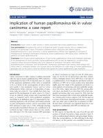
Abdominal sonogram before operation shows intrauterine gestational sac containing foetal pole with positive foetal cardiac motion with a normal spectral trace on pulse DopplerFigure 1
Abdominal sonogram before operation shows intrauterine gestational sac containing foetal pole with positive foetal cardiac
motion with a normal spectral trace on pulse Doppler.
Journal of Medical Case Reports 2008, 2:390 />Page 3 of 5
(page number not for citation purposes)
was positive with a normal tracing by pulse Doppler (Fig-
ures 1 and 2).
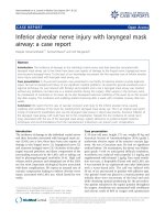
There was also an echo complex mass in the left side of the
pelvis (Figure 2). The pelvic cavity, particularly in the left
lower quadrant, was full of echo complex images. The
boundary of the ovaries and tubes, particularly in the left,
was obscure. These findings demonstrated first an IUP
with a ruptured tubal pregnancy and if not, then an IUP
with a ruptured ovarian cyst.
Her haemoglobin concentration had dropped to 8.8 g/dL
and hematocrit to 27%. Because of the clinical presenta-
tion, laboratory and sonographic findings, the patient was
taken directly to the operating room. She was transfused
with three units of whole blood. An emergency laparot-
omy was done under general anaesthesia that revealed
1500 mL of old blood and abundant clots and a ruptured
middle left tubal pregnancy.
A left salpingectomy was performed. The histopathologi-
cal examination of tissue confirmed a left tubal ectopic
pregnancy which was ruptured at the ampullary portion.
Postoperatively her course was uneventful, and she was
discharged in good general condition on the third day
after the operation. Two weeks after surgery, a live IUP
with a CRL equivalent to 9 weeks gestation was visualized
on a transabdominal ultrasound and which also showed
a marked trophoblastic flow on colour Doppler (Figure
3).
The pregnancy continued without any significant compli-
cation. She was successfully delivered of a male infant at
37 weeks gestation by CS (due to spontaneous onset of
Abdominal sonogram before operation shows intrauterine gestational sac with Yolk sac and an echo complex mass in the left site of the pelvis (arrows)Figure 2
Abdominal sonogram before operation shows intrauterine gestational sac with Yolk sac and an echo complex mass in the left
site of the pelvis (arrows).
Journal of Medical Case Reports 2008, 2:390 />Page 4 of 5
(page number not for citation purposes)
labour and contracted pelvis), the birth weight was 3100
g and her postnatal recovery was unremarkable.
Discussion
HTP was first described by Duverney in 1708 [3,7]. Now-
adays, the use of ART and fertility agents such as CC can
increase a patient's risk of a HTP probably due to the com-
bined effects of hyperstimulation and the subsequent,
simultaneous transfer of several embryos into the uterus
with retrograde flow into the fallopian tubes. Indeed, any
factor predisposing a patient to an increased risk of
ectopic pregnancy (EP) and/or multiple gestations may
contribute to HTP [3,7-9]. In our patient, pregnancy also
occurred in association with ovulation induction by CC.
The majority of HTP cases are diagnosed late. Significant
morbidity and occasional mortality have been reported as
a result of a delay in diagnosis [3]. As no single investiga-
tion can predict the presence of a HTP, it should be sus-
pected in any patient who presents with lower abdominal
pain in the early phase of an obvious IUP following fertil-
ity treatment [7,10].
Often, abdominal and pelvic USG fails to show the EP or
is misinterpreted because of the awareness of an existing
Two weeks after the operation, an abdominal sonogram shows the IUP at 9 weeks gestation and the power Doppler sonogram demonstrates colour signals at the site of the foetal heart (arrow) and retroplacental vessels (arrows)Figure 3
Two weeks after the operation, an abdominal sonogram shows the IUP at 9 weeks gestation and the power Doppler sonogram
demonstrates colour signals at the site of the foetal heart (arrow) and retroplacental vessels (arrows).
Publish with BioMed Central and every
scientist can read your work free of charge
"BioMed Central will be the most significant development for
disseminating the results of biomedical research in our lifetime."
Sir Paul Nurse, Cancer Research UK
Your research papers will be:
available free of charge to the entire biomedical community
peer reviewed and published immediately upon acceptance
cited in PubMed and archived on PubMed Central
yours — you keep the copyright
Submit your manuscript here:
/>BioMedcentral
Journal of Medical Case Reports 2008, 2:390 />Page 5 of 5
(page number not for citation purposes)
IUP [3,9] but demonstration of an IUP is no longer a reli-
able indicator for excluding an EP [3,5].
Most ultrasonographic reports make no mention of a
search for coexistent EP when evaluating intrauterine ges-
tation, because a HTP is still thought to be extremely rare
and for this reason, almost all EPs are diagnosed by
excluding an IUP [8].
Our case also presented early in the pregnancy with a his-
tory of nausea, scant vaginal bleeding and lower abdomi-
nal pain. These symptoms are common in IUP. There was
also a delay in the detection of the EP component, there-
fore diagnosis was not made until an EP rupture had
occurred and the patient developed haemoperitoneum
and instability of her vital signs. Although the primary
USG helped to confirm the presence of an IUP, it failed to
identify the EP, while a HTP as a cause for abdominal pain
should have been suspected immediately in our case.
The management of HTP remains controversial. Surgical
therapy has been the traditional mainstay but involves
surgical and anaesthetic risks to both the mother and IUP
[9]. Studies suggest that laparoscopic management is pre-
ferred over laparotomy in patients with a suspected EP,
and with a documented IUP because of minimal manipu-
lation of the uterus [7].
A non-surgical approach can be used safely and effectively
to manage patients who are clinically stable and where a
HTP is recognized relatively early in gestation. The suc-
cessful non-surgical management of six cases of HTP using
potassium chloride (KCl) injection into the tubal EP has
been reported [9]. In our case, if EP had been diagnosed
early, then it might have been possible to complete the
surgery with the laparoscope, but because of hemody-
namic instability in our case, an urgent laparotomy was
arranged.
Conclusion
We can conclude that HTP must always be considered in
patients presenting with abdominopelvic pain in the face
of a documented IUP, because the presence of an IUP can
no longer be considered reassuring and a HTP has to be
ruled out. Thus, we recommend that all patients shown
on USG to have an IUP should be given a comprehensive
pelvic ultrasound so that the possibility of a simultaneous
HTP may be excluded. We also emphasize the need for
prompt and immediate action at the first sign which indi-
cates a HTP, to avoid missing this potentially life-threat-
ening condition.
Consent
Written informed consent was obtained from the patient
for publication of this case report and accompanying
images. A copy of the written consent is available for
review by the Editor-in-Chief of this journal.
Competing interests
The authors declare that they have no competing interests.
Authors' contributions
AH interpreted the patient's sonographic findings, sug-
gested a heterotopic pregnancy. EK searched the literature,
drafted the manuscript and revised the manuscript. SM
was the surgeon of the patient (laparotomy and C/S), clin-
ical assessor. AH, EK and SM authors read and approval
the final manuscript.
Acknowledgements
We would like to thank Lois Green, June Vasic and Alison Bunting for their
help with language revision in the manuscript.
References
1. Dumesic DA, Damario MA, Session DR: Interstitial heterotopic
pregnancy in a woman conceiving by in vitro fertilization
after bilateral salpingectomy. Mayo Clin Proc 2001, 76:90-92.
2. Maalt ME, Murad Nand Dabbas M: Advanced heterotopic preg-
nancy. J Obstet Gynaecol 1999, 19:677-678.
3. Mistry BM, Balasubramaniam S, Silverman R, Sakabu SA, Troop BR:
Heterotopic pregnancy presenting as an acute abdomen: A
diagnostic masquerader. Am Surg 2000, 66(3):307-308.
4. Dessole S, Ruiu GA, Cherchi PL: Coexistence of a heterotopic
pregnancy associated with a homolateral ovarian cyst in a
patient submitted to elective abortion. Gynecol Obstet Invest
2000, 49:277-278.
5. Hill J: Assisted reproduction and the multiple pregnancy:
increasing the risks for heterotopic pregnancy. J Diagn Med
Sonogr 2003, 19:258-260.
6. Bello G, Schonholz D, Moshirpur J, Jeng DY, Berkowitz RL: Com-
bined pregnancy: the Mount Sinai Experience. Obstet Gynecol
Surv 1986, 41:603-613.
7. Perkins JD, Mitchell MR: Heterotopic pregnancy in a large
inner-city hospital: a report of two cases. J Natl Med Assoc 2004,
96:363-366.
8. Mishra A, Youssefzadeh D, Parente JT: Heterotopic pregnancy.
Female Patient 1998, 23:39-42.
9. Scheiber MD, Cedars MI: Successful non-surgical management
of a heterotopic an abdominal pregnancy following embryo
transfer with cryopreserved-thawed embryos. Hum Reprod
1999, 14:1375-1377.
10. Archibong EI, Etuk SJ: Case report, Heterotopic pregnancy fol-
lowing induction of ovulation. Trop J Obstet Gynecol 2002,
19:115-116.