Báo cáo y học: "A giant colonic diverticulum presenting as a ''''phantom mass'''': a case report" pptx
Bạn đang xem bản rút gọn của tài liệu. Xem và tải ngay bản đầy đủ của tài liệu tại đây (739.52 KB, 3 trang )
BioMed Central
Page 1 of 3
(page number not for citation purposes)
Journal of Medical Case Reports
Open Access
Case report
A giant colonic diverticulum presenting as a 'phantom mass': a case
report
Ayman S Abdelrazeq*
1
, Anwar E Owais
2
, Munther I Aldoori
3
and
Ian D Botterill
4
Address:
1
North Cheshire NHS Trust Hospitals, Lovely Lane, Warrington, WA5 1QG, UK,
2
Department of General Surgery, Scarborough General
Hospital, Scarborough, UK,
3
General surgery, Huddersfield Royal Infirmary, Huddersfield, UK and
4
Department of Colorectal Surgery, St. James's
University Hospital, Leeds, UK
Email: Ayman S Abdelrazeq* - ; Anwar E Owais - ;
Munther I Aldoori - ; Ian D Botterill -
* Corresponding author
Abstract
Introduction: Diverticulosis coli is the most common disease of the colon in Western countries.
Giant colonic diverticulum, defined as a colonic diverticulum measuring 4 cm in size or larger,
represents an unusual manifestation of this common clinical entity.
Case presentation: A 68-year-old Caucasian British woman with a history of intermittent lower
abdominal mass, leg swelling and focal neurological symptoms underwent extensive non-diagnostic
investigations over a significant period under a number of disciplines. The reason for a diagnosis
being elusive in part related to the fact that the mass was never found on clinical and ultrasound
examination. As a result, the patient's validity was questioned. Ultimately, this 'phantom-mass' was
diagnosed as a giant colonic diverticulum causing intermittent compression of the iliac vein and
obturator nerve.
Conclusion: Intermittent compression of the iliac vein and the obturator nerve by a colonic
diverticulum has not previously been reported. A giant colonic diverticulum presenting as an
intermittent mass is very rare. This case also illustrates two factors. First, the patient is often right.
Second, the optimal mode of investigation for any proven or described abdominal mass with
referred symptoms is cross-sectional imaging, typically a computed tomography scan, irrespective
whether the mass or symptoms are constant or intermittent.
Introduction
Giant colonic diverticulum (GCD) defined as a colonic
diverticulum measuring 4 cm in size or larger is a rare dis-
ease [1]. It is nearly always associated with diverticular dis-
ease.
Several theories have been proposed to explain its aetiol-
ogy: a unidirectional flap-valve mechanism at the base of
the diverticulum allowing bowel gas and debris to enter
but not to leave the diverticulum; gas-forming organisms
distend a diverticulum after its mouth has become oblit-
erated; an organized abscess that develops around an
infected pre-existing pseudodiverticulum; or a true con-
genital duplication [1,2]. The exact aetiology remains
unknown.
Published: 27 January 2009
Journal of Medical Case Reports 2009, 3:29 doi:10.1186/1752-1947-3-29
Received: 7 March 2008
Accepted: 27 January 2009
This article is available from: />© 2009 Abdelrazeq et al; licensee BioMed Central Ltd.
This is an Open Access article distributed under the terms of the Creative Commons Attribution License ( />),
which permits unrestricted use, distribution, and reproduction in any medium, provided the original work is properly cited.
Journal of Medical Case Reports 2009, 3:29 />Page 2 of 3
(page number not for citation purposes)
Here we report a patient with GCD who presented with
some peculiarities, which we believe will be valuable in
further explaining the mechanism behind GCD, and may
help in the prompt diagnosis and treatment of future sim-
ilar cases.
Case presentation
A 68-year-old Caucasian British woman presented with a
history of an intermittent large painless mass in her lower
abdomen. The mass was more pronounced before defeca-
tion and was associated with paraesthesia in the medial
side of the left thigh and swelling of the left lower limb.
Following defecation, the mass disappeared with instant
relief of the associated symptoms. She had no other asso-
ciated complaints and clinical examination was normal.
Blood tests and abdominal ultrasound were normal.
Colonoscopy showed sigmoid diverticulosis. She was
reassured and discharged.
The patient's symptoms recurred and she was referred for
a second opinion from a gastroenterologist, a urologist,
and a neurologist. Clinical and neurological examination
was again confirmed to be normal. An upper gastrointes-
tinal series, intravenous pyelogram, cystoscopy, MRI
spine and venous duplex scan were all reported as unre-
markable, though at the time of these tests, the patient
was asymptomatic. The patient was referred to a psychia-
trist and subsequently commenced on various sedatives
and antidepressants with no improvement.
Two years later, she was referred to our team with similar
ongoing complaints. Clinical examination remained
unremarkable. Barium enema confirmed uncomplicated
diverticular disease. Shortly following barium enema she
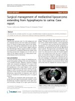
developed severe lower abdominal pain and fever. Com-
puterized tomography demonstrated a thick-walled, air
filled cavity closely related to the sigmoid colon, compat-
ible with a GCD (Figure 1).
At laparotomy, an 8 cm mass was found to originate from
the antimesenteric border of the sigmoid colon. It was
adherent to the iliac vessels and lateral pelvic wall. Diver-
ticulosis involving mainly the sigmoid colon was noted.
Sigmoid colectomy was performed. She had an uncompli-
cated recovery. At 12-month follow-up, she remained
asymptomatic.
Pathology confirmed a GCD with diverticulitis and peri-
tonitis. This diverticulum was 8 cm across with a 5 mm
luminal orifice and was filled with air and faecal debris; a
flap-valve effect was noted at the opening of the colon.
[Uninflamed simple diverticula were also noted.
Discussion
Diverticulosis coli is the most common disease of the
colon in Western countries; GCD represents an unusual
manifestation of this common clinical entity [3].
Histologically, there are three different types of GCD with
similar clinical presentations [2]. The first type is a
pseudo-diverticulum which gradually increases in size;
remnants of muscularis mucosa and muscularis propria
may be found in its wall. In most cases, the mucosa is not
completely intact. This type of GCD could be explained by
the flap-valve or gas-forming organisms theories.
If no mucosal remnants are found, the GCD is considered
inflammatory (type 2), which is actually a result of a local
perforation of the mucosa with an abscess cavity that
remains in contact with the lumen of the colon (organ-
ized abscess theory). The wall of this diverticulum con-
tains reactive scar tissue only. The third type is a true
diverticulum, in which the wall contains all of the layers
of normal bowel (congenital duplication theory).
In this patient, the diverticulum gradually inflated
because of a flap-valve action of the tiny opening from the
bowel, allowing gas and debris to enter, but not escape,
during periods of straining. The failure of barium to enter
the GCD attests to the small size of the opening. The fact
that this GCD could expand and shrink indicates that its
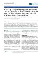
Axial computed tomography image with oral and intravenous contrast at the level of S1 demonstrating a giant colonic diverticulum*Figure 1
Axial computed tomography image with oral and
intravenous contrast at the level of S1 demonstrating
a giant colonic diverticulum*. The wall of the diverticu-
lum and the surrounding fat are thickened denoting recent
inflammation. Note the relationship to the left external and
internal iliac vessels (arrows A and B, respectively). The
obturator nerve (L2–4) emerges from the medial aspect of
the psoas muscle and runs downwards and forwards, deep
into [the internal iliac vessels.
A
B
*
Journal of Medical Case Reports 2009, 3:29 />Page 3 of 3
(page number not for citation purposes)
wall had elastic properties. When fully expanded, it pro-
duced compressive symptoms.
Symptoms of GCD and their duration are variable; often
the patient's complaint can be ascribed to the associated
diverticulitis [4]. A palpable abdominal mass in a patient
with diverticulitis is almost invariably a paracolic inflam-
matory mass related to acute diverticulitis. Presentation of
colonic diverticula as a palpable abdominal mass in the
absence of acute diverticulitis is extremely rare [5]. The
intermittent appearance of a large painless mass in our
patient was probably due to filling of the GCD with gas
and faecal material whose expulsion led to the disappear-
ance of the mass. The associated symptoms are related to
pressure on the left iliac vein and the left obturator nerve.
Such a presentation of GCD as a longstanding non-
inflammatory intermittent abdominal mass is distinctly
unusual. Intermittent compression of the iliac vein or the
obturator nerve as a presentation of GCD has not previ-
ously been reported.
Plain film diagnosis of GCD can be made in the presence
of a persistent smooth-walled gas containing structure,
'balloon sign', adjacent to the colon with or without air
fluid level [6].
Diagnostic colonoscopy is not considered to be helpful
[7] except in cases with a large ostium where an incidental
diagnosis of GCD is possible [8]. The use of barium
enema did not yield a positive diagnosis in this patient,
although a communication with the colon can be demon-
strated on contrast studies in 25% to 66% of GCD cases
[7].
Ultrasound examination does not seem to be helpful in
detecting a non-complicated GCD [7]. In our patient, we
were falsely reassured by a normal ultrasound. Computer-
ized tomography and magnetic resonance imaging are
useful in defining the GCD and its relation to surrounding
structures [9]. The intermittent nature of this patient's
symptoms made diagnosis difficult. Arguably, an earlier
CT scan may have helped. The final diagnosis was only
made when barium inspissated in the neck of the GCD
and provoked local diverticulitis which merited immedi-
ate CT evaluation.
Conclusion
This case of GCD which presented initially with a long-
standing non-inflammatory intermittent abdominal mass
is distinctly unusual. Intermittent compression of the iliac
vein or the obturator nerve as a presentation of GCD has
not previously been reported.
This case also illustrates two factors. First, the patient is
often right. Second, a proven or described abdominal
mass with or without referred symptoms should prompt
cross-sectional imaging, typically a CT scan, irrespective of
whether the mass or symptoms are constant or intermit-
tent.
Abbreviations
CT: computed tomography; GCD: giant colonic diverticu-
lum; MRI: magnetic resonance imaging
Consent
Written informed consent was obtained from the patient
for publication of this case report and any accompanying
images. A copy of the written consent is available for
review by the Editor-in-Chief of this journal.
Competing interests
The authors declare that they have no competing interests.
Authors' contributions
AA and AO identified the association between the GCD
and the patient's unique presentation, reviewed the litera-
ture and drafted the manuscript. MA and IB contributed to
drafting the manuscript and critically revised the discus-
sion and conclusions. All authors have read and approved
the manuscript.
References
1. Choong CK, Frizelle FA: Giant colonic diverticulum: report of
four cases and review of the literature. Dis Colon Rectum 1998,
41:1178-1185.
2. McNutt R, Schmitt D, Schulte W: Giant colonic diverticula –
three distinct entities. Report of a case. Dis Colon Rectum 1988,
31:624-628.
3. Kuganeswaran E, Fisher JK: Giant sigmoid diverticulum: rare
manifestation of diverticular disease. South Med J 1998,
91:952-955.
4. Naber A, Sliutz AM, Freitas H: Giant diverticulum of sigmoid
colon. Br J Surg 1995, 82:985.
5. Gallagher JJ, Welch JP: Giant diverticula of the sigmoid colon: a
review of differential diagnosis and operative management.
Arch Surg 1972, 114:1079-1083.
6. Rosenberg RF, Naidich JB: Plain film recognition of giant colonic
diverticulum. Am J Gastroenterol 1981, 76:59-69.
7. Steenvoorde P, Vogelaar FJ, Oskam J, Tollenaar R: Giant colonic
diverticula. Review of diagnostic and therapeutic options.
Dig Surg 2004, 21:1-6.
8. Mehta DC, Baum JA, Dave PB, Gumaste VV: Giant colonic diver-
ticulum: report of two cases and endoscopic recognition. Am
J Gastroenterol 1996, 91:1269-1271.
9. Smith TR, Tyler IM: CT demonstration of a giant colonic diver-
ticulum. Gastrointest Radiol 1987, 12:73-75.