Báo cáo y học: "Temporal fossa arachnoid cyst presenting with bilateral subdural hematoma following trauma: two case reports" pptx
Bạn đang xem bản rút gọn của tài liệu. Xem và tải ngay bản đầy đủ của tài liệu tại đây (848.08 KB, 5 trang )
BioMed Central
Page 1 of 5
(page number not for citation purposes)
Journal of Medical Case Reports
Open Access
Case report
Temporal fossa arachnoid cyst presenting with bilateral subdural
hematoma following trauma: two case reports
Promod Pillai*
1
, Sajesh K Menon
2
, Raju P Manjooran
2
, Rajiv Kariyattil
2
,
Ashok B Pillai
2
and Dilip Panikar
2
Address:
1
Department of Neurological Surgery, the Ohio State University Medical Center, Hamilton Hall, Neil Avenue, Columbus, Ohio 43210,
USA and
2
Department of Neurosurgery, Amrita Institute of Medical Sciences, Kochi, India
Email: Promod Pillai* - ; Sajesh K Menon - ;
Raju P Manjooran - ; Rajiv Kariyattil - ; Ashok B Pillai - ;
Dilip Panikar -
* Corresponding author
Abstract
Introduction: Intracranial arachnoid cysts are considered to be congenital malformations with a
predilection for the temporal fossa. They are often asymptomatic but can sometimes be
symptomatic due to enlargement or hemorrhage. There are multiple case reports of arachnoid
cysts becoming symptomatic with hemorrhagic complications following head trauma. In such cases,
the bleeding is often confined to the side ipsilateral to the arachnoid cyst. Occurrence of
contralateral subdural hematomas in patients with temporal fossa arachnoid cysts has rarely been
observed and is reported less frequently in the medical literature.
Case presentation: We report two cases of people (a 23-year-old man and a 41-year-old man)
with temporal fossa arachnoid cysts complicated by a subdural hematoma following head injury.
Both patients developed a subdural hematoma contralateral to the side of a temporal fossa
arachnoid cyst. It is likely that lack of adequate intracranial cushioning in the presence of an
intracranial arachnoid cyst may result in injury not only to ipsilateral but also to contralateral
bridging veins, following head trauma.
Conclusion: It is important to identify and report such rare complications with intracranial
arachnoid cysts, so that asymptomatic patients with an intracranial arachnoid cyst can be counseled
about such possibilities following head trauma.
Introduction
Arachnoid cysts are believed to be developmental anoma-
lies and are often documented as incidental findings on
imaging [1]. A common location is at the temporal fossa
[2,3]. Occasionally, these cysts become symptomatic with
hemorrhagic complications, often precipitated by head
trauma. Hemorrhagic complications are often confined to
the side ipsilateral to the location of the arachnoid cyst,
and the occurrence of contralateral hemorrhage has been
documented on occasion in the literature. We report two
patients with temporal fossa arachnoid cysts who experi-
enced contralateral subdural hematomas following head
trauma.
Published: 9 February 2009
Journal of Medical Case Reports 2009, 3:53 doi:10.1186/1752-1947-3-53
Received: 15 February 2008
Accepted: 9 February 2009
This article is available from: />© 2009 Pillai et al; licensee BioMed Central Ltd.
This is an Open Access article distributed under the terms of the Creative Commons Attribution License ( />),
which permits unrestricted use, distribution, and reproduction in any medium, provided the original work is properly cited.
Journal of Medical Case Reports 2009, 3:53 />Page 2 of 5
(page number not for citation purposes)
Case presentation
Case 1
A previously asymptomatic 23-year-old man was exam-
ined in the emergency services unit of the referring hospi-
tal following a fall while riding a bicycle. He reported no
loss of consciousness and no external injuries but was
complaining of mild headache and nausea. On examina-
tion, the patient was fully conscious and demonstrated no
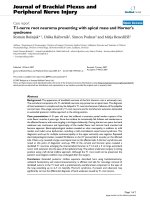
focal neurological deficit. On admission, a computer tom-
ography (CT) scan of his head revealed a giant arachnoid
cyst, Galassi type II [4], occupying the left middle cranial
fossa and extending into the sylvian fissure (Figure 1).
There was no brain parenchymal injury or intracranial
hemorrhage. Incidentally, the patient also presented with
a mega cisterna magna. He was subsequently discharged.
Four weeks later, he presented to our institution with
increasing headaches, nausea and vomiting. On admis-
sion, the patient was conscious and without any focal
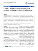
neurological deficits. A repeat CT scan of his head revealed
a bilateral mixed density subdural hematoma with a
mixed density mass lesion within the left middle cranial
fossa extending into the sylvian fissure (Figure 2), replac-
ing the previously documented cerebrospinal fluid (CSF)
density lesion. The patient underwent a left-sided craniot-
omy for evacuation of the subdural hematoma as well as
the intracystic hematoma and cyst fenestration into the
basal cisterns. Right frontal and parietal burr holes were
also performed to evacuate the right-sided subdural
hematoma. The patient tolerated the procedure well and
recovered completely.
Case 2
A 41-year-old man presented to our out-patient unit with
an increasingly severe headache and nausea of 2 weeks'
duration. He recalled sustaining a trivial fall about 12
weeks prior to presentation, with no loss of consciousness
or any external injuries. On admission, the patient was
conscious, with no focal neurological deficits. A cranial
CT scan revealed a Galassi Type I arachnoid cyst occupy-
ing the right temporal fossa (Figure 3A,B) with an ipsilat-
eral subdural hygroma and a contralateral subdural
hematoma (Figure 3C,D). The hematoma and hygroma
were evacuated by bilaterally placed burr holes, followed
by an uneventful recovery.
Discussion
With the advent of neuroimaging, there has been an
increased incidence of detection of incidental asympto-
Computed tomographic scan of the head without contrast immediately following the trauma, showing a giant arachnoid cyst in the middle cranial fossaFigure 1
Computed tomographic scan of the head without contrast immediately following the trauma, showing a giant
arachnoid cyst in the middle cranial fossa.
Journal of Medical Case Reports 2009, 3:53 />Page 3 of 5
(page number not for citation purposes)
matic arachnoid cysts. Arachnoid cysts most frequently
occur in the middle fossa, followed by the posterior fossa,
convexity, and suprasellar regions. Although these lesions
are considered congenital, the exact etiology is still not
clear.
Hemorrhage into an arachnoid cyst and the associated
subdural hematoma following head trauma are well doc-
umented, although the mechanism and true incidence are
not clearly understood. The annual risk for hemorrhage in
patients with a middle cranial fossa cyst probably remains
below 0.1% [2]. In a recent study by Wester et al., the inci-
dence of chronic subdural or intracystic haematomas was
reported as 4.6% of all patients with intracranial arach-
noid cyst referred for treatment [3]. We propose two
mechanisms leading to formation of subdural hemor-
rhage. First, the cyst membrane is loosely attached to the
convexity dura. The mechanical forces that are sustained
during a moderate head trauma can cause the cyst mem-
brane to be detached from the dura and thus cause a
bleeding episode. Second, the parietal cyst membrane
also covers the area where the bridging Sylvian veins, or
the veins that traverse the membrane unsupported by
brain tissue, enter into the dural venous sinuses behind
the sphenoid ridge. Even a moderate manipulation of the
parietal membrane can disrupt these veins, leading to
bleeding into subdural space [3]. Parsch and colleagues
suggest an approximately 5-fold greater prevalence
(2.43% versus 0.46%) of arachnoid cysts of the middle
fossa in patients with chronic subdural hematomas than
in the general population who undergo magnetic reso-
nance imaging [5]. A middle cranial fossa arachnoid cyst
is now recognized as one of the causes of chronic subdural
hematomas after head injury, especially in young people,
as the cysts appear to be more susceptible to hemorrhagic
complications, including subdural and intracystic
hematomas [1-8]. The membrane is vascular, and bridg-
ing veins are often observed traversing the cyst wall. This
could in part explain the liability of intracystic subdural
bleeding in these patients [5,6,9].
The post-traumatic hemorrhagic complication in a setting
of a temporal fossa arachnoid cyst is often confined to the
side ipsilateral to the cyst, and contralateral subdural
hematoma is not well documented in the literature [2,8].
Occurrence of contralateral subdural hematomas with
arachnoid cysts was previously reported by Mori et al.
(two cases) and Parsch et al. (one case) [5,8]. Both of our
Computed tomographic scan of the head without contrast 3 weeks after the traumaFigure 2
Computed tomographic scan of the head without contrast 3 weeks after the trauma. Figure 2A shows an intra-
cystic hematoma. Figure 2B shows a bilateral subdural hematoma of mixed density.
Journal of Medical Case Reports 2009, 3:53 />Page 4 of 5
(page number not for citation purposes)
Computed tomographic scan of the head without contrast showing an arachnoid cyst in the right temporal fossaFigure 3
Computed tomographic scan of the head without contrast showing an arachnoid cyst in the right temporal
fossa. A, B) an ipsilateral subdural hygroma and C, D) a contralateral subdural hematoma.
Publish with BioMed Central and every
scientist can read your work free of charge
"BioMed Central will be the most significant development for
disseminating the results of biomedical research in our lifetime."
Sir Paul Nurse, Cancer Research UK
Your research papers will be:
available free of charge to the entire biomedical community
peer reviewed and published immediately upon acceptance
cited in PubMed and archived on PubMed Central
yours — you keep the copyright
Submit your manuscript here:
/>BioMedcentral
Journal of Medical Case Reports 2009, 3:53 />Page 5 of 5
(page number not for citation purposes)
patients were previously asymptomatic and had subdural
hematomas contralateral to the arachnoid cyst following
the head injury. This finding reinforces the notion that an
arachnoid cyst, being a large fluid-filled lesion, is less
compliant than normal brain parenchyma, making both
ipsilateral and contralateral bridging veins prone to
injury. Even though it is rare, there have been reports of
ruptured arachnoid cyst, presenting with a subdural CSF
collection without evidence of hemorrhage [9-11]. The
sudden collapse of the cyst can cause a sudden shift of the
brain, which, along with the force of the trauma, can lead
to stretching, and tearing of bridging veins on the oppo-
site side. This could explain the occurrence of contralat-
eral subdural hematoma as in both our cases. Thus, we
should inform patients with arachnoid cysts and their
families of the possibility of such complications and
advise care to avoid head injury in daily life, regardless of
the size and symptoms of the cyst.
Conclusion
Although many arachnoid cysts are incidentally detected
and require no intervention, some of them are sympto-
matic. A contralateral subdural hematoma following head
trauma may result from inadequate intracranial cushion-
ing provided by the arachnoid cyst, which makes both
ipsilateral and contralateral bridging veins prone to
injury. It is important to identify and report such rare
complications with intracranial arachnoid cysts, so that
the asymptomatic patient with an intracranial arachnoid
cyst can be counseled about such possibilities following
head trauma.
Abbreviations
CT: computer tomography; CSF: cerebrospinal fluid.
Consent
Written informed consent was obtained from the patients
for publication of this case report and accompanying
images. A copy of the written consent is available for
review by the Editor-in-Chief of this journal.
Competing interests
The authors declare that they have no competing interests.
Authors' contributions
PP, SKM, RPM, RK and ABP all contributed to the patients'
management, the conception of the manuscript, acquisi-
tion and interpretation of data, and drafting and revision
of the manuscript. DP was also involved in critically revis-
ing the manuscript and gave the final approval of the
manuscript.
Acknowledgements
The authors thank Dr E Antonio Chiocca MD, Ph.D. for critically evaluating
the manuscript and for giving constructive suggestions. The authors greatly
appreciate the editorial assistance of Rosalyn Annette Uhrig MA in the
preparation of this manuscript.
References
1. Helland CA, Wester K: A population-based study of intracra-
nial arachnoid cysts – Clinical and radiological outcome fol-
lowing surgical cyst decompression in adults. J Neurol
Neurosurg Psychiatry 2007, 78(10):1129-1135.
2. Iaconetta G, Esposito M, Maiuri F, Cappabianca P: Arachnoid cyst
with intracystic haemorrhage and subdural haematoma:
case report and literature review. Neurol Sci 2006,
26(6):451-455.
3. Wester K, Helland CA: How often do chronic extra-cerebral
haematomas occur in patients with intracranial arachnoid
cysts? J Neurol Neurosurg Psychiatry 2008, 79(1):72-75.
4. Galassi E, Piazza G, Gaist G, Frank F: Arachnoid cyst of the middle
cranial fossa: a clinical and radiological study of 25 cases
treated surgically. Surg Neurol 1980, 14(3):211-219.
5. Parsch CS, Krauss J, Hofmann E, Meixensberger J, Roosen K: Arach-
noid cysts associated with subdural hematomas and hygro-
mas: analysis of 16 cases, long-term follow-up, and review of
the literature. Neurosurgery 1997, 40(3):483-490.
6. De K, Berry K, Denniston S: Haemorrhage into an arachnoid
cyst: a serious complication of minor head trauma. Emerg
Med J 2002, 19:365-366.
7. Demetriades AK, McEvoy AW, Kitchen ND: Subdural hae-
matoma associated with an arachnoid cyst after repetitive
minor heading injury in ball games. Br J Sports Med 2004,
38(4):E8-4.
8. Mori K, Yamamoto T, Horinaka N, Maeda M: Arachnoid cyst is a
risk factor for chronic subdural hematoma in juveniles:
twelve cases of chronic subdural hematoma associated with
arachnoid cyst. J Neurotrauma 2002, 19(9):1017-1027.
9. Sener RN: Arachnoid cysts associated with post-traumatic
and spontaneous rupture into the subdural space. Comput
Med Imaging Graph 1997, 21(6):341-344.
10. Donaldson JW, Edwards-Brown M, Luerssen TG: Arachnoid cyst
rupture with concurrent subdural hygroma. Pediatr Neurosurg
2000, 32(3):137-139.
11. Gelabert-González M, Fernández-Villa J, Cutrín-Prieto J, Garcìa Allut
A, Martínez-Rumbo R: Arachnoid cyst rupture with subdural
hygroma: report of three cases and literature review. Childs
Nerv Syst 2002, 18(11):609-613.