báo cáo khoa học: " The buccal minor salivary glands as starting point for a metastasizing adenocarcinoma – report of a case" pptx
Bạn đang xem bản rút gọn của tài liệu. Xem và tải ngay bản đầy đủ của tài liệu tại đây (1.22 MB, 5 trang )
BioMed Central
Page 1 of 5
(page number not for citation purposes)
Head & Face Medicine
Open Access
Case report
The buccal minor salivary glands as starting point for a
metastasizing adenocarcinoma – report of a case
Tobias Ettl
1
, Johannes Kleinheinz*
2,5
, Ravi Mehrotra
3
, Stephan Schwarz
4
,
Torsten Eugen Reichert
1
and Oliver Driemel
1
Address:
1
Department of Oral and Maxillofacial Surgery, Regensburg University, Germany,
2
Department of Oral and Maxillofacial Surgery,
Muenster University, Germany,
3
Department of Pathology, Moti Lal Nehru Medical College, Allahabad University, India,
4
Department of
Pathology, Erlangen University, Germany and
5
Department of Cranio-Maxillofacial Surgery, University Hospital Muenster, Waldeyerstr. 30, D-
48149, Muenster, Germany
Email: Tobias Ettl - ; Johannes Kleinheinz* - ; Ravi Mehrotra - ;
Stephan Schwarz - ; Torsten Eugen Reichert - ;
Oliver Driemel -
* Corresponding author
Abstract
Background: With the 2005 WHO classification of salivary gland tumours and its increasingly
recognized diagnostic entities, the frequency of adenocarcinoma (NOS) has decreased significantly.
Case presentation: This paper describes a fast growing adenocarcinoma (NOS), originating from
the minor salivary glands of the left buccal mucosa with a rapid onset of multiple local and distant
metastases, especially in the lung. A lung primary was unlikely as the tumour was characterized by
positivity for cytokeratin 20 and negativity for the thyroid transcription factor-1 protein (TTF-1) in
immunohistochemistry.
Conclusion: A rare case of an adenocarcinoma (NOS) of the minor salivary glands with a rapid
development and an unfavourable clinical course is reported. It shows that additional
immunohistochemical analysis can decisively contribute to determine the site of the primary
tumour in cases with unknown primary.
Background
Epithelial tumours arising in the intra-oral minor salivary
glands account for 9–23% of all salivary gland tumours
[1,2] and of these, carcinomas are responsible for about
40–54% [3-5]. Adenocarcinoma not otherwise specified
(NOS) is a malignant neoplasm of the salivary glands
with ductal, glandular or secretory differentiation that
cannot be attributed to any other currently recognized
type of salivary gland carcinoma [6,7]. With the 2005
WHO classification of salivary gland tumours and its
increasingly recognized diagnostic entities, frequency of
adenocarcinoma (NOS) has decreased significantly [7].
This article describes a fast growing adenocarcinoma
(NOS), originating in the left buccal mucosa with a rapid
onset of multiple local and distant metastases. Immuno-
histochemistry was found to be useful in confirming a sal-
ivary gland origin.
Case presentation
A 68-year old female patient with a painless swelling of
the left buccal mucosa was referred to our department. An
initial incisional biopsy of the lesion was inconclusive
Published: 30 July 2008
Head & Face Medicine 2008, 4:16 doi:10.1186/1746-160X-4-16
Received: 17 May 2008
Accepted: 30 July 2008
This article is available from: />© 2008 Ettl et al; licensee BioMed Central Ltd.
This is an Open Access article distributed under the terms of the Creative Commons Attribution License ( />),
which permits unrestricted use, distribution, and reproduction in any medium, provided the original work is properly cited.
Head & Face Medicine 2008, 4:16 />Page 2 of 5
(page number not for citation purposes)
and magnetic resonance imaging (MRI) of the head and
neck diagnosed a benign appearing connective tissue
tumour, arising without local invasion.
Detailed medical history pointed to a more than three
months consisting, rapidly enlarging mass in the patient's
left buccal mucosa, which provoked pain while using her
dentures. The patient further complained of lack of appe-
tite, sleeping disturbance and weight loss of 11 kilograms
(15% of body weight) over the last five months. Tobacco
and alcohol abuse was excluded.
Intraoral examination revealed an asymptomatic, solid,
firm, exophytic and endophytic growing tumour of the
left buccal mucosa (Fig 1). The tumour was fixed to adja-
cent structures and extended caudal to the mandible.
Examination of the patient did not reveal facial paralysis,
paraesthesia and palpable regional lymphadenopathy.
Haematologic parameters were all within normal range.
For further elucidation, a deeper biopsy was performed.
During surgery, the tumour could hardly be separated
from the surrounding connective soft tissue and adjacent
alveolar bone. The retromolar alveolar crest appeared dis-
integrated and was suspicious of bone invasion, so a spec-
imen of the alveolar bone was taken as well.
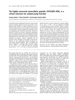
Histopathological analysis of the specimen, supported by
immunohistochemistry (CK7 and CK20 positive; CK5/6,
Aktin and HER 2 negative) allowed the diagnosis of a
poorly differentiated adenocarcinoma (NOS) of the
minor salivary glands (Fig 2a–c).
Positron-emission tomography with 'low dose CT' (PET-
CT), computerised tomography (CT head and neck, chest,
pelvis and abdomen) and bone scan showed the tumour
in the left buccal area and an additional circular mass in
the hilum of the left lung, a tumour of the left kidney, as
well as multiple pulmonary, cervical lymph nodes and
osseous (skull, spine, rib, pelvis) masses (Fig 3, 4, 5, 6).
Bronchoscopic biopsy of the hilum mass also identified a
poorly differentiated adenocarcinoma (NOS). Since the
immunohistochemical analysis was negative for the thy-
roid transcription factor-1 protein (TTF-1) (Fig 2d) and
was positive for cytokeratin 20, a primary adenocarci-
noma of the lung was unlikely and the tumour was finally
attributed to the minor salivary glands as site of origin.
Due to the extent of the disease, palliative chemotherapy
was initiated.
Discussion
Data concerning the relative frequency of adenocarci-
noma (NOS) vary from 1.2% to 17.8% of all salivary
gland carcinomas [6,8], since in previous classifications
tumours, which are currently established as more specific
histologies like salivary duct carcinoma, epithelial-
myoepithelial carcinoma or polymorphous low-grade
adenocarcinoma, were often categorized as adenocarci-
noma (NOS) [6,8]. About 40% of adenocarcinomas
(NOS) are located in the minor salivary glands [7], with a
relative frequency of 4.3%–10.3% of all minor gland car-
cinomas [3-5]. The palate is the most commonly involved
site (39%–75%), followed by the lips and the buccal
mucosa, as described in the case report [3,4]. In most
Intraoral finding after initial biopsy: Exophytic and endophytic growing tumour of the left buccal mucosa (3 × 2 × 1.5 cm
3
) with indiscernible bordersFigure 1
Intraoral finding after initial biopsy: Exophytic and
endophytic growing tumour of the left buccal
mucosa (3 × 2 × 1.5 cm
3
) with indiscernible borders.
HistopathologyFigure 2
Histopathology. a: Tumour with solid and invasive growth
pattern surrounded by desmoplastic connective tissue (H&E,
40×). b: in detail: Hyperchromatic, pleomorphic nuclei with
necrosis and numerous mitoses (H&E, 200×). c: Positive
immunohistochemical staining for Cytokeratin 7 (CK7,
200×). d: Negativity for the thyroid transcription factor 1
(TTF-1, 200×).
Head & Face Medicine 2008, 4:16 />Page 3 of 5
(page number not for citation purposes)
cases, the lesion presents as a firm, solid and painless
mass, which may be characterized by ulceration and fixa-
tion to the surrounding soft tissues. Mechanical irritation
like friction from the patient's denture may evoke tender-
ness.
In general there are various differential diagnoses for a
buccal swelling comprising both benign and malignant
neoplasia. Tumours may originate from the squamous
epithelium (papilloma, squamous cell carcinoma), the
soft tissue (fibromatosis, nodular fasciitis, malignant
fibrous histiocytoma, fibrosarcoma, leiomyoma, leiomy-
osarcoma, lipoma, liposarcoma, neurofibroma, schwan-
noma, malignant peripheral nerve sheath tumour,
hemangioma, angiosarcoma) and from salivary glands
(pleomorphic adenoma, adenoid cystic carcinoma etc.)
[9,10]. In view of the fact that the majority of Non-Hodg-
kin's lymphomas affecting the oral cavity present as a sub-
mucosal mass, this differential diagnosis should also be
taken into account, although the hard palate and the gin-
giva are the most common intraoral sites of occurrence
[10]. Oral metastatic lesions can also be the initial appear-
ance of undiagnosed primary malignancies. Because of
the rapid growth of the tumour, its firm appearance and
spread to adjacent structures, its intraoperatively obvious
bony invasion and considering the patient's history (lack
of appetite and weight loss), a malignancy was the most
likely diagnosis in the present case.
Microscopically, adenocarcinoma (NOS) is characterized
by a variable spectrum of different architectural patterns,
which may include glandular, papillary, cystic, cribriform
or solid structures [6]. Tumours with considerable hetero-
geneity of growth patterns, which cannot clearly be attrib-
Computerized tomography (CT) with contrast medium (CM): Axial image of the head and neck: Tumour (4 × 5 cm
2
) of the left buccal soft tissues with central necrotic and partly calcified components and resorption of the left mandibleFigure 3
Computerized tomography (CT) with contrast medium
(CM): Axial image of the head and neck: Tumour (4 × 5 cm
2
)
of the left buccal soft tissues with central necrotic and partly
calcified components and resorption of the left mandible.
Chest: Left hilar mass (4.5 × 4.4 cm
2
)Figure 4
Chest: Left hilar mass (4.5 × 4.4 cm
2
). Local infiltration into
mediastinum; additional mass on the left side.
Abdomen: Left renal tumour (4.6 × 4.1 cm
2
)Figure 5
Abdomen: Left renal tumour (4.6 × 4.1 cm
2
).
Head & Face Medicine 2008, 4:16 />Page 4 of 5
(page number not for citation purposes)
uted to well known entities of adenocarcinoma should
best be classified as adenocarcinomas (NOS). According
to the most recent WHO classification, tumours showing
a high morphologic heterogeneity, a low mitotic rate and
slight nuclear atypia can better be assessed as polymor-
phous low-grade adenocarcinoma. Hence, the majority of
adenocarcinomas will be of high malignancy grade, as in
this case, characterized by hyperchromatic and pleomor-
phic nuclei, necrosis and high mitotic rate [7]. Adenocar-
cinomas with overt presence of ductal structures should
better be classified as salivary duct carcinoma (SDC) than
as adenocarcinoma NOS, but the distinction might be
arbitrary. Immunohistochemistry may help, as more than
90% of SDCs are specifically positive for androgen recep-
tors (AR) and because most of these carcinomas show
positive staining for HER-2/neu (c-erbB-2) [11].
Cytokeratins (CK) are distinctive intermediate filaments,
which are confined to epithelia and indicate the tissue of
origin in malignant transformation and metastasis [12].
They may also be useful in the determination of the pri-
mary site. While CK 5/6 is common in squamous epithe-
lia, the expression of CK 7 and CK 20 is distinctive in
glandular epithelia. This may include tumours like color-
ectal, pancreatic or bronchioloalveolar adenocarcinoma
as well as adenocarcinomas of the salivary glands [13].
Since the patient in this case report presented with an
additional adenocarcinoma of the lung, the primary site
of the carcinoma had to be elucidated, especially oral
metastasis by a lung primary had to be excluded. The thy-
roid transcription factor 1 (TTF-1) is a specific marker of
the thyroid gland and the epithelia of the lung, regulating
the expression of surfactant in the latter organ [14,15].
Evidence of antibodies to TTF-1 may identify the lung as
the primary site of origin in adenocarcinoma with
unknown primary. In the reported case TTF-1 turned out
to be negative. Together with the positivity for CK20
which is usually negative in primary adenocarcinomas of
the lung, a salivary gland origin was most likely. Immuno-
histochemistry might also aid in the differential diagnosis
of salivary gland carcinoma types. In the present case the
tumour cells were negative for CK5/6, a marker of basal
cells, myoepithelial cells and squamous epithelium
excluding a variety of carcinoma types: mucoepidermoid
carcinoma, squamous cell carcinoma and myoepithelial
carcinoma.
The overall prognosis of adenocarcinoma (NOS) depends
on clinical stage and malignancy grade. For stage I a 10-
year survival rate of 75% has been reported by Spiro et al
[16], dropping to 36% for stage II, irrespective of grade.
According to the same study 15-year survival rates for low-
, intermediate- and high-grade adenocarcinoma are 54%,
31% and 3% respectively [16]. However, this study most
likely includes tumours, which are today, further subclas-
sified. Tumour site has also been mentioned to govern the
prognosis. Carcinomas of the oral cavity are reported to
have a more favourable outcome (76% at 10 years) than
those of the parotid (26% at 10 years) or the submandib-
ular glands [17]. In a study of 54 patients with adenocar-
cinoma (NOS) of the major and minor salivary glands,
cervical lymph node metastases were recorded in 23% of
the patients and distant metastases developed in 37% of
these patients [17].
Conclusion
Although incidence of the adenocarcinoma (NOS) is
decreasing with the establishment of new neoplastic enti-
ties of the salivary glands, this carcinoma still occurs and
should be taken into account in case of intraoral mucosal
tumours with indiscernible borders. High-grade malig-
nancies arising in the minor glands may show a rapid
Bone scanFigure 6
Bone scan. a: Total body, b: Head-neck-SPECT-image: For
metastasis suggestive accumulation of 99mTc in the calvar-
ium, the left mandible, the second rib, the second lumbar
vertebral body and the left hip.
Publish with Bio Med Central and every
scientist can read your work free of charge
"BioMed Central will be the most significant development for
disseminating the results of biomedical research in our lifetime."
Sir Paul Nurse, Cancer Research UK
Your research papers will be:
available free of charge to the entire biomedical community
peer reviewed and published immediately upon acceptance
cited in PubMed and archived on PubMed Central
yours — you keep the copyright
Submit your manuscript here:
/>BioMedcentral
Head & Face Medicine 2008, 4:16 />Page 5 of 5
(page number not for citation purposes)
growth and early metastases to lymph nodes and distant
organs. Additional immunohistochemical analysis can
decisively contribute to determine the site of the primary
tumour.
Competing interests
The authors declare that they have no competing interests.
Authors' contributions
TE drafted the manuscript. JK helped to the critical review
of the article. RM helped to the critical review of the arti-
cle. SS performed the histopathological investigations.
TER helped to the critical review of the manuscript. OD
performed the surgical procedure, helped to draft the
manuscript, helped to the critical review of the manu-
script.
Consent section
Written informed consent was obtained from the patient
for publication of this case report and accompanying
images. A copy of the written consent is available for
review by the Editor-In-Chief of this journal.
References
1. Auclair PL, Ellis GL: Adenocarcinoma, not otherwise specified.
In Surgical pathology of the salivary glands Edited by: Ellis GL, Auclair PL,
Gnepp DR. Philadelphia: Saunders; 1991:318-332.
2. Eveson JW, Cawson RA: Salivary gland tumours. A review of
2410 cases with particular reference to histological types,
site, age and sex distribution. J Pathol 1985, 146:51-58.
3. Buchner A, Merrell PW, Carpenter WM: Relative frequency of
intra-oral minor salivary gland tumors: a study of 380 cases
from northern California and comparison to reports from
other parts of the world. J Oral Pathol Med 2007, 36:207-214.
4. Wang D, Li Y, He H, Liu L, Wu L, He Z: Intraoral minor salivary
gland tumors in a Chinese population: a retrospective study
on 737 cases. Oral Surg Oral Med Oral Pathol Oral Radiol Endod 2007,
104:94-100.
5. Yih W-Y, Kratochvil FJ, Stewart JCB: Intraoral minor salivary
gland neoplasms: Review of 213 cases. Oral Maxillofac Surg 2005,
63:805-810.
6. Li J, Wang BY, Nelson M, Li L, Hu Y, Urken ML, Brandwein-Gensler
M: Salivary adenocarcinoma, not otherwise specified. Arch
Pathol Lab Med 2004, 128:1385-94.
7. Auclair P, Wal JE Van der: Salivary Glands: Adenocarcinoma
NOS. In World Health Organization Classification of Tumours. Pathology
and Genetics of the Head and Neck Tumours Edited by: Barnes L, Eveson
JW, Reichart P, Sidransky D. Lyon: IARC; 2005:238-239.
8. Batsakis JG, El-Naggar AK, Luna MA: "Adenocarcinoma, not oth-
erwise specified": A diminishing group of salivary carcino-
mas. Ann Otol Rhinol Laryngol 1992, 101:102-104.
9. Philipsen HP: Benign soft tissue tumors of the oral cavity. DZZ
2001, 56:11-5.
10. Ramani P, Chandrasekar T, Anuja N, Muthusekar R, Sherlin HJ,
Kulkarni A: A swelling in the buccal mucosa with intracranial
involvement. Oral Surg Oral Med Oral Pathol Oral Radiol Endod 2007,
103:308-13.
11. Di Palma S, Simpson RHW, Skalova A, Leivo I: Major and minor sal-
ivary glands. Salivary duct carcinoma. In Pathology of the head
and neck Edited by: Cardesa A, Slootweg PJ. Berlin Heidelberg New
York: Springer; 2006:154-155.
12. Chu P, Wu E, Weiss LM: Cytokeratin 7 and cytokeratin 20
expression in epithelial neoplasms: a survey of 435 cases.
Mod Pathol 2000, 13:962-72.
13. Meer S, Altini M: CK7+/CK20 – immunoexpression profile is
typical of salivary gland neoplasia. Histopathology 2007,
51:26-32.
14. Chang YL, Lee YC, Liao WY, Wu CT: The utility and limitation
of thyroid transcription factor-1 protein in primary and met-
astatic pulmonary neoplasms. Lung Cancer 2004, 44:149-57.
15. Chhieng DC, Cangiarella JF, Zakowski MF, Goswami S, Cohen J-M,
Yee HT: Use of thyroid transcription factor 1, PE-10, and
Cytokeratins 7 and 20 in discriminating between primary
lung carcinomas and metastatic lesions in fine-needle aspira-
tion biopsy specimens. Cancer 2001, 93(5):330-6.
16. Spiro RH, Huvos AG, Strong EW: Adenocarcinoma of salivary
origin: Clinicopathologic study of 204 patients. Am J Surg 1982,
144:423-31.
17. Matsuba HM, Mauney M, Simpson JR, Thawley SE, Pikul FJ: Adeno-
carcinomas of major and minor salivary gland origin: A his-
topathologic review of treatment failure patterns.
Laryngoscope 1988, 98:784-8.