báo cáo khoa học: " Ectopic internal carotid artery presenting as an oropharyngeal mass" docx
Bạn đang xem bản rút gọn của tài liệu. Xem và tải ngay bản đầy đủ của tài liệu tại đây (337.41 KB, 4 trang )
BioMed Central
Page 1 of 4
(page number not for citation purposes)
Head & Face Medicine
Open Access
Case report
Ectopic internal carotid artery presenting as an oropharyngeal mass
Emmanuel P Prokopakis
1
, Constantinos A Bourolias
1
, Argyro J Bizaki
1
,
Spyros K Karampekios
2
, George A Velegrakis
1
and John G Bizakis*
1
Address:
1
Department of Otolaryngology, University of Crete School of Medicine, Heraklion, Crete, Greece and
2
Department of Radiology,
University of Crete School of Medicine, Heraklion, Crete, Greece
Email: Emmanuel P Prokopakis - ; Constantinos A Bourolias - ;
Argyro J Bizaki - ; Spyros K Karampekios - ; George A Velegrakis - ;
John G Bizakis* -
* Corresponding author
Abstract
Ectopic internal carotid artery (ICA) is a very rare variation. The major congenital abnormalities of
the ICA can be classified as agenesis, aplasia and hypoplasia, and they can be unilateral or bilateral.
Anomalies of the neck artery may be vascular neoplasms or ectopic position. Carotid angiograms
provide absolute confirmation of an aberrant carotid artery, while EcoColorDoppler (ECD) gives
also important information about the evaluation of carotid vassels. Nevertheless Computed
Tomography (CT) and Magnetic Resonance Imaging (MRI) of the neck provide spatial information
about the adjacent pharyngeal anatomy and are less invasive than angiogram. Injuries to the ICA
during simple pharyngeal surgical procedures can be catastrophic due to the risk of massive
bleeding. We report a case of a 56 year-old male patient suffering from dysphagia associated with
aberrant ICA manifesting itself as a pulsative protruding of the left lateral wall of the oropharynx.
Background
The congenitally tortuous internal carotid artery (ICA) is
an uncommon but important anomaly for the otolaryn-
gologist, to recognize. Numerous descriptions of the
anomalies of the greatest vessels of the head and neck, as
well as of the ICA have been presented in the literature.
The deformities of the ICA have been reported with a large
variability of pattern and degree. Some of them determine
a dislocation of the ICA that can be found at the level of
the pharyngeal wall in some cases. Because of this disloca-
tion, the ICA may cause a widening of the retropharyngeal
and lateropharyngeal soft tissues. The ectopic ICA poses a
risk during both major oropharyngeal tumor resection
and less extensive procedures, such as tonsillectomy, ade-
noidectomy, and uvulopalatopharyngoplasty. We report a
case of a 56 year-old male patient suffering from dys-
phagia associated with aberrant ICA manifesting itself as a
pulsative protruding of the left lateral wall of the orophar-
ynx.
Case presentation
A 56 year-old male patient was admitted to our service
with dysphagia, and malaise that had progressed over the
last week. Oral examination revealed an edema at the gin-
gival and the soft palate area, as well as a redness and pul-
sative protruding of the left lateral wall of the oropharynx.
The rest clinical evaluations, as well as the blood tests
were normal. Because of the palatal edema, he was admin-
istered methylprednisolone per os. No other medication
was given.
Published: 26 August 2008
Head & Face Medicine 2008, 4:20 doi:10.1186/1746-160X-4-20
Received: 23 February 2007
Accepted: 26 August 2008
This article is available from: />© 2008 Prokopakis et al; licensee BioMed Central Ltd.
This is an Open Access article distributed under the terms of the Creative Commons Attribution License ( />),
which permits unrestricted use, distribution, and reproduction in any medium, provided the original work is properly cited.
Head & Face Medicine 2008, 4:20 />Page 2 of 4
(page number not for citation purposes)
A Computed Tomography (CT) of the neck was then per-
formed, which revealed the helicoids, ectopic course of
the right internal carotid artery (ICA) at the level of the
oropharynx (figure 1a). Multiplanar reconstruction at the
coronal plane demonstrates an angiographic appearance
of the vessels of the neck, showing the ectopic portion of
the right ICA (figure 1b).
The abnormal extension of the ICA subsequently was con-
firmed by Magnetic Resolution Angiography (MRA) of the
neck (figure 2). This abnormal course of the ICA was
responsible for the gross appearance at the posterior wall
of the oropharynx.
Conclusion
Ectopic internal artery is a very rare variation. The venous
anomalies are relatively more frequent than arterials [1].
The ICA originates from the third aortic arch, and it
remains controversial whether the common and external
carotids have the same third aortic arch origin or they
originate from the aortic sac [2-5]. The ICA irrigates most
of the cerebral hemispheres and the orbits, and contrib-
utes with ramifications to the frontonasal area.
The ICA ascends within the carotid sheath towards the
scull base. It is first crossed laterally by the hypoglossal
nerve as this nerve passes forward from its position
behind the internal carotid. ICA then crosses the occipital
artery, as this artery passes posteriorly from its origination
of the external carotid artery. Near the skull base the ICA
crosses laterally towards the posterior belly of the digastric
muscle and the muscle attached to the styloid process. Lat-
erally to the carotid canal is the deep lobe of the parotid
gland. Medially to the carotid are the retropharyngeal
space and the superior constrictor muscle.
Other vital structures located close to the ICA, are the
internal jugular vein, the cranial nerves IX to XII, and the
external carotid artery. Inferiorly the internal jugular vein
lies laterally to the ICA. The glossopharyngeal nerve
passes forward between the internal and external carotid
artery at the bifurcation. The hypoglossal nerve passes for-
ward laterally to the internal carotid artery just above the
bifurcation. The external carotid artery travels anterior to
the ICA throughout its entire course.
The major congenital abnormalities of the ICA can be
classified as agenesis, aplasia and hypoplasia, and they
can be unilateral or bilateral. Absence of the ICA is
referred to as agenesis or aplasia [6].
Anomalies of ICA in the neck may be vascular neoplasms
or ectopic position. Vascular neoplasms are more com-
mon in children, but two relatively rare neoplasms that
occur in the adults are the angiosarcoma and hemangi-
opericytoma.
The ectopic carotid artery usually occurs in the temporal
bone [1]. Angulations of the ICA is a rare condition, while
the variations in the course of the carotid artery are
divided into two distinct categories: tortuosity and kink-
ing [7]. Elongation, redundancy, undulation, and a S-
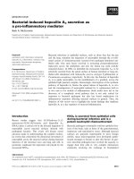
a. CT scan of the neck, following contrast administrationFigure 1
a. CT scan of the neck, following contrast administration. Axial section of the level of the oropharynx, demonstrates
the horizontal extension of the right ICA towards the midline and behind the oropharynx. b. Multiplanar reconstruction at the
coronal plane demonstrates an angiographic appearance of the vessels of the neck, showing the ectopic portion of the right
ICA.
Head & Face Medicine 2008, 4:20 />Page 3 of 4
(page number not for citation purposes)
shaped curve are classified as tortuosity, while any sharp
bend in the vessel is classified as kinking. The causes of
this malformation are atherosclerosis as observed in our
patient, and congenital deformity. The mean age at diag-
nosis is 58 years, and the patients are usually asympto-
matic.
While the reports of fatal posttonsillectomy hemorrhage
and the dissections of Kelly clearly describe the unusual
laterally placed of the ICA, midline carotid arteries are
even less commonly reported [8]. Kelly noted that only
four of his 150 patients had posterior pharyngeal wall pul-
sation. In addition, there are two reports of cases of pro-
fuse postadenoidectomy hemorrhage due to laceration of
a midline ICA. Mc Kenzie et al described two fatal cases
coarsening ICA injuries during adenoidectomy, one of
which resulted in complete arterial ablation [9]. Bergqvist
described a visible ICA in the nasopharynx that had not
been detected preoperatively but was seen intraopera-
tively after an adenoidectomy had been performed [10].
Ectopic ICAs should be differentiated from other vascular
lesions, such as angiosarcoma and hemangiopericytoma.
Peritonsillar abscess, masses as lymphomas, and other
tumors must be take under consideration, when a panic-
ula in the oropharynx is detected.
We prefer the use of CT or MRI since they are less invasive
than angiogram and provide spatial information about
the adjacent pharyngeal anatomy. In MRA the resolution
of details is not as precise as in angiograms and imaging
artifacts due to turbulent flow or patient movement may
be a major limitation. Another one examination for the
evaluation of carotid vessels is the EcoColorDoppler
(ECD), which is easy to perform, and gives quick and
important information that MRI and CT do not provide
(velocimetry, haemodynamics) [11].
Transposition of the ICA bulging the posterior pharyngeal
wall constitutes a risk factor for impressive intraoperative
and postoperative hemorrhage in surgical procedure such
as adenoidectomy, tonsillectomy, uvulopalatopharyngo-
plasty and incision of peritonsillar abscess, which are
often performed by young and inexperienced ENT doc-
tors. The surgeon should be careful in performing routine
surgical procedures in the area of the upper pharynx,
which generally represent the most frequent interventions
carried out by inexperienced surgeons as the first steps of
their surgical training. The hidden presence of an asymp-
tomatic anomaly of the internal carotid artery may cause
impressive and life-threatening hemorrhage. In the litera-
ture is reported a massive blood loss during tonsillectomy
Magnetic Resolution Angiography after gadolinium administration shows the helicoids-ectopic course of the right ICA, immedi-ately after the carotid bulbFigure 2
Magnetic Resolution Angiography after gadolinium administration shows the helicoids-ectopic course of the
right ICA, immediately after the carotid bulb. Notice also, the significant stenosis of the controlateral left ICA.
Publish with Bio Med Central and every
scientist can read your work free of charge
"BioMed Central will be the most significant development for
disseminating the results of biomedical research in our lifetime."
Sir Paul Nurse, Cancer Research UK
Your research papers will be:
available free of charge to the entire biomedical community
peer reviewed and published immediately upon acceptance
cited in PubMed and archived on PubMed Central
yours — you keep the copyright
Submit your manuscript here:
/>BioMedcentral
Head & Face Medicine 2008, 4:20 />Page 4 of 4
(page number not for citation purposes)
in a child with congenital vascular malformation of the
lips and the oropharynx [12].
In our case the referring physician thought that panicula
in the lateral wall of oropharynx was edema. The otolaryn-
gologists surgeons must use caution in evaluating patients
with masses in the pharynx and augment a careful and
complete head and neck examination with appropriate
imaging studies before operating. A thorough ocular and
digital exploration of the pharynx for arterial pulsations
should never be omitted.
Acknowledgements
Publication of the manuscript was consented by the patient.
References
1. Glasscock ME III, Dickins JRE, Jackson CG, Wiet rj: : Vascular
anomalies of the middle ear. Laryngoscope 1980, 90:77-88.
2. Moore KL: The developing human, Clinically Oriented Embri-
ology W.B. Saunders Company. Philadelphia 1973:256-257.
3. Quint DJ, Boulos RS, Spera TD: Congenital absence of the cervi-
cal and petrous internal carotid artery with intracavernous
anastomosis. Am J Neurosci 1989, 10:435-439.
4. Puzzolo D, Micali A: Embryological considerations on a multi-
ple vascular anomaly in a child. Ital J Anat Embryol 1995,
100:125-33.
5. Quint DJ, Silbergleit R, Young WC: Absence of the carotid canals
at skull base. Radiology 1992, 182:477-481.
6. Clarós P, Bandos R, Gilea I, Clarós A Jr, Capdevila A, García
Rodríguez J, Clarós A: Major congenital anomalies of the inter-
nal carotid artery: agenesis, aplasia and hypoplasia. Int J Pedi-
atr Otor 1999, 49:69-76.
7. Leipzig TJ, Dohrmann GJ: The tortuous or kinked carotid artery:
pathogenesis and clinical consideration-a historical review.
Surg Neurol 1986, 25:478-486.
8. Kelly AB: Tortuosity of the internal carotid artery in the rela-
tion to the pharynx. J Laryngol Otol 1925, 40:15-23.
9. Mc Kenzie W, Woolf CI: Carotid abnormalities and adenoid
surgery. J Laryngol Otol 1959, 73:596-602.
10. Bergqvist B: Anomalies in the course of arteria carotid inter-
nal in the upper region of the pharynx. Acta Otolaryngol 1946,
34:246-255.
11. Docimo L, Papagno P, Topatino A, Sparavigna L, Di Sapio M, Amoroso
V, Verde I, Capuano P, Manzi F, Docimo G, Rizzo R: Eco-color-Dop-
pler venous catheterization of internal jugular vein in obese
patient. Ann Ital Chir 2006, 77(2):123-6.
12. Foley PJ, Beste DJ, Farber NE: Massive blood loss during tonsil-
lectomy in a child with congenital venous malformation. Pae-
diatr Anaesth 1997, 7:243-246.