báo cáo khoa học: " In vitro evaluation of various bioabsorbable and nonresorbable barrier membranes for guided tissue regeneration" pptx
Bạn đang xem bản rút gọn của tài liệu. Xem và tải ngay bản đầy đủ của tài liệu tại đây (833.99 KB, 8 trang )
BioMed Central
Page 1 of 8
(page number not for citation purposes)
Head & Face Medicine
Open Access
Research
In vitro evaluation of various bioabsorbable and nonresorbable
barrier membranes for guided tissue regeneration
Adrian Kasaj*
1
, Christoph Reichert
1
, Hermann Götz
2
, Bernd Röhrig
3
,
Ralf Smeets
4
and Brita Willershausen
1
Address:
1
Department of Operative Dentistry and Periodontology, Johannes Gutenberg University, Mainz, Germany,
2
Institute of Applied
Structure and Microanalysis, Medical Faculty, Johannes Gutenberg University, Mainz, Germany,
3
Institute for Medical Biostatistics, Epidemiology
and Informatics, Johannes Gutenberg University, Mainz, Germany and
4
Department of Oral and Maxillofacial Surgery, Aachen University,
Germany
Email: Adrian Kasaj* - ; Christoph Reichert - ; Hermann Götz - ;
Bernd Röhrig - ; Ralf Smeets - ; Brita Willershausen -
* Corresponding author
Abstract
Background: Different types of bioabsorbable and nonresorbable membranes have been widely
used for guided tissue regeneration (GTR) with its ultimate goal of regenerating lost periodontal
structures. The purpose of the present study was to evaluate the biological effects of various
bioabsorbable and nonresorbable membranes in cultures of primary human gingival fibroblasts
(HGF), periodontal ligament fibroblasts (PDLF) and human osteoblast-like (HOB) cells in vitro.
Methods: Three commercially available collagen membranes [TutoDent
®
(TD), Resodont
®
(RD)
and BioGide
®
(BG)] as well as three nonresorbable polytetrafluoroethylene (PTFE) membranes
[ACE (AC), Cytoplast
®
(CT) and TefGen-FD
®
(TG)] were tested. Cells plated on culture dishes
(CD) served as positive controls. The effect of the barrier membranes on HGF, PDLF as well as
HOB cells was assessed by the Alamar Blue fluorometric proliferation assay after 1, 2.5, 4, 24 and
48 h time periods. The structural and morphological properties of the membranes were evaluated
by scanning electron microscopy (SEM).
Results: The results showed that of the six barriers tested, TD and RD demonstrated the highest
rate of HGF proliferation at both earlier (1 h) and later (48 h) time periods (P < 0.001) compared
to all other tested barriers and CD. Similarly, TD, RD and BG had significantly higher numbers of
cells at all time periods when compared with the positive control in PDLF culture (P ≤ 0.001). In
HOB cell culture, the highest rate of cell proliferation was also calculated for TD at all time periods
(P < 0.001). SEM observations demonstrated a microporous structure of all collagen membranes,
with a compact top surface and a porous bottom surface, whereas the nonresorbable PTFE
membranes demonstrated a homogenous structure with a symmetric dense skin layer.
Conclusion: Results from the present study suggested that GTR membrane materials, per se, may
influence cell proliferation in the process of periodontal tissue/bone regeneration. Among the six
membranes examined, the bioabsorbable membranes demonstrated to be more suitable to
stimulate cellular proliferation compared to nonresorbable PTFE membranes.
Published: 14 October 2008
Head & Face Medicine 2008, 4:22 doi:10.1186/1746-160X-4-22
Received: 1 August 2008
Accepted: 14 October 2008
This article is available from: />© 2008 Kasaj et al; licensee BioMed Central Ltd.
This is an Open Access article distributed under the terms of the Creative Commons Attribution License ( />),
which permits unrestricted use, distribution, and reproduction in any medium, provided the original work is properly cited.
Head & Face Medicine 2008, 4:22 />Page 2 of 8
(page number not for citation purposes)
Background
The final goal of periodontal therapy is to control perio-
dontal tissue inflammation and to produce predictable
regeneration of periodontium lost as a result of periodon-
tal disease. In order to promote the regeneration of the
periodontium the appropriate positioning of cells capable
of synthesizing collagen, cementum and bone is required.
The procedure of guided tissue regeneration (GTR) was
developed to ensure that regenerative potential cells such
as periodontal ligament (PDL) cells, bone cells, and
cementoblasts selectively repopulate the periodontal
wound area by using a physical barrier to exclude the
unwanted re-growth of the gingival epithelium and con-
nective tissue cells [1,2]. Various types of materials have
been tested for their effectiveness as barriers including
millipore filters, expanded polytetrafluoroethylene
(ePTFE) membranes, collagen membranes, and polylactid
acid membranes [1,3,4]. Several clinical studies have
demonstrated significant reductions in periodontal prob-
ing depth and gains in clinical attachment level following
GTR therapy using bioabsorbable and nonresorbable bar-
rier membranes [5-7]. However, several problems have
been associated with the use of nonresorbable barrier
mebranes, especially the need for a second-step surgery to
remove the membrane. Furthermore, early spontaneous
exposure to the oral environment and subsequent bacte-
rial colonization have been reported to be common prob-
lems of nonresorbable membranes resulting in lower
probing attachment level gains in intrabony defects [8]. In
order to overcome these issues, a variety of bioabsorbable
materials, such as polylactid and polyglycolic acids or col-
lagen have been used as membrane barriers [9]. Barrier
materials derived from type I and III porcine or bovine
collagen demonstrated their usefulness in GTR procedures
[10-12]. However, several complications such as early
membrane degradation, epithelial downgrowth and pre-
mature loss of the material were reported following the
use of collagen materials [1]. Furthermore, a recent in vitro
study has pointed out that native as well as cross-linked
membranes derived from bovine or porcine type I and III
collagens limited attachment and proliferation of human
PDL cells and human SaOs-2 osteoblasts as compared to
cells plated on culture dishes [13]. Although, the use of
collagen membranes seems to be a commonly used pro-
cedure, it still remains unknown how these barriers, per
se, affect the cells around the periodontium. In vitro assays
with human PDL cells, gingival fibroblasts and human
osteoblast-like cells suggest a proper model for studying
the interactions of these cells with biomaterials.
The use of radioisotopes (e.g.,
51
Cr) or radiolabelled bio-
chemicals (e.g.,
3
H-thymidine) have been widely used in
cell proliferation studies [14,15]. However, the main
drawbacks of these techniques are the potentially hazard-
ous radioactivity and the labor intensiveness. In this
study, the proliferation rate and viability of cells was
assessed by means of the non-radioactive and non toxic
Alamar Blue (AB) assay.
The purpose of the present investigation was to determine
the biological effects of various commercially available
bioabsorbable membranes made of collagen and nonre-
sorbable membranes in cultures of human gingival
fibroblasts, periodontal ligament fibroblasts and human
osteoblast-like cells. In particular, we assessed the prolif-
eration rate/cell viability and the morphology of the
membranes by scanning electron microscopy (SEM).
Methods
Membranes examined
Six commercially available membranes with different
compositions and structures were examined in this study:
(1) ACE (AC) (non-textured polytetrafluoroethylene
(PTFE); ACE Surgical Supply Co., Brockton, USA), (2)
Cytoplast
®
Regentex GBR-200 (CT) (high-density poly-
tetrafluoroethylene (d-PTFE); Oraltronics
®
Dental
Implant Technology GmbH, Bremen Germany), (3) Tef-
Gen-FD
®
(TG) (nano-porous polytetrafluoroethylene (n-
PTFE); Lifecore Biomedical GmbH, Alfter, Germany), as
well as the bioabsorbable barriers (4) Resodont
®
(RD)
(equine type I collagen; Resorba
®
, Nurnberg, Germany),
(5) BioGide
®
(BG) (porcine type I and III collagen;
Geistlich Biomaterials, Wolhusen, Switzerland), (6)
TutoDent
®
(TD) (bovine type I collagen; Tutogen Medical
GmbH, Neunkirchen, Germany).
Cell cultures
Periodontal and gingival fibroblasts were obtained from
healthy human periodontal tissues isolated from third
molars extracted for orthodontic reasons in three young
volunteers (two males and one female aged from 14 to 18
years). Prior to extraction, patients were informed about
the study and agreed to experimental use of the extracted
teeth. PDL fibroblasts were obtained from the PDL
remaining attached to extracted molars, whereas gingival
fibroblasts were obtained from loose gingival tissue that
was free of epithelium and associated alveolar bone. Gin-
gival and PDL fibroblasts from each subject were cultured
under identical conditions. In brief, tissue explants were
maintained in DMEM (Invitrogen, Carlsbad, CA, USA)
containing 1% penicillin/streptomycin (Invitrogen,
Carlsbad, CA, USA), 1% fungizone (Sigma, St. Louis, MO,
USA) and 10% fetal bovine serum (FBS; PAA, Pasching,
Austria). Within 3 weeks the tissue explants were success-
fully forming primary cultures with a sufficient number of
new cells. Cultures were incubated in a humidified atmos-
phere of 5% CO
2
and 95% air. Tissue culture medium was
changed every 2 days until confluence was reached and
cells were passaged at a 1 : 2 split ratio following trypsini-
zation with 0.05% trypsin (Invitrogen, Carlsbad, CA,
Head & Face Medicine 2008, 4:22 />Page 3 of 8
(page number not for citation purposes)
USA). Cell cultures were also tested regularly to be free of
mycoplasma and cell growth was monitored by phase-
contrast microscopy. In order to investigate whether the
cells were not merely gingival fibroblasts, cells were tested
for alkaline phosphatase (ALP). Since the cell lysates of
the various PDL fibroblast isolations yielded a strong and
over multiple cell passages stable ALP signal as compared
to the gingival fibroblasts, it was assumed that the cells
were indeed periodontal fibroblasts. The PDL and gingi-
val fibroblasts were used for the experiments between the
fourth and ninth passages. All experiments were per-
formed in triplicate using cells prepared from three differ-
ent donors.
Primary human osteoblasts (HOB) were purchased from
PromoCell
®
(Heidelberg, Germany) and cultured as rec-
ommended by the supplier in Osteoblast Growth
Medium (PromoCell) encompassing 10% foetal calf
serum. The cells were originally isolated from human
trabecular bone obtained during hip replacement surger-
ies. HOB cells were used in 4–9 passage in experiments.
Each of the barrier membranes was trimmed to an approx-
imate size of 3 × 3 mm, immersed in cell culture medium
for 5 minutes and adapted on the floor of the wells with a
double-faced adhesive tape. Two inserts for each mem-
brane were used for one assay. In order to ensure repro-
ducibility, all experiments were repeated thrice with three
replicates each. In case of the bilayered RD, BG and TD
membranes, cells were cultivated on the porous surface.
Cells plated on culture dishes (CD) served as positive con-
trols.
AlamarBlue™ proliferation assay
Former experiments (data not shown) were carried out to
measure Alamar Blue (AB) reduction over time. The aim
was to determine optimal seeding density and culture
period. HGF, PDLF and HOB cells were trypsinized after
serum starvation and suspended into standard culture
medium with 10% FBS. HGF and PDLF were seeded into
a 96-well plate with a density of 2,5 × 10
3
/well and further
incubated under standard cultivation conditions (37°C,
95% air, 5% CO
2
). After an initial 4 h incubation to allow
cellular attachment for HGF and PDLF, AB solution was
added directly in a final concentration of 10% and the
plate was further incubated. Optical density of the plate
was measured at a wavelength of 560/20 and 620/40 nm
with a fluorescence reader (FLx800 Microplate Fluores-
cence Reader, BioTek Instruments, Vermont, USA) at 1,
2.5, 4, 24 and 48 h after adding AB. The logarithmic sig-
nals were converted to values on a linear scale and
expressed as relative fluorescence units (RFU) to calculate
mean fluorescence. As a negative control, AB was added to
the medium without cells. The same experimental setup
was determined for HOB cells in the same density of 2,5
× 10
3
/well but with an initial adhesion time of 24 h. All
samples were tested in triplicate.
SEM examination
The scanning electron microscope (SEM) was used to
study the structure and surface morphology of the mem-
branes. Images were obtained by detecting the signal of
secondary electrons emitted by the sample when hit by
the incident electron beam.
Statistical analysis
All statistical analyses were performed using statistical
software SPSS
®
(Version 12.0, for Windows, Chicago, IL,
USA). Statistical analysis was performed for each cell
group (HGF, PDLF and HOB) separately. To figure out
netto fluorescence the autofluorescence of the tested
materials was substracted from the raw data of AB. Mean
and standard deviation (SD) were calculated for each
group. Proliferation for all groups and points of time was
shown graphically with a plot (abscissa: point of time,
ordinate: proliferation). In order to find the best mem-
brane, all six relevant membranes were compared to the
control (CD). If a relevant membrane was in the statistical
test significant better than CD, a post-test was performed.
If more than two membranes were selected a post-hoc
Scheffé test was performed. All statistical tests included all
points of time and a General Linear Model (GLM) with
repeated measures was used. The outcome of a statistical
test was considered to be significant when P < 0.05.
Results
During the experimental period, there was no evidence
indicating any bacterial or fungal contamination of the
well chambers. The effect of the barrier membranes on
HGF, PDLF and HOB cell proliferation was counted by
the AB fluorometric proliferation assay after 1, 2.5, 4, 24
and 48 h time periods in vitro. The rate of cell proliferation
with time was different among the membranes examined.
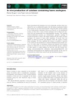
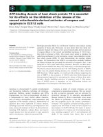
Of the six barriers tested, TD and RD demonstrated the
highest rate of HGF proliferation at both earlier (1 h) and
later (48 h) time periods compared to CD (P < 0.001). In
comparison with the positive control, BG, TG, CT and AC
showed statistically fewer cells (P < 0.05) at all points of
time. Furthermore, TD showed significantly increased
number of cells at 1, 2.5, 4, 24 and 48 h compared to RD
(P < 0.001). Cell proliferation at 48 h was as follows: TD
(3064.3 ± 29.3) > RD (1724.3 ± 22.1) > CD (1358.7 ±
29.1) > CT (1196.7 ± 4.2) > AC (1171.7 ± 13.8) > TG
(1156.3 ± 5.8) > BG (1033.7 ± 7.4) (Fig. 1).
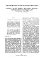
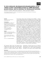
In PDLF culture, TD, RD and BG had significantly higher
numbers of cells at all time periods when compared with
the positive control (P ≤ 0.001). The nonresorbable mem-
branes TG, CT and AC demonstrated significantly fewer
cells compared to CD and all the tested collagen mem-
Head & Face Medicine 2008, 4:22 />Page 4 of 8
(page number not for citation purposes)
branes at all points of time (P < 0.001). Furthermore, RD
and BG exhibited significantly fewer cells than TD at all
time periods (P < 0.001). After 48 h cell proliferation in
PDLF culture was as follows: TD (2791.7 ± 15.5) > RD
(1726.3 ± 8.3) > CD (1432.3 ± 35.8) > BG (1399.0 ± 2.6)
> AC (1342.7 ± 25.0) > CT (1316.0 ± 27.0) > TG (1167.7
± 20.1) (Fig. 2).
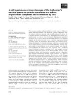
In HOB cell culture, TD, RD, TG and AC had significantly
higher numbers of cells at all time periods when com-
pared with the positive control (P < 0.05). The highest rate
of cell proliferation was calculated for TD at all time peri-
ods. This was followed by RD, AC and TG with statistically
significant fewer cells (P < 0.001). BG showed the least
number of cells among all membranes, both at 24 h and
48 h. At 48 h following cell counts were calculated: TD
(2389.7 ± 18.6) > AC (1903.0 ± 34.6) > RD (1809.0 ± 9.0)
> CT (1739.0 ± 38.6) > TG (1738.7 ± 20.4) > CD (1447.0
± 13.7) > BG (1405.7 ± 5.9) (Fig. 3).
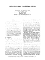
SEM observations showed that all collagen membranes
were microporous, with a compact top surface and a
porous bottom surface (Figs. 4a–c). In contrast, the non-
resorbable PTFE membranes demonstrated a homoge-
nous structure with a symmetric dense skin layer (Figs.4d–
f).
Discussion
The principle of guided tissue regeneration (GTR) is uti-
lized to exlude epithelium from the root surfaces and to
promote selective repopulation of the root surface by
multipotential cells. The main goal of the present study
was to investigate the compatibility of various barrier
membranes in human cell cultures, which are comparable
to the regenerative cells of the periodontium. Further-
more, barrier membrane surfaces were examined by SEM.
The proliferative capacity of primary human periodontal
and gingival fibroblasts as well as human osteoblast-like
cells were examined by the fluorometric AB assay. AB con-
Effects of various membranes on proliferation of human gingival fibroblasts (HGF) after 1, 2.5, 4, 24 and 48 hFigure 1
Effects of various membranes on proliferation of human gingival fibroblasts (HGF) after 1, 2.5, 4, 24 and 48 h.
Cells were incubated in the presence of 10% Alamar Blue. Fluorescence was measured in a microplate fluorescence reader,
and is presented as relative fluorescence units (RFU). CD: culture dishes; BG: BioGide
®
; RD: Resodont
®
; TD: TutoDent
®
; TG:
TefGen-FD
®
; CT: Cytoplast
®
; AC: ACE.
Head & Face Medicine 2008, 4:22 />Page 5 of 8
(page number not for citation purposes)
tains an oxidation-reduction indicator that both fluo-
resces and changes color in response to the chemical
reduction by cell metabolism. The AB assay is considered
superior to other cell viability assays, because it is non-
toxic to cells and does not necessitate killing the cells dur-
ing the assay procedure [16]. Moreover, the AB assay is
comparable in sensitivity to the thymidine incorporation
and tetrazolium reduction assays for the measurement of
cell proliferation [17]. Previously, this assay has been used
for measuring the proliferation of human lymphocytes
[16], primary rat hepatocytes [18] and human fibroblasts
cells [19].
Within the limits of this in vitro study, the number of pro-
liferated gingival fibroblasts was the highest on the bioab-
sorbable collagen membrane TD, followed by RD. Similar
results were noted for the mean number of proliferated
PDL fibroblasts, which was greatest on TD, followed by
RD and BG. The mean number of HOB cells was also
greatest on TD, followed by RD, AC and TG. Thus, it may
be assumed that the tested collagen membranes enhanced
cell proliferation of human gingival and periodontal liga-
ment fibroblasts and human osteoblast-like cells, whereas
nonresorbable PTFE membranes limited cell prolifera-
tion. These findings correspond well with data from pre-
vious studies evaluating the growth of HGF, PDLF and
HOB cells on various GTR membranes [20-22]. Locci et al.
[20] demonstrated that matrix membranes composed of
collagen and chondroitin glycosaminoglycan enhanced
cellular proliferation and extracellular macromolecule
accumulation. In addition, it was found that PTFE mem-
branes inhibited gingival fibroblast DNA synthesis and
caused a marked decrease in synthesis of extracellular col-
lagen and glycosaminoglycan, the major components of
extracellular matrix. The authors proposed that collagen
might be more suitable than PTFE membranes to achieve
periodontal regeneration. Indeed, it is well known that
collagen favors the adhesion to the substrate of various
cell types, permits the in vitro maintenance of cells over a
long period of time and stimulates cell proliferation [23].
Alpar et al. [21] evaluated the cytocompatibility of resorb-
able and nonresorbable membranes in human periodon-
tal ligament fibroblast and osteoblast-like cell cultures. It
was reported that the collagen barriers exhibited high
Number of periodontal ligament cells (PDLF) on various membranes examined after 1, 2.5, 4, 24 and 48 hFigure 2
Number of periodontal ligament cells (PDLF) on various membranes examined after 1, 2.5, 4, 24 and 48 h.
Abbrevations are specified in the legend of Figure 1.
Head & Face Medicine 2008, 4:22 />Page 6 of 8
(page number not for citation purposes)
cytocompatibility, whereas PTFE and polylactic acid
membranes induced slight to moderate cytotoxic reac-
tions. Marinucci et al. [22] investigated cell proliferation
on human osteoblasts and found that collagen stimulated
DNA synthesis more than ePTFE. In contradiction to our
data, Rothamel et al. [13] noted that the mean number of
human PDL fibroblasts and human osteosarcoma-derived
SaOs-
2
cells was the highest on CD as compared to four
collagen membranes. It was reported that TD and BG
exhibited significantly fewer cells in PDLF and SaOs-
2
cul-
ture in comparison with the positive control. However,
discrepancies noted in these results may be explained by
differences in cell characteristics as well as the different
assays used to measure proliferative activity. Further stud-
ies are needed to clarify which specific factor has more
effect on cell proliferation. In this context, it has to be
pointed out that there are no previously published data
using HGF, PDLF as well as HOB cells simultaneously to
evaluate the growth of these cells on various membranes.
Our data indicated that the nonresorbable PTFE mem-
branes limited cell proliferation. This findings correspond
well with the results of Payne et al. [24]. They demon-
strated that ePTFE membranes inhibited migration of
human gingival fibroblasts and induced cell death. These
observations indicate that those materials may be respon-
sible for impaired tissue integration in vivo in comparison
to collagen membranes. Although minimal tissue integra-
tion to ePTFE membranes may be an advantage for mem-
brane retrieval, it may also create potential problems for
initial clot formation, wound stabilization and mem-
brane stability.
Although TD, RD and BG were all belonging to collagen
devices, cell proliferation was different on these mem-
branes. Thus, cell proliferation of HGF, PDLF and HOB on
BG was less compared to the other two collagen barriers
TD and RD throughout the experimental period. The dif-
ference in surface topography, surface characteristics and
pore sizes may account for the different effects on cell pro-
liferation. These findings corroborate with our SEM obser-
vations demonstrating varieties in the porous structure
and surface roughness between the different collagen
membranes. Moreover, the discrepancies noted between
Effects of various bioabsorbable and nonresorbable membranes on proliferation of human osteoblast-like (HOB) cells after 1, 2.5, 4, 24 and 48 h of incubationFigure 3
Effects of various bioabsorbable and nonresorbable membranes on proliferation of human osteoblast-like
(HOB) cells after 1, 2.5, 4, 24 and 48 h of incubation. Abbrevations are given in the legend of Figure 1.
Head & Face Medicine 2008, 4:22 />Page 7 of 8
(page number not for citation purposes)
the collagen membranes may be explained by differences
in dissolution of the membrane material as suggested by
Zhao et al. [25]. They evaluated histologically different
biodegradable and non-biodegradable membranes
implanted subcutaneously in rats and found that BG was
dissolved in the early phase with a profound giant cell and
inflammatory reaction. These findings imply that BG
might inhibit regeneration of periodontal tissues due to
the early fragmentation and the inflammatory reaction of
the material. Further confirmation of this hypothesis is
required.
One must be cautious when interpreting results obtained
by using in vitro experimental model, since it can not rec-
reate the complex interactions of cells in vivo. Further lim-
itations in this study include the short study period.
Future studies should include a longer follow-up period.
Within the limits of the present study, it was concluded
that GTR membrane materials, per se, may influence cell
proliferation in the process of periodontal tissue/bone
regeneration. Among the six membranes examined, the
bioabsorbable membranes demonstrated to be more suit-
able to stimulate cellular proliferation compared to non-
resorbable membranes.
Competing interests
The authors declare that they have no competing interests.
Authors' contributions
The study design was established by BW and AK, who also
wrote the manuscript. CR carried out the in-vitro experi-
ments. The SEM analyses were undertaken by HG. BR per-
formed the data management and data analysis. RS
carried out the manuscript editing and manuscript review.
All authors read and approved the final version of the
manuscript.
Acknowledgements
The authors would like to thank Cornelia Metz from the Department of
Operative Dentistry and Periodontology, University Hospital Mainz, Ger-
many, for her excellent technical assistance during the whole project.
This project was supported by a grant (MAIFOR 135/2007) from the Uni-
versity Mainz, medical section, for the promotion of medical research, Ger-
many.
References
1. Gottlow J: Guided tissue regeneration using bioresorbable
and non-resorbable devices: initial healing and long-term
results. J Periodontol 1993, 64(11 Suppl):1157-1165.
2. Tatakis DN, Promsudthi A, Wikesjö UM: Devices for periodontal
regeneration. Periodontol 2000 1999, 19:59-73.
3. Teparat T, Solt CW, Claman LJ, Beck FM: Clinical comparison of
bioabsorbable barriers with non-resorbable barriers in
Surface SEM micrographs of the examined bioabsorbable and nonresorbable membranes: (a) TutoDent
®
, (b) Resodont
®
, (c) BioGide
®
, (d) TefGen-FD
®
, (e) Cytoplast
®
, and (f) ACEFigure 4
Surface SEM micrographs of the examined bioabsorbable and nonresorbable membranes: (a) TutoDent
®
, (b)
Resodont
®
, (c) BioGide
®
, (d) TefGen-FD
®
, (e) Cytoplast
®
, and (f) ACE.
Publish with Bio Med Central and every
scientist can read your work free of charge
"BioMed Central will be the most significant development for
disseminating the results of biomedical research in our lifetime."
Sir Paul Nurse, Cancer Research UK
Your research papers will be:
available free of charge to the entire biomedical community
peer reviewed and published immediately upon acceptance
cited in PubMed and archived on PubMed Central
yours — you keep the copyright
Submit your manuscript here:
/>BioMedcentral
Head & Face Medicine 2008, 4:22 />Page 8 of 8
(page number not for citation purposes)
guided tissue regeneration in the treatment of human
intrabony defects. J Periodontol 1998, 69:632-641.
4. Giardino R, Fini M, Nicoli Aldini N, Giavaresi G, Rocca M: Polylac-
tide bioabsorbable polymers for guided tissue regeneration.
J Trauma-Injury Infection Crit Care 1999, 47:303-308.
5. Cortellini P, Prato GP, Tonetti MS: Periodontal regeneration of
human intrabony defects with bioresorbable membranes. A
controlled clinical trial. J Periodontol 1996, 67:217-223.
6. Weigel C, Brägger U, Hämmerle CH, Mombelli A, Lang NP: Mainte-
nance of new attachment 1 and 4 years following guided tis-
sue regeneration (GTR). J Clin Periodontol 1995, 22:661-669.
7. Machtei EE, Grossi SG, Dunford R, Zambon JJ, Genco RJ: Long-term
stability of Class II furcation defects treated with barrier
membranes. J Periodontol 1996, 67:523-527.
8. Selvig KA, Kersten BG, Chamberlain AD, Wikesjö UM, Nilveus RE:
Regenerative surgery of intrabony periodontal defects using
ePTFE barrier membranes: scanning electron microscopic
evaluation of retrieved membranes versus clinical healing. J
Periodontol 1992, 63:974-978.
9. Lorenzoni M, Pertl C, Keil C, Wegscheider WA: Treatment of
peri-implant defects with guided bone regeneration: a com-
parative clinical study with various membranes and bone
grafts. Int J Oral Maxillofac Implants 1998, 13:639-646.
10. Minabe M, Kodama T, Kogou T, Tamura T, Hori T, Watanabe Y, Miy-
ata T: Different cross-linked types of collagen implanted in rat
palatal gingiva. J Periodontol 1989, 60:35-43.
11. Christgau M, Bader N, Schmalz G, Hiller KA, Wenzel A: Postoper-
ative exposure of bioresorbable GTR membranes: effect on
healing results. Clin Oral Investig 1997, 1:109-118.
12. Eickholz P, Lenhard M, Benn DK, Staehle HJ: Periodontal surgery
of vertical bony defects with or without synthetic bioabsorb-
able barriers. 12-month results. J Periodontol 1998,
69:1210-1217.
13. Rothamel D, Schwarz F, Sculean A, Herten M, Scherbaum W, Becker
J: Biocompatibility of various collagen membranes in cul-
tures of human PDL fibroblasts and human osteoblast-like
cells. Clin Oral Implants Res
2004, 15(4):443-449.
14. Schlager SI, Adams AC: Use of dyes and radioisotopic markers
in cytotoxicity tests. Methods Enzymol 1983, 93:233-245.
15. Bilir A, Altinoz MA, Attar E, Erkan M, Aydiner A: Acetaminophen
modulations of chemotherapy efficacy in MDAH 2774
human endometrioid ovarian cancer cells in vitro. Neoplasma
2002, 49:38-42.
16. Ahmed SA, Gogal RM Jr, Walsh JE: A new rapid and simple non-
radioactive assay to monitor and determine the prolifera-
tion of lymphocytes: an alternative to [3H] thymidine incor-
poration assay. J Immunol Methods 1994, 170:211-224.
17. Page B, Page M, Noel C: A new fluorimetric assay for cytotoxic-
ity measurements in vitro. Int J Oncol 1993, 3:473-476.
18. O'Brien J, Wilson I, Orton T, Pognan F: Investigation of the AB
(resazurin) fluorescent dye for the assessment of mamma-
lian cell cytotoxicity. Eur J Biochem 2000, 267:5421-5426.
19. Saunders RE, Gough JE, Derby B: Delivery of human fibroblast
cells by piezoelectric drop-on-demand inkjet printing. Bioma-
terials 2008, 29:193-203.
20. Locci P, Calvitti M, Belcastro S, Pugliese M, Guerra M, Marinucci L,
Staffolani N, Becchetti E: Phenotype expression of gingival
fibroblasts cultured on membranes used in guided tissue
regeneration. J Periodontol 1997, 68:857-863.
21. Alpar B, Leyhausen G, Gunay H, Geurtsen W: Compatibility of
resorbable and nonresorbable guided tissue regeneration
membranes in cultures of primary human periodontal liga-
ment fibroblasts and human osteoblast-like cells. Clin Oral
Investig 2000, 4:219-225.
22. Marinucci L, Lilli C, Baroni T, Becchetti E, Belcastro S, Calducci C,
Locci P: In vitro comparison of bioabsorbable and non-resorb-
able membranes in bone regeneration. J Periodontol 2001,
72:753-759.
23. Royce PM, Barnes MJ: Interaction of embryonic chick calvarial
bone cells with collagen substrata; attachment characteris-
tics and growth behaviour.
Connect Tissue Res 1988, 17:55-70.
24. Payne JM, Cobb CM, Rapley JW, Killoy WJ, Spencer P: Migration of
human gingival fibroblasts over guided tissue regeneration
barrier materials. J Periodontol 1996, 67:236-244.
25. Zhao S, Pinholt EM, Madsen JE, Donath K: Histological evaluation
of different biodegradable and non-biodegradable mem-
branes implanted subcutaneously in rats. J Craniomaxillofac Surg
2000, 28:116-122.