báo cáo khoa học: " Behavior of osteoblastic cells cultured on titanium and structured zirconia surfaces" ppsx
Bạn đang xem bản rút gọn của tài liệu. Xem và tải ngay bản đầy đủ của tài liệu tại đây (494.59 KB, 9 trang )
BioMed Central
Page 1 of 9
(page number not for citation purposes)
Head & Face Medicine
Open Access
Research
Behavior of osteoblastic cells cultured on titanium and structured
zirconia surfaces
Rita Depprich
1
, Michelle Ommerborn*
2
, Holger Zipprich
3
,
Christian Naujoks
†1
, Jörg Handschel
†1
, Hans-Peter Wiesmann
4
,
Norbert R Kübler
1
and Ulrich Meyer
1
Address:
1
Department of Cranio- and Maxillofacial Surgery, Heinrich-Heine-University, Düsseldorf, Germany,
2
Department of Operative and
Preventive Dentistry and Endodontics, Heinrich-Heine-University, Düsseldorf, Germany,
3
Department of Prosthetic Dentistry, Section of Materials
Sciences, Johann Wolfgang Goethe University, Frankfurt, Germany and
4
Department of Cranio- and Maxillofacial Surgery, Westphalian Wilhelms-
University, Münster, Germany
Email: Rita Depprich - ; Michelle Ommerborn* - ;
Holger Zipprich - ; Christian Naujoks - ;
Jörg Handschel - ; Hans-Peter Wiesmann - ;
Norbert R Kübler - ; Ulrich Meyer -
* Corresponding author †Equal contributors
Abstract
Background: Osseointegration is crucial for the long-term success of dental implants and depends
on the tissue reaction at the tissue-implant interface. Mechanical properties and biocompatibility
make zirconia a suitable material for dental implants, although surface processings are still
problematic. The aim of the present study was to compare osteoblast behavior on structured
zirconia and titanium surfaces under standardized conditions.
Methods: The surface characteristics were determined by scanning electron microscopy (SEM).
In primary bovine osteoblasts attachment kinetics, proliferation rate and synthesis of bone-
associated proteins were tested on different surfaces.
Results: The results demonstrated that the proliferation rate of cells was significantly higher on
zirconia surfaces than on titanium surfaces (p < 0.05; Student's t-test). In contrast, attachment and
adhesion strength of the primary cells was significant higher on titanium surfaces (p < 0.05; U test).
No significant differences were found in the synthesis of bone-specific proteins. Ultrastructural
analysis revealed phenotypic features of osteoblast-like cells on both zirconia and titanium surfaces.
Conclusion: The study demonstrates distinct effects of the surface composition on osteoblasts in
culture. Zirconia improves cell proliferation significantly during the first days of culture, but it does
not improve attachment and adhesion strength. Both materials do not differ with respect to protein
synthesis or ultrastructural appearance of osteoblasts. Zirconium oxide may therefore be a suitable
material for dental implants.
Published: 8 December 2008
Head & Face Medicine 2008, 4:29 doi:10.1186/1746-160X-4-29
Received: 27 October 2008
Accepted: 8 December 2008
This article is available from: />© 2008 Depprich et al; licensee BioMed Central Ltd.
This is an Open Access article distributed under the terms of the Creative Commons Attribution License ( />),
which permits unrestricted use, distribution, and reproduction in any medium, provided the original work is properly cited.
Head & Face Medicine 2008, 4:29 />Page 2 of 9
(page number not for citation purposes)
Background
The objective of implantology is to design devices that
induce controlled, guided, and rapid integration into sur-
rounding tissues [1]. Events leading to integration of an
implant, and ultimately to success or failure of the device,
take place largely at the tissue-implant interface, and oste-
oblasts covering the implant surface are the crucial cell
type that regulate the tissue response at the biomaterial
surface [2]. Based on the results of numerous in vitro stud-
ies, it is now well understood that surface morphology
decisively determines the cellular behavior of osteoblasts
[2-4].
Titanium (Ti) and titanium alloys are widely used as
implant materials due to their excellent biocompatibility.
Many surface modifications have been developed to
improve cell reactions on the surface. In addition to exist-
ing titanium implants bearing machined or plasma-
sprayed surfaces, there is a great number of implants on
the market which offer surfaces altererd by grit blasting
and/or acid etching. Zirconia (zirconium dioxide, ZrO
2
) is
a bio-inert non-resorbable metal oxide that offers
improved mechanical properties compared to other
ceramic biomaterials, i.e. alumina. It has a good chemical
and dimensional stability, and a high strength and tough-
ness [5]. Tetragonal zirconia polycrystals (TZP) are used
for manufacturing femoral heads for total hip replace-
ments since the late 1980s [6]. Because of the tooth-like
colour, the excellent biocompatibility and mechanical
properties, ambitious efforts were made to introduce zir-
conia for applications in dentistry. Successful use of zirco-
nia for treatment of non-vital teeth [7,8], crown and
bridge restorations [9] and ceramic abutments [10] are
reported. Zirconia is also a desirable alternative material
to titanium for the fabrication of dental implants.
Titanium has a superior corrosion resistance because of its
characteristic oxide layer, however, accumulation of tita-
nium in the inner organs and lymph nodes after implan-
tation has been reported [11]. Galvanic side effects after
contact with saliva and fluoride were also described [12].
Although allergic reactions to titanium are very rare, cellu-
lar sensitization has been demonstrated [13,14]. The
main disadvantage of the biomaterial titanium is its dark
grayish colour. Unfavorable soft tissue conditions or
retraction of the gingiva may lead to aesthetic impair-
ment, especially when the maxillary incisors are involved
[15]. The clinical use of zirconia is limited, because fabri-
cation of surface modifications is difficult and smooth
implant surfaces are not beneficial for osseointegration,
due to a poor interaction with tissues [1].
Some animal experiments and numerous case reports
demonstrated osseointegration of zirconia implants simi-
lar to that of titanium implants, suggesting that zirconia
might be a suitable implant material [16-19]. However,
data evaluating the role of surface topography on the
response of osteoblasts at zirconia interfaces are rare [20].
Cell reactions on surfaces are strongly dependent on the
culture system that is used [21]. Since most of the widely
used osteosarcoma cell lines do not demonstrate a com-
plete pattern of osteoblastic features in vitro, the use of pri-
mary non-transformed cells seems to be superior for
assessing of osteoblast reactions on biomaterial surfaces
[2]. Therefore, the aim of this study was to compare oste-
oblast behavior on structured zirconia and titanium sur-
faces under standardized conditions using primary bovine
osteoblasts. Attachment kinetics, proliferation rate, and
synthesis of bone-associated proteins on both surfaces
were examined and compared between each other.
Methods
A modified (acid-etched) zirconia implant surface was
compared to an acid-etched titanium surface. Standard
24-well tissue culture plates (polystyrene) were used as
control surface. Zircona disks (12 mm diameter, 1 mm
thick) were made of yttrium-stabilized tetragonal poly-
crystals and titanium disks (13 mm diameter, 1.5 mm
thick) were made of commercially pure titanium. Both
materials were supplied by Konus Dental Implants (Bin-
gen, Germany). To evaluate the surfaces of zirconia and
titanium disks, scanning electron microscopy (SEM) was
performed using a a JEOL 6300F (JEOL, Eching, Ger-
many) high-resolution field emission scanning electron
microscope equipped with a EDX analysis system. The zir-
conia and titanium disks were carefully washed in diluted
water, rinsed thoroughly in 70% ethanol, and ultrasoni-
cally cleaned for 20 min in absolute alcohol. Finally, the
samples were air dried and maintained under sterile con-
ditions after gamma ray sterilization.
Primary osteoblast cell culture
Primary bovine osteoblasts were used in this study. Extrac-
tion and cultivation were performed following the
instructions of Jones et al. [22]. Under sterile conditions
periosteum was removed from the bovine metacarpus.
The periosteum was cultured at 37°C in an atmosphere of
5% CO
2
and 100% humidity for 4–5 weeks in high-
growth enhancement medium (High GEM, Flow Labora-
tories, Rickmansworth, UK) containing 10% fetal bovine
serum (FBS, Gibco Laboratories Grand Island, NY, USA).
Media were changed weekly. Osteoblastic differentiation
was tested by detection of osteocalcin/osteonectin and
high alkaline phosphatase activity. When the cells reached
confluence they were harvested (20 min incubation at
37°C with 0.4 g collagenase, 98.8 mg HAM's F10 in 10 ml
HEPES (2-[4-(2-hydroxyethyl)-1-piperazinyl]ethanesul-
fonic acid); repeated washing with phosphate-buffered
saline (PBS); subsequent incubation for 15 min with 300
mg ethylenediaminetetraacetic acid (EDTA)-Na, 200 mg
Head & Face Medicine 2008, 4:29 />Page 3 of 9
(page number not for citation purposes)
KCl, 8 g NaCl, 1 g NaHCO
3
, 50 mg NaH
2
PO
4
and 1 g glu-
cose/l) and centrifuged. The pellets were resuspended
with buffer and the cell numbers were counted in a cell
counter (CASY
®
I Modell TT, Schärfe System, Reutlingen,
Germany).
Cell proliferation
Cell proliferation was measured after 1, 3 and 5 days,
respectively. Cells were marked with fluorescent dye
(Vybrant
®
CM DiI, Molecular Probes, Netherlands) and
10.000/cm
2
osteoblasts were seeded into 24-well plates
on the zirconia/titanium disks or the well plate. The
experiments were repeated at least three times. Osteob-
lasts were fixed in methanol and stained with methylene
blue and azure blue according to the method described by
Richardson. Morphometric evaluation of cells was per-
formed by means of light microscopy. To determine the
cell number digital photos were taken under standardized
conditions and counted using the software program Anal-
ysis 3.0 (Olympus Soft Imaging System, Münster, Ger-
many).
Cell detachment
To determine cell adhesion on the surface of the different
materials, 60.000/cm
2
primary osteoblasts were seeded
into 24-well plates on the zirconia/titanium disks or the
well plate. After incubation for 24 hrs at 37°C, 500 μl of a
trypsin-containing solution (0.25% diluted 1:2 in PBS)
was added and 400 μl aliquots of the cell suspension were
taken after a contact time of 5, 15, 25, and 35 min. Cell
numbers were determined by the use of a cell counter. As
control, the remaining of the 500 μl was removed from
the wells and 500 μl trypsin (0.25% solution, non-
diluted) was added to detach the remaining cells. After 5
min contact time and washing with PBS, aliquots of the
cell suspension (400 μl) were taken and the cell number
counted.
Immunocytochemistry
To test for osteoblastic differentiation, expression of colla-
gen I, osteocalcin and osteonectin was assessed by means
of immunocytochemistry. 60.000 osteoblasts/cm
2
were
seeded into 24-well plates on the zirconia/titanium disks
and into 6-well plates on polystytol. After incubation for
7, 14, or 28 days at 37°C in an atmosphere of 5% CO
2
in
the High GEM medium, primary antibodies were used
according to the manufacturers' instructions: rabbit poly-
clonal anti-collagen I (Biotrend, Cologne, Germany),
Mouse monoclonal anti-osteocalcin (TaKaRa Bio,
MoBiTec, Goettingen, Germany) and rabbit polyclonal
anti-osteonectin (SPARC; Chemicon Millipore GmbH,
Schwalbach, Germany). Alexa Flour 488-labelled second-
ary antibodies were purchased from MoBiTec (Goettin-
gen, Germany) and used according to the manufacturers'
instructions. Digital images were taken under standard-
ized conditions using a fluorescence microscope and
processed using the software program Analysis 3.0.
Scanning electron microscopy (SEM)
Cell morphology was investigated after 2 hrs, 4 hrs and 7
days. Primary osteoblasts were seeded at a density of
15.000/cm
2
on zirconia/titanium disks and for control on
smooth titanium disks and incubated for 2 hrs or 4 hrs at
37°C in an atmosphere of 5% CO
2
in the High GEM
medium. To investigate confluent cells after 7 days,
40.000/cm
2
osteoblasts were seeded on the zirconia/tita-
nium disks and incubated under the same conditions.
Cells were fixed in 2.5% glutaraldehyde for 3 hrs and then
washed with PBS. After sputtering with gold (Bal-tec Ag,
Balzers, Liechtenstein) the samples were investigated
using the scanning electron microscope JEOL 6300F
(JEOL, Eching, Germany).
Statistical analysis
Statistical analyses were performed using Student's t-tests
and Mann-Whitney U tests. A p < 0.05 was considered sig-
nificant. Experiments were repeated three-fold.
Results
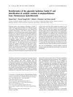
Surface topography
Scanning electron microscopy demonstrated noticeable
differences between zirconia and titanium surfaces by
SEM revealed (Figure 1). The titanium surface was rough
and contained many pores and grooves of different size
which were regularly distributed over the whole surface.
In contrast, the zirconia surface appeared smooth with
only a few pores.
Energy-dispersion X-ray analysis
Energy-dispersion X-ray analysis confirmed the character-
istic element composition of commercial pure titanium
and zirconium dioxide. Titanium disks were composed of
the elements titanium and oxygen but also traces of sili-
cium and carbon were detected. Zirconia consisted of zir-
conium (Zr) and oxygen (O), but also hafnium (Hf) was
found frequently associated with ZrO
2
.
Cell proliferation
Cell proliferation was assessed on the different surfaces.
We found an increase in cell number on all surfaces over
the observation period (Figure 2). At day 1 cell prolifera-
tion was significantly higher on zirconia surfaces as com-
pared to polystyrene controll surfaces (p = 0.000) but was
similar to titanium surfaces (p = 0.158). At day 3 cell
growth was significantly higher on the zirconia surfaces
than on polystyrene (p = 0.037) and titanium surfaces (p
= 0.002). At day 5 cell proliferation was continued to be
significantly higher on zirconia surfaces than on titanium
(p = 0.001) or polystyrene surfaces (p = 0.001).
Head & Face Medicine 2008, 4:29 />Page 4 of 9
(page number not for citation purposes)
Cell detachment
Results revealed that at every time of the assessment fewer
cells were detached from titanium surfaces compared to
zirconia or polystyrol surfaces. The number of detached
cells from titanium surfaces remained constant at a low
level over the whole period of investigation. In contrast,
detached cells from zirconia surfaces doubled from 5 to
15 min, but remained constant thereafter. A minor
Scanning electron micrographs of a zirconia disk (left) showing occasionally pores on the smooth surface and a titanium disk (right) with rough surface and frequent pores and grooves of different size (2 kV, magnification 500-fold)Figure 1
Scanning electron micrographs of a zirconia disk (left) showing occasionally pores on the smooth surface and a titanium disk
(right) with rough surface and frequent pores and grooves of different size (2 kV, magnification 500-fold).
zirconia
titanium
Cell proliferation rates of osteoblasts on differently coated surfaces at day 1, 3 and 5, respectivelyFigure 2
Cell proliferation rates of osteoblasts on differently coated surfaces at day 1, 3 and 5, respectively. Increase in cell number was
detected on all surfaces over the observation period. Significantly higher cell proliferation was observed on zirconia surfaces on
day 1, 3 and 5 compared to titanium and polystyrene surfaces. Statistical differences (p < 0.05) as calculated by Student's t-tests
are marked with arrows.
cell proliferation - day 1
0
20
40
60
80
100
120
140
160
180
200
1
number of osteoblast
polystyrene titanium zirconia
cell proliferation - day 3
0
100
200
300
400
500
1
number of osteoblasts
polystyrene titanium zirconia
cell proliferation - day 5
0
100
200
300
400
500
600
700
1
number of osteoblasts
polystyrene titanium zirconia
**
**
***
*
**
Head & Face Medicine 2008, 4:29 />Page 5 of 9
(page number not for citation purposes)
increase of detached cells was found in the polystyrol con-
trol group, and after 35 min the number of detached cells
had quadrupled. Statistical analysis confirmed significant
higher cell detachment rates from zirconia surfaces as
compared to titanium surfaces after 5 min (p = 0.047), 15
min (p = 0.009) and 25 min (p = 0.009) but not after 35
min (p = 0.1). Differences between zirconia and control
group were not significant (p < 0.05) at any time of assess-
ment.
Immunocytochemical analysis
After 7 days expression of collagen I, osteocalcin and
osteonectin were evident on all different surfaces exam-
ined. Cells were uniformly distributed throughout the
material surface and positive immunolabeling was
detected on zirconia, titanium and polystyrol surfaces.
Lower expression of osteocalcin compared to collagen I
and osteonectin was observed on all different surfaces
(Figure 3). After 14 days of culture, up-regulated expres-
sion of reticular collagen I expression was evident espe-
cially on the titanium and zirconia surfaces, whereas
osteocalcin and osteonectin expression showed no detect-
able differences on the investigated surfaces. Expression of
characteristic bone derived proteins was still detectable
after 28 days on all samples and showed no significant
differences between titanium, zirconia and polystyrol sur-
faces except of a minimally denser accumulation of colla-
gen I found on zirconia surfaces as compared to titanium
surfaces (Figure 4).
Scanning electron microscopy (SEM)
The SEM analysis performed on osteoblast-seeded sam-
ples after 2 hrs showed typically flat polygonal cells regu-
larly distributed on the titanium and on the zirconia
surfaces. Development of radiate cell filopodia was appar-
ent. After 4 hrs of culture, cell morphology on both sur-
faces showed no significant differences and was similar to
that after 2 hrs. Cell filopodia exploring the surface could
be demonstrated in fixed cells. After 7 days a mosaic-
shaped confluent cell layer had formed on zircona and
titanium surfaces (Figure 5). No ultrastructural signs of
apoptotic fibroblast-shaped cells were detected. Signifi-
cant differences could not be found.
Immunocytochemical analysis of characteristic bone derived proteinsFigure 3
Immunocytochemical analysis of characteristic bone derived proteins. After 7 days extracellular expression of collagen I and
osteonectin is evident on all different surfaces examined. Scattered expression of osteocalcin is demonstrated (magnification
20-fold).
osteonectin osteocalcin
colla
g
en I
titanium
20x 20x
20x
20x
zir conia
20x
20x
20x
polystyrene
20x20x
Head & Face Medicine 2008, 4:29 />Page 6 of 9
(page number not for citation purposes)
Discussion
Substratum composition and microtopography are
important factors influencing growth and differentiation
of osteoblasts [23]. The results of this study confirm pre-
vious observations that osteoblast-like cells react sensitive
to surface roughness and material composition [24,25].
It was shown that osteoblast-like cells (MG63) grown on
rough (titanium) surfaces exhibited reduced cell prolifer-
ation rate but increased alkaline phosphatase-specific
activity and osteocalcin production [23,26,27]. In this
study primary bovine osteoblasts were used as a culture
model, because most transformed osteosarcoma cell lines
do not demonstrate a complete pattern of in vitro differen-
tiation. Substrate-dependent cell reactions are generally
difficult to assess in cells derived from the osteoblastic lin-
eage. Until now no study showed the reactions of primary
osteoblasts on modified zircona surfaces and only a few
studies focussed on cellular reactions of different osteob-
last-like cells on zircona implant materials. Aldini et al.
analysed in vitro and in vivo the reactions of osteoblast-like
cells on zirconia surfaces that were either uncoated or
coated with biological glass. Viability and metabolism of
human osteoblast-like cells (HOS/TE85) were not
affected by the presence of material extract in the culture
[28]. Ko et al. also used HOS cells to investigate the initial
bone cell response to pure titanium and zirconia/alumina
composite ceramics ((Y, Nb)-TZP/alumina) and detected
high cell proliferation rates and alkaline phosphatase
activity at day 8. However expression of osteonectin
showed no differences between titanium and ceramic
materials [29]. Recently published studies analysed reac-
tions of osteoblast-like cells (MG63) on zirconia surfaces
using microarray techniques [30-32].
A specific pattern of differently regulated genes was
detected. Bächle et al. [33]compared the growth of osteob-
last-like osteosarcoma cells (CAL 72) on zirconia ceramics
with different surface modifications to SLA titanium sur-
faces. After 3 days significantly lower proliferation rates
After 28 days expression of collagen I, osteocalcin and osteonectin is still evident on all different surfaces examinedFigure 4
After 28 days expression of collagen I, osteocalcin and osteonectin is still evident on all different surfaces examined. Minimally
denser accumulation of reticular collagen fibrils on zirconia surfaces as compared to titanium surfaces are observed (magnifica-
tion 20-fold).
colla
g
en I
osteocalcin
osteonectin
titanium
20x 20x 20x
zir conia
20x 20x 20x
polystyr ene
20x 20x
20x
Head & Face Medicine 2008, 4:29 />Page 7 of 9
(page number not for citation purposes)
were detected on the machined zirconia surface. After 6
and 12 days these differences were no longer detectable.
After 12 days fully cell-covered areas were less frequently
found on airborne particle-abraded and acid-etched zirco-
nia surfaces, while high cell growth rates were observed on
polystyrene surfaces. The authors concluded that cell mor-
phology and cell-covered surface area were not affected by
the type of substrate and that roughened zirconia is an
appropriate substrate for the proliferation and spreading
of osteoblastic cells.
Recently Rothamel and coworkers [19] investigated the
biocompatibility and osseointegration of structured zirco-
nia implants in vitro and in vivo. The growth of osteoblast-
like SAOS-2 cells was significantly better on the machined
zirconia surfaces compared to sand-blasted zirconia and
polished titanium surfaces. The authors emphazised that
manufacturing and cleaning processes may have an
impact on the biocompatibilty of rough zirconia surfaces.
Hoffmann et al. [34] observed a high degree of bone
apposition on zirconia and titanium implants with com-
parable results for the two tested materials in a histologic
evaluation in rabbits.
The results of our study showed cell growth and expres-
sion of characteristic bone proteins on all investigated sur-
faces. SEM observations demonstrated appropriate
adhesion and spreading of cells on both zirconia and tita-
nium surfaces. These results implicate a high biocompati-
bility of the used zirconia material. According to previous
observations [25,35,36], cell proliferation rates were
higher on smoother zirconia surfaces than on rougher
titanium surfaces, suggesting that rough surfaces have no
benefical effect on primary osteoblasts. This observation
is in contrast to the widely used osteosarcoma cell lines
MG 63 [3,27,36].
Ponader et al. [35] reported on higher growth rates of pri-
mary osteoblasts on compact smooth as compared to
rough textured titanium surfaces but did not find effects of
surface roughness on expression of osteogenic genes.
According to these results, no different expression of oste-
oblast proteins on the zirconia or titanium surfaces was
observed in this study. Fillies et al. [25] demonstrated
increased synthesis of bone-specific matrix proteins, while
other studies showed reduced alkaline phosphatase-spe-
cific activity in primary osteoblasts on rough surfaces [36].
Guizzardi et al. [37] detected no influence of surface
topography on expression of characteristic osteoblast pro-
teins. These controversial results underscore the complex-
ity of osteoblast reactions on surface composition and
topography. Hao et al. showed that an increased surface
energy of magnesia-partially stabilized zirconia (MgO-
PSZ) bioceramic after CO
2
laser treatment resulted in
higher initial cell attachment and enhanced cell growth of
human foetal osteoblast cells (hFOB) [21,38].
In contrast to other authors [25,36], in the presented
study increased cell attachment was detected on rough
titanium surfaces as compared to smoother zirconia sur-
faces. Molecules involved in cell adhesion include extra-
cellular matrix proteins, transmembrane receptors, and
intracellular cytoskeletal components [33]. Zirconia
ceramics are assumed to promote less intensive protein
Osteoblasts after 7 days incubation showing a dense confluent cell layer on both zircona (left) and titanium surfaces (2 kV, mag-nification 100-fold)Figure 5
Osteoblasts after 7 days incubation showing a dense confluent cell layer on both zircona (left) and titanium surfaces (2 kV, mag-
nification 100-fold).
zirconia
titanium
Head & Face Medicine 2008, 4:29 />Page 8 of 9
(page number not for citation purposes)
adsorption as compared to titanium and, in particular,
polystyrene, and protein adsorption is a crucial factor for
the initial cell adhesion on artificial surfaces [19]. The
high cell detachment from the zirconia surfaces could also
be due to the surface topography, because the zirconia
surfaces showed less pores and irregularities than the tita-
nium surfaces and osteoblasts prefer attaching into deep
lying areas [35]. Further studies need to be conducted to
investigate the complexity of osteoblast reactions on sur-
face composition and topography of zirconia ceramics.
Conclusion
The present study showed that primary bovine osteoblasts
are able to attach, proliferate and differentiate on modi-
fied zirconia surfaces in vitro, suggesting that the ceramic
material may also have beneficial effects on biocomparti-
bility and osseointegration when used in patients.
Competing interests
The authors declare that they have no competing interests.
Authors' contributions
RD suggested the original idea for the study, supervised
the study and did the statistical analysis, interpreted the
data, reviewed and contributed to the writing of all itera-
tions of the paper, including the final version of the man-
uscript. MO, CN, JH, HPW, UM participated in
discussions on the undertaking of the study, interpreted
the data, reviewed the paper for content, and reviewed
and contributed to the writing of all iterations of the
paper, including the final version of the manuscript. HZ
and NRK participated in the early preparation of the man-
uscript and contributed to write the revised version of the
article. All authors read and approved the final manu-
script.
Acknowledgements
This study was supported by the University of Düsseldorf. The disks were
donated by Konus Dental Implants (Bingen, Germany).
References
1. Puleo DA, Thomas MV: Implant surfaces. Dent Clin North Am 2006,
50:323-38.
2. Meyer U, Buchter A, Wiesmann HP, Joos U, Jones DB: Basic reac-
tions of osteoblasts on structured material surfaces. Eur Cell
Mater 2005, 9:39-49.
3. Anselme K, Linez P, Bigerelle M, Le Maguer D, Le Maguer A, Hardouin
P, Hildebrand HF, Iost A, Leroy JM: The relative influence of the
topography and chemistry of TiAl6V4 surfaces on osteoblas-
tic cell behaviour. Biomaterials 2000, 21:1567-77.
4. Deligianni DD, Katsala N, Ladas S, Sotiropoulou D, Amedee J, Missir-
lis YF: Effect of surface roughness of the titanium alloy Ti-6Al-
4V on human bone marrow cell response and on protein
adsorption. Biomaterials 2001, 22:1241-51.
5. Piconi C, Maccauro G, Muratori F, Brach del Prever E: Alumina and
zirconia ceramics in joint replacements. Journal of Applied Bio-
materials & Biomechanics 2003, 1:19-32.
6. Christel P, Meunier A, Dorlot JM, Crolet JM, Witvoet J, Sedel L, Bou-
tin P: Biomechanical compatibility and design of ceramic
implants for orthopedic surgery. Ann N Y Acad Sci 1988,
523:234-56.
7. Meyenberg KH, Luthy H, Scharer P: Zirconia posts: a new all-
ceramic concept for nonvital abutment teeth. J Esthet Dent
1995, 7:73-80.
8. Kakehashi Y, Luthy H, Naef R, Wohlwend A, Scharer P: A new all-
ceramic post and core system: clinical, technical, and in vitro
results. Int J Periodontics Restorative Dent 1998, 18:586-93.
9. Tinschert J, Natt G, Mohrbotter N, Spiekermann H, Schulze KA: Life-
time of alumina- and zirconia ceramics used for crown and
bridge restorations. J Biomed Mater Res B Appl Biomater 2007,
80:317-21.
10. Yildirim M, Edelhoff D, Hanisch O, Spiekermann H: Ceramic abut-
ments – a new era in achieving optimal esthetics in implant
dentistry. Int J Periodontics Restorative Dent
2000, 20:81-91.
11. Schliephake H, Neukam FW, Urban R: Titanbelastung parenchy-
matöser Organe nach Insertion von Titanschraubenimplan-
taten. Erste Ergebnisse. Z Zahnärztl Implantol 1989, 5:180-184.
12. Tschernitschek H, Borchers L, Geurtsen W: Nonalloyed titanium
as a bioinert metal – a review. Quintessence Int 2005, 36:523-30.
13. Valentine-Thon E, Schiwara HW: Validity of MELISA for metal
sensitivity testing. Neuro Endocrinol Lett 2003, 24:57-64.
14. Yamauchi R, Morita A, Tsuji T: Pacemaker dermatitis from tita-
nium. Contact Dermatitis 2000, 42:52-3.
15. Heydecke G, Kohal R, Glaser R: Optimal esthetics in single-
tooth replacement with the Re-Implant system: a case
report. Int J Prosthodont 1999, 12:184-9.
16. Akagawa Y, Hosokawa R, Sato Y, Kamayama K: Comparison
between freestanding and tooth-connected partially stabi-
lized zirconia implants after two years' function in monkeys:
a clinical and histologic study. J Prosthet Dent 1998, 80:551-8.
17. Akagawa Y, Ichikawa Y, Nikai H, Tsuru H: Interface histology of
unloaded and early loaded partially stabilized zirconia
endosseous implant in initial bone healing. J Prosthet Dent 1993,
69:599-604.
18. Kohal RJ, Klaus G: Eine vollkeramische Implantatversorgung
als Einzelzahnersatz. Zahnärztl Mitt 2003, 16:1952-1956.
19. Rothamel D, Ferrari D, Herten M, Schwarz F, Becker J: Biokompat-
ibilität und Hartgewebsintegration einphasiger oberflächen-
strukturierter Zirkoniumoxidimplantate – Eine kombinierte
in-vitro- und in-vivo-Studie. Implantologie 2007, 15:405-414.
20. Pearce AI, Richards RG, Milz S, Schneider E, Pearce SG: Animal
models for implant biomaterial research in bone: a review.
Eur Cell Mater 2007, 13:1-10.
21. Hao L, Lawrence J, Chian KS: Osteoblast cell adhesion on a laser
modified zirconia based bioceramic.
J Mater Sci Mater Med
2005, 16:719-26.
22. Jones DB, Nolte H, Scholubbers JG, Turner E, Veltel D: Biochemical
signal transduction of mechanical strain in osteoblast-like
cells. Biomaterials 1991, 12:101-10.
23. Martin JY, Schwartz Z, Hummert TW, Schraub DM, Simpson J, J Lank-
ford Jr, Dean DD, Cochran DL, Boyan BD: Effect of titanium sur-
face roughness on proliferation, differentiation, and protein
synthesis of human osteoblast-like cells (MG63). J Biomed
Mater Res 1995, 29:389-401.
24. Lincks J, Boyan BD, Blanchard CR, Lohmann CH, Liu Y, Cochran DL,
Dean DD, Schwartz Z: Response of MG63 osteoblast-like cells
to titanium and titanium alloy is dependent on surface
roughness and composition. Biomaterials 1998, 19:2219-32.
25. Fillies T, Wiesmann HP, Sommer D, Joos U, Meyer U: [Osteoblast
reaction on SLA and microgrooved implant surfaces]. Mund
Kiefer Gesichtschir 2005, 9:24-8.
26. Boyan BD, Batzer R, Kieswetter K, Liu Y, Cochran DL, Szmuckler-
Moncler S, Dean DD, Schwartz Z: Titanium surface roughness
alters responsiveness of MG63 osteoblast-like cells to 1
alpha,25-(OH)2D3. J Biomed Mater Res 1998, 39:77-85.
27. Kieswetter K, Schwartz Z, Hummert TW, Cochran DL, Simpson J,
Dean DD, Boyan BD: Surface roughness modulates the local
production of growth factors and cytokines by osteoblast-
like MG-63 cells. J Biomed Mater Res 1996, 32:55-63.
28. Ko HC, Han JS, Bachle M, Jang JH, Shin SW, Kim DJ: Initial osteob-
last-like cell response to pure titanium and zirconia/alumina
ceramics. Dent Mater 2007, 23:1349-55.
29. Aldini NN, Fini M, Giavaresi G, Torricelli P, Martini L, Giardino R,
Ravaglioli A, Krajewski A, Mazzocchi M, Dubini B, et al.: Improve-
ment in zirconia osseointegration by means of a biological
glass coating: An in vitro and in vivo investigation. J Biomed
Mater Res 2002, 61:282-9.
Publish with BioMed Central and every
scientist can read your work free of charge
"BioMed Central will be the most significant development for
disseminating the results of biomedical research in our lifetime."
Sir Paul Nurse, Cancer Research UK
Your research papers will be:
available free of charge to the entire biomedical community
peer reviewed and published immediately upon acceptance
cited in PubMed and archived on PubMed Central
yours — you keep the copyright
Submit your manuscript here:
/>BioMedcentral
Head & Face Medicine 2008, 4:29 />Page 9 of 9
(page number not for citation purposes)
30. Sollazzo V, Palmieri A, Pezzetti F, Bignozzi CA, Argazzi R, Massari L,
Brunelli G, Carinci F: Genetic effect of zirconium oxide coating
on osteoblast-like cells. J Biomed Mater Res B Appl Biomater 2008,
84:550-8.
31. Palmieri A, Pezzetti F, Brunelli G, Muzio LL, Scarano A, Scapoli L, Mar-
tinelli M, Arlotti M, Guerzoni L, Rubini C, et al.: Short-period
Effects of Zirconia and Titanium on Osteoblast MicroRNAs.
Clin Implant Dent Relat Res 2008.
32. Palmieri A, Pezzetti F, Brunelli G, Zollino I, Lo Muzio L, Martinelli M,
Scapoli L, Arlotti M, Masiero E, Carinci F: Zirconium oxide regu-
lates RNA interfering of osteoblast-like cells. J Mater Sci Mater
Med 2008.
33. Bachle M, Butz F, Hubner U, Bakalinis E, Kohal RJ: Behavior of
CAL72 osteoblast-like cells cultured on zirconia ceramics
with different surface topographies. Clin Oral Implants Res 2007,
18:53-9.
34. Hoffmann O, Angelov N, Gallez F, Jung RE, Weber FE: The zirconia
implant-bone interface: a preliminary histologic evaluation
in rabbits. Int J Oral Maxillofac Implants 2008, 23:691-5.
35. Ponader S, Vairaktaris E, Heinl P, Wilmowsky CV, Rottmair A,
Korner C, Singer RF, Holst S, Schlegel KA, Neukam FW, et al.:
Effects of topographical surface modifications of electron
beam melted Ti-6Al-4V titanium on human fetal osteob-
lasts. J Biomed Mater Res A 2007, 84(4):1111-1119.
36. Lohmann CH, Bonewald LF, Sisk MA, Sylvia VL, Cochran DL, Dean
DD, Boyan BD, Schwartz Z: Maturation state determines the
response of osteogenic cells to surface roughness and 1,25-
dihydroxyvitamin D3. J Bone Miner Res 2000, 15:1169-80.
37. Guizzardi S, Galli C, Martini D, Belletti S, Tinti A, Raspanti M, Taddei
P, Ruggeri A, Scandroglio R: Different titanium surface treat-
ment influences human mandibular osteoblast response. J
Periodontol 2004, 75:273-82.
38. Hao L, Lawrence J, Chian KS: Effects of CO2 laser irradiation on
the surface properties of magnesia-partially stabilised zirco-
nia (MgO-PSZ) bioceramic and the subsequent improve-
ments in human osteoblast cell adhesion. J Biomater Appl 2004,
19:
81-105.