báo cáo khoa học: " Penetrating eyelid injury: a case report and review of literature" ppt
Bạn đang xem bản rút gọn của tài liệu. Xem và tải ngay bản đầy đủ của tài liệu tại đây (262.31 KB, 4 trang )
BioMed Central
Page 1 of 4
(page number not for citation purposes)
Head & Face Medicine
Open Access
Case report
Penetrating eyelid injury: a case report and review of literature
Ehab Wasfi
1
, B Kendrick
1
, T Yasen
1
, Priya Varma
2
and Alaa A Abd-Elsayed*
3
Address:
1
Department of Ophthalmology, Accident and Emergency Department, Great Western Hospital, Swindon, UK,
2
Anesthesiology
Outcomes Research, Anesthesiology Department, Cleveland Clinic, Cleveland, USA and
3
Public Health and Community Medicine Department,
Faculty of Medicine, Assiut University, Assiut, Egypt
Email: Ehab Wasfi - ; B Kendrick - ; T Yasen - ;
Priya Varma - ; Alaa A Abd-Elsayed* -
* Corresponding author
Abstract
Introduction: In literature, many different types of foreign objects have been found to have
caused eye injuries. These objects can range from organic to inorganic matter such as glass, wood,
pencil, nails and fishhooks. Once the injury is recognized, removal of the foreign body and technique
used in the management of the injury is very important to reduce further ocular damage. This case
report investigates an injury caused by an object similar to a fishhook that pierced into the eyelid
in the opposite direction to normal.
Case presentation: A 19 year old man presented with a one hour history of the right upper
eyelid injury from a wire fence. The loose end of the wire penetrated the full thickness of the eyelid
in the direction opposite to the normal. The wire passed from under the eyelid, through the centre
of the upper lid, to the external surface. After the application of topical anesthetic drops, the eye
could be opened manually, the lid averted, and the wire passed out through the defect. No
complications were observed. Post removal, the acuity increased to 6/9 and there was no
intraocular penetration. Full recovery was observed as well.
Conclusion: A severe eyelid penetrating injury can be uncomplicated with a full recovery when
there is no intraocular penetration. It is also possible to have an injury pass under the lower margin
of the lid and penetrate from inside to out, with no associated corneal injury.
Introduction
Orbital injury may be caused by several types of foreign
bodies such as organic and inorganic matter, non-autoge-
nous surgical implants and allograft, and surgical hard-
ware and materials utilized in reconstructive surgery. In
eye injury patients, the nature of the foreign body deter-
mines the clinical behavior; inert objects such as steel and
glass may not cause significant inflammation to warrant
their removal. Removal of organic foreign bodies, how-
ever, is mandatory since these objects usually lead to sec-
ondary infection [1].
Once the injury has occurred, the eye should be examined
very gently without putting any pressure on the globe.
Prolapsed of the intraocular contents and irreversible
damage can be caused if the eye and orbit are not exam-
ined carefully. Signs to look for include a distorted pupil,
cataract, prolapsed black uveal tissue on the ocular sur-
face, and vitreous hemorrhage. The pupil should be
dilated (if there is no head injury) and a thorough search
made for an intraocular foreign body [2].
Published: 14 January 2009
Head & Face Medicine 2009, 5:2 doi:10.1186/1746-160X-5-2
Received: 7 May 2008
Accepted: 14 January 2009
This article is available from: />© 2009 Wasfi et al; licensee BioMed Central Ltd.
This is an Open Access article distributed under the terms of the Creative Commons Attribution License ( />),
which permits unrestricted use, distribution, and reproduction in any medium, provided the original work is properly cited.
Head & Face Medicine 2009, 5:2 />Page 2 of 4
(page number not for citation purposes)
In this case report, upper eyelid injury is of interest. Ocu-
lar fishhook injuries can cause potentially devastating
ocular trauma. Aiello et al reported five cases of penetrating
ocular fishhook injuries and showed that with appropri-
ated surgical techniques excellent visual outcome can be
achieved in these cases. Appropriate techniques have to be
employed to remove the fishhook and avoid major dam-
age to the eyelid anatomy [3].
Case Presentation
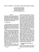
A 19 year old man presented to the Accident and Emer-
gency Department with a one hour history of the right
upper eyelid injury from a wire fence, (figure 1). The
patient was walking across an allotment when he fell onto
a damaged fence. The loose end of the wire penetrated the
full thickness of the right upper eyelid. The patient was
unable to extricate himself, requiring the Fire Brigade to
cut him free. Of relevant past history, there was an injury
to the same eyelid from a coat hanger two years earlier.
Upon gentle examination with no external pressure, the
patient was unable to open the eye himself. The wire
passed from under the eyelid, through the centre of the
upper lid, to the external surface. Approximately 15 mm
of wire was superficial to the lid margin; the cut end was
approximately 90 mm and taped to the cheek for security.
The patient had eaten ninety minutes previously so he was
unfit for a general anesthetic. The decision was made to
infiltrate with local anesthetic and remove the foreign
body. This was complicated by the patient's inebriation
and needle phobia.
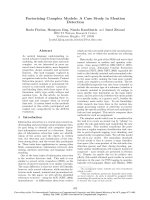
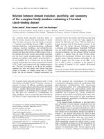
1% Lignocaine was infiltrated in to the upper lid, the lid
averted, and the wire passed out through the defect, (fig-
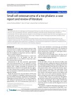
ure 2 and 3). Post removal, the acuity increased to 6/9 and
there was no intraocular penetration, (figure 4). No
abnormalities were detected in the anterior or posterior
segments and intraocular pressure was within the normal
range.
After the application of topical anesthetic drops, the eye
could be opened manually. Acuity in the right was count-
ing fingers. The anterior chamber was formed and there
were no pupil abnormalities. It was difficult to assess
whether there was any intraocular penetration. In addi-
tion, the injured region was examined for any remains or
other possible foreign bodies.
At follow up the next day acuity was still maintained and
one week later, after a full course of antibiotics, examina-
tion was unremarkable with equal acuity bilateral
Discussion
A variety of orbital foreign bodies have been reported in
the literature to have penetrated the eyelids. These include
glass, stone, metal, wood, graphite, button, faucet handle,
fish jaw, iron hat peg, chopstick, pencil, large wooden
plank, pocket knife, meat hook, and pitchfork [4]. Fur-
thermore, removal of such foreign bodies and the appro-
priate technique used is important in the management of
the injury otherwise it could lead to vision loss, corneal
scaring, retinal detachment and endophthalmitis [5].
A review of the appropriate literature demonstrated that
penetrating eyelid injury, particularly from fishhooks, was
common, with a range of removal techniques available
such as retrograde, needle cover, advance and cut, string
yank and vertical eyelid-splitting [5]. There were no
reports found of penetration from anything with a greater
Patient eye on arrivalFigure 1
Patient eye on arrival.
Removal of the wire from the patients' eyelidFigure 2
Removal of the wire from the patients' eyelid.
Head & Face Medicine 2009, 5:2 />Page 3 of 4
(page number not for citation purposes)
caliber or injuries which penetrated in the opposite direc-
tion to normal.
The unusual aspects of this presentation were firstly the
nature of the injury, in that it was sustained from a fall on
to a sharp object rather than a moving foreign body. Sec-
ondly there was enough force to penetrate the lid but
essentially left the globe without injury. Lastly, the direc-
tion of the penetration was unusual as it passed from the
under to the external surface.
Moreover, topical anesthesia has been shown to be safe
and effective [6] especially in this case where the patient
had expressed needle phobia. As a result, the decision to
infiltrate local anesthetic is more appropriate as the
advantages of local anesthesia include immediate onset,
short duration of action, rapid return of visual function,
and avoidance of the attendant risks of general anesthesia.
These advantages determine a shorter hospital stay, more
rapid resumption of a regular diet and normal insulin or
oral therapy, and ambulation for the patient [7]. Plus, it
has been found that this method of anesthesia is particu-
larly useful in patients who have distressing fears of injec-
tion and in whom poor cooperation renders the patient
vulnerable to needle related injuries [8].
In addition to removing the fishhook, post- removal
wound care is also of interest. After removal of the fish-
hook, the wound should be explored for possible foreign
bodies. It is usually sufficient to leave the wound open,
and then apply an antibiotic ointment and a simple dress-
ing. Tetanus toxoid should be administered to persons for
whom more than five years has elapsed since their last tet-
anus booster [9]. In this case, the patient had a recent his-
tory (less than five years) of tetanus booster as a result; he
did not require a tetanus shot.
This case demonstrates that a severe eyelid penetrating
injury can be uncomplicated with a full recovery when
there is not intraocular penetration. It is also possible to
have an injury pass under the lower margin of the lid and
penetrate from inside to out, with no associated corneal
injury.
Conclusion
A severe eyelid penetrating injury can be uncomplicated
with a full recovery when there is no intraocular penetra-
tion. It is also possible to have an injury pass under the
lower margin of the lid and penetrate from inside to out,
with no associated corneal injury.
Consent
Written informed consent was obtained from our patient
for publication of this case report and the accompanying
images. A copy of the written consent is available for
review by the Editor-in-Chief of this journal.
Competing interests
The authors declare that they have no competing interests.
Authors' contributions
EW, BK, TY carried out the patient diagnosis, investiga-
tion, follow up and management. PV participated in writ-
ing the final manuscript. AAA-E participated in patient
management, general coordination, drafting of the manu-
script, writing the final manuscript and provided impor-
tant suggestions.
The wire completely removed from the eyelidFigure 3
The wire completely removed from the eyelid.
Patients eye after complete removal of the wireFigure 4
Patients eye after complete removal of the wire.
Publish with BioMed Central and every
scientist can read your work free of charge
"BioMed Central will be the most significant development for
disseminating the results of biomedical research in our lifetime."
Sir Paul Nurse, Cancer Research UK
Your research papers will be:
available free of charge to the entire biomedical community
peer reviewed and published immediately upon acceptance
cited in PubMed and archived on PubMed Central
yours — you keep the copyright
Submit your manuscript here:
/>BioMedcentral
Head & Face Medicine 2009, 5:2 />Page 4 of 4
(page number not for citation purposes)
All authors read and approved the final manuscript.
References
1. Karcioglu Z, Nasr A: Diagnosis and management of orbital
inflammation and infections secondary to foreign bodies: a
clinical review. Orbit Opthalmology 1998, 17(4):247-269.
2. Khaw P, Shah P, Elkington A: ABC of eyes, injury to the eye. BMJ
2004, 328(7430):36-38.
3. Srinivasan S, Macleod S: Fish hook injury to the eyelid. Indian J
Ophthalmol 2001, 49:115-6.
4. Liu D, Al Shail E: Retained orbital wooden foreign body a sur-
gical technique and rationale. Ophthalmology 2002, 109:393-399.
5. Fuentes-Mallozzi D, Méndez-Orozco C: Eyelid fish-hook injury:
case report. Bol Med Hosp Infant Mex 2005:6.
6. Karp C, Cox T, Wagoner D, Ariyasu R, Jacobs S: Intracameral
anesthesia: a report by the American Academy of Ophthal-
mology. American Academy of Ophthalmology 2001, 108:1704-1710.
7. Boscia F, La Tegola M, Columbo G, i Alessio G, Sborgia C: Com-
bined topical anesthesia and sedation for open-globe injuries
in selected patients. American Academy of Ophthalmology 2003,
110:1555-1559.
8. Li R, Lai J, Ng J, Law R, Lau E, Lam D: Efficacy of Lignocaine 2% gel
in chalazion surgery. British J of Opthalmol 2003, 87:157-159.
9. Gammons M, Jackson E: Fish hook removal. Am Fam Physician
2001, 63:2231-2236.