báo cáo khoa học: " Surface electromyography as a screening method for evaluation of dysphagia and odynophagia" ppt
Bạn đang xem bản rút gọn của tài liệu. Xem và tải ngay bản đầy đủ của tài liệu tại đây (1.06 MB, 11 trang )
BioMed Central
Page 1 of 11
(page number not for citation purposes)
Head & Face Medicine
Open Access
Review
Surface electromyography as a screening method for evaluation of
dysphagia and odynophagia
Michael Vaiman* and Ephraim Eviatar
Address: Department of Otolaryngology, Assaf Harofe Medical Center, Affiliated to Sackler Faculty of Medicine, Tel Aviv University, Israel
Email: Michael Vaiman* - ; Ephraim Eviatar -
* Corresponding author
Abstract
Objective: Patients suspected of having swallowing disorders, could highly benefit from simple
diagnostic screening before being referred to specialist evaluations. The article analyzes various
instrumental methods of dysphagia assessment, introduces surface electromyography (sEMG) to
carry out rapid assessment of such patients, and debates proposed suggestions for sEMG screening
protocol in order to identify abnormal deglutition.
Data sources: Subject related books and articles from 1813 to 2007 were obtained through
library search, MEDLINE (1949–2007) and EMBASE (1975–2007).
Methods: Specifics steps for establishing the protocol for applying the technique for screening
purposes (e.g., evaluation of specific muscles), the requirements for diagnostic sEMG equipment,
the sEMG technique itself, and defining the tests suitable for assessing deglutition (e.g., saliva,
normal, and excessive swallows and uninterrupted drinking of water) are presented in detail. SEMG
is compared with other techniques in terms of cost, timing, involvement of radiation, etc.
Results: According to the published data, SEMG of swallowing is a simple and reliable method for
screening and preliminary differentiation among dysphagia and odynophagia of various origins. This
noninvasive radiation-free examination has a low level of discomfort, and is simple, time-saving and
inexpensive to perform. The major weakness of the method seems to be inability for precise
diagnostic of neurologically induced dysphagia.
Conclusion: With standardization of the technique and an established normative database, sEMG
might serve as a reliable screening method for optimal patient management but cannot serve for
proper investigation of neurogenic dysphagia.
Introduction
Swallowing disorders comprise an interdisciplinary phe-
nomenon. Practitioners in various fields of medicine,
such as otorhinolaryngology, neurology, general medi-
cine, gastroenterology, head and neck surgery, dentistry
and facial surgery, pediatrics and psychiatry deal with
these disorders regularly, but family doctors and emer-
gency department personnel might well be the first physi-
cians to evaluate these patients.
Basic terminology
Dysphagia is a difficult or abnormal swallowing, which
can include nasopharyngeal regurgitation and aspiration
[1]. It is also defined as any defect in the intake or trans-
Published: 20 February 2009
Head & Face Medicine 2009, 5:9 doi:10.1186/1746-160X-5-9
Received: 26 November 2008
Accepted: 20 February 2009
This article is available from: />© 2009 Vaiman and Eviatar; licensee BioMed Central Ltd.
This is an Open Access article distributed under the terms of the Creative Commons Attribution License ( />),
which permits unrestricted use, distribution, and reproduction in any medium, provided the original work is properly cited.
Head & Face Medicine 2009, 5:9 />Page 2 of 11
(page number not for citation purposes)
port of saliva, liquids and food necessary for the mainte-
nance of life. Odynophagia is a painful swallowing.
Odynophagia is not a constant pain in the throat but
rather pain on swallowing (swallow-evoked pain) [2]. It
was F. Magendie who, in 1813, established the concept of
three stages in the act of swallowing: oral, pharyngeal, and
oesophageal [3,4]. Since the 1980s, the act of swallowing
has sometimes been described as consisting of four stages,
with the oral stage being divided into oral initial (for sol-
ids – oral preparation stage) and oral final stages [5]. In
case of liquid swallowing, during the oral initial stage the
water intake takes place and a labial seal occurs. The oral
final stage occurs when the tongue squeezes the liquid
volume against the hard palate so that it is propelled past
the anterior faucial arches. At this stage the automatic
reflexive gesture of swallowing is triggered, but the oral
stage itself remains under complete conscious control.
During the pharyngeal stage the liquid volume is trans-
ferred from the level of the faucial arches through the
pharynx to the cricopharyngeal sphincter at the rostral
aspect of the esophagus. This stage is often described as a
reflex, i.e. a person cannot stop swallowing in the middle
of the process. In the oesophageal stage of the swallow,
the water volume is transferred in a continuation of the
peristaltic movement from the cricopharyngeal to the gas-
tro-oesophageal sphincter at the entrance to the stomach.
Background
The above described combination of voluntary and invol-
untary stages in deglutition makes evaluation of swallow-
ing pathophysiology a difficult and sometimes timely and
expensive process. Different diagnostic techniques were
proposed for it. 20 years ago, in 1988, in a general descrip-
tion of evaluation of swallowing pathophysiology, the list
of these techniques was as follows: still X-ray, computer-
ized axial tomography (CAT), magnetic resonance imag-
ing (MRI), indirect laryngoscopy, pharyngeal manometry,
scintigraphy, ultrasound, videofluoroscopy [6]. Electro-
myography (EMG) was not in this list. 10 years ago, in
1998, the list was somewhat changed and included bar-
ium esophagram, air contrast esophagram, manometry,
manofluorography, flexible endscopic evaluation of swal-
lowing with sensory testing (FEESST), bolus scintigraphy,
ultrasonography, videofluoroscopic swallowing study
(VFSS), and videoendoscopic swallowing study (VESS)
[7]. Once again, EMG was not mentioned.
At that moment, disadvantages for each of the above men-
tioned methods were well known. In brief, they are:
barium esophagram – uses x-irradiation, needs a facility
and personnel, no dynamic futures;
air contrast esophagram – the same as above [8,9];
manometry – cannot diagnose visible lesions, unpleasant,
can be unvalid because of movement of larynx [10,11];
manofluorography – not widely available, costly [12,13];
FEESST – requires complicated equipment, not wide-
spread, expensive [14,15];
bolus scintigraphy – cannot see patient anatomy, uses iso-
topes [7];
ultrasonography – segmental, cannot present panoramic
anatomic detail, expensive [7];
VESS – the phases of swallowing cannot be seen directly,
and also aspiration that occurs during the swallow, the
function of the cricopharyngeus muscle cannot be directly
assessed [16].
VFSS – relies on x-irradiation, needs radiology equipment
and personnel, expensive [7];
Currently VFSS is the most commonly used tool in the
assessment of oropharyngeal dysphagia, and it is consid-
ered the gold standard in the dysphagia workup. But in
addition to above mentioned drawbacks, VFSS does not
always identify neuromuscular abnormalities in pharyn-
geal or laryngeal physiology.
In addition to the above, one can see that all these meth-
ods hardly can be used for rapid screening purposes.
Comparison between swallowing studies options in
respect to radiation, timing and cost is shown in the Table
1. While cost of the procedures varies from country to
country, simplified "inexpensive – moderate – expensive"
scale is used. Total time needed per person per procedure
is a combination of the time needed for preparation of a
patient, time needed for preparation of an instrument,
actual time of a procedure, and the time needed for data
interpretation and report. The timing also depends of how
well the personnel are trained.
Do we need dysphagia screening?
Dysphagia occurs in approximately 14% of patients in
acute care setting an up to 50% of patients in nursing
homes [17]. Its prevalence is related to the fact that dys-
phagia often is present in patients who have sudden-onset
ENT or neurologic disorders [18], head and neck cancers,
chronic neurodegenerative diseases, and patients with
general medical problems, but in general it is indeed an
interdisciplinary phenomenon. Even practitioners in the
field of pediatrics [19,20] deal with these disorders. How-
ever, more than 75% of cases of oropharyngeal dysphagia
are caused by ENT problems or neuromuscular disorders
[21]. To carry out the rapid assessment of patients with
Head & Face Medicine 2009, 5:9 />Page 3 of 11
(page number not for citation purposes)
dysphagia and/or odynophagia requires a simple screen-
ing diagnostic tool to be used before extensive clinical and
instrumental examination will be performed by a special-
ist. The diagnostic tool should be
• reliable
• preferably noninvasive
• preferably radiation-free
• inexpensive
• time saving
• providing both qualitative and quantitative data
• simple and easy to operate
In general, aim of a screening procedure is to separate
patients into a "passed"- vs. "failed" group, specifically
into groups with normal and abnormal deglutition.
Screening so far is a filter to decide whether further clinical
diagnostics or treatment is necessary. In case of dysphagia,
however, the second step after normal-abnormal screen-
ing is needed. This step might answer the question to what
specialist a dysphagic patient should be referred to: neu-
rologist? ENT-physician? dentist? gastroenterologist? psy-
chiatrist? This second screening stage is actually more
practicable comparing with the first passed-failed one
because initial dysphagia detection starts with a history
taking.
Swallowing is a muscular process. Its mechanism, by
which food is transmitted to the stomach, is a complex
action involving 26 muscles and five cranial nerves [22].
This fact suggests that surface electromyography (SEMG)
might be a valuable method to be used for screening pur-
poses and early diagnostics of dysphagia and
odynophagia complaints. Indeed, surface SEMG provides
information on the timing of selected muscle contraction
patterns during swallowing [23-25], amplitude of electric
activity of the muscles [26], and can be easily learned by
the personnel [27,28]. Some suggestions have been
already made for using SEMG for screening purposes in
neurogenic dysphagia [29].
Despite numerous studies of SEMG activity of face and
neck muscles during swallowing made in the 1990s [23-
30], lack of agreement arose both in some of the basic
aspects common to all subjects and in establishing nor-
mal limits of this activity beyond which the act becomes
pathological. That is why the first step was to establish a
normative database for the phenomenon, and it was
reported in numerous studies [31-36]. A review of 440
healthy adults (230F, 210M, age mean 31.8 years) in one
study and 300 adults (170F, 130M, age mean 33.9 years)
in another study provided data which we used as a basis
for comparison of swallowing performance both within
and between patients. Quick reference simplified set of
Table 1: Comparison between swallowing studies options in respect to radiation, timing and cost
Procedure Radiation Time Cost
Barium Esophagram Yes Moderate Moderate
Air Contrast Esophagram Yes Moderate Moderate
Manometry No Time-consuming Inexpensive
Manofluorography Yes Time-consuming Expensive
FEESST No Time-consuming Expensive
Bolus Scintigraphy Yes Moderate Moderate
Ultrasonography No Moderate Expensive
VESS No Time-consuming Inexpensive
VFSS Yes Time-consuming Expensive
Surface EMG No Time-saving Inexpensive
* Goes in two stages, the examination is videotaped and then again analyzed.
** in addition to radiologist, speech pathologists must be present.
Head & Face Medicine 2009, 5:9 />Page 4 of 11
(page number not for citation purposes)
normative data for electric activity obtained by surface
EMG for masseter (MS) and submental muscle group +
platisma (SUB) during various tests is presented in the
Table 2[33].
Up to date, however, we face various technical approaches
to SEMG usage in evaluation of swallowing process. While
the normative database for SEMG assessment of degluti-
tion has been established, the next step might be estab-
lishment of standards for this diagnostic procedure, i.e.
the protocol. Large variation in examination techniques,
strategies, interpretations and diagnostic criteria have
been found among electromyographers and it is suggested
that the value of SEMG studies of deglutition may be fur-
ther improved by international standardization.
Surface EMG as a screening method
SEMG is to be clearly separated from needle electrode
examination EMG [37]. Needle electrodes should be used
within the neck area with great precaution and this proce-
dure will be neither time-saving, nor noninvasive. The
needle EMG report includes tabulated nerve conduction
studies, which are not needed at the screening stage of
investigation.
Reliability of the method was proved in a series of
research using SEMG for diagnostic purposes in oral
[38,39], pharyngeal [40-43] and esophageal [44] diseases.
These investigations suggested that different diseases have
specific SEMG patterns, both in timing and amplitude of
the record. The graphic record itself has visible peculiari-
ties specific for each disease. Filtered SEMG provides a
simple EKG-looking line easy to analyze and interpret.
If a diagnostic method is offered to be used in different
areas of medicine but for one purpose, the standard pro-
tocol is a must. The need for established protocols in
SEMG investigations is well understood [45-48], and sev-
eral attempts already have been made [49,50]. To our
knowledge, no such protocols were proposed for SEMG
evaluation of dysphagia and odynophagia. SEMG evalua-
tion of deglutition is not a new diagnostic method. Nev-
ertheless, lack of standard requirements decrease the
outcome of this investigation technique significantly. In
the case of SEMG evaluation of deglutition the protocol
might be based on:
• protocol application
• protocol requirements for diagnostic equipment
• protocol technique
• protocol tests
• normative database and standard analysis
Table 2: Quick reference simplified set of normative data for electric activity obtained by surface EMG for masseter and submental
group + platisma during various tests, in μV
Saliva swallow
masseter range 18–30: 4.5 – 15.9 31–70: 5.54 – 12.1 70+: 2.94–22.42
Submental range 18–30: 13.4–59.72 31–70: 9.52 – 49.5 70+: 10.2–42.32
Normal swallow
masseter range 18–60:2.2–31.0 61–70: 1.97 – 27.69 70+: 3.77–20.0
submental range 18–30:11.4–63.41 31–50: 12.58–51.6 51–70+: 7.4 – 44.8
20 cc excessive swallow
masseter range 18–40:1.5–37.0 41–70:1.2 – 29.4 70+: 4.65–21.13
submental range 18–30:19.28–50.80 31–70+: 12.1 – 47.44
100 cc drinking
masseter mean (real) 18–70: 0.8 – 6.2 70+: 1.0 – 7.84
submental mean (real) 18–60: 3.5 – 11.5 61 – 70+: 4.25 – 16.25
[age: normal values]
Head & Face Medicine 2009, 5:9 />Page 5 of 11
(page number not for citation purposes)
Suggestions for protocol application
As it was mentioned above, the act of swallowing is
described as consisting of four stages, as the oral stage was
divided into oral initial (for solids – oral preparation
stage) and oral final stages [5]. This staging can be helpful
in diagnostic evaluation of disorders providing dysphagia
and odynophagia. Each of these stages can be impaired
and the screening evaluation should be capable to indi-
cate the impaired stage. Surface SEMG recording cannot
trace oesophageal activity and only initial oesophageal
stage can be recorded and evaluated. SEMG does not
detect silent breathing.
Protocol equipment
Surface SEMG devices are non-invasive, radiation-free,
generally inexpensive and as simple as standard EKG
equipment [51]. Four-channel computer-based SEMG
unit equipped with surface electrodes is usually enough.
(Fig. 1). Two-channel unit is inadequate and eight-chan-
nel unit being useful in scientific research is not conven-
ient for rapid screening procedure. Standard surface
electrodes AE-131 and AE-178 are sufficient. (Electrode
material: silver coated; shape: discs; size: diameter 11 mm;
the inter-electrode distance: 10 mm). However other sur-
face electrodes with similar characteristics can be applied
[52]. SEMG recordings are to be preferably performed by
a unit which has a wide bandpass filter, a bandwidth
(RMS) of 25–450 Hz and a 60 Hz notch filter. The system
uses an active electrode consisting of a compact sensor
assembly that includes a miniaturized instrument pream-
plifier. Locating the amplifier at the electrode site allows
cancellation of artifacts and boosting of the signal before
it is transferred down the electrode cable. The integration
period of the sSEMG signal at the hardware level is 25 mil-
liseconds. This has very little effect on the shape of the sig-
nal [53]. For the software, the underlying sampling speed
is 100 Hz (i.e., 100 samples per second). The recordings
of this high-speed sampling are then averaged, based on
the selected sampling rate. Each sSEMG recording should
be full-wave rectified and low-passed filtered (the remote
preamplifier, low-pass cutoff = 30 Hz). The computer pro-
gram should be able at least to calculate the mean, stand-
ard deviation, minimum, maximum, and range of muscle
activity during each trial, as well as its duration. Muscle
activity (SEMG) is quantified in microvolts (μV).
Any other SEMG device with similar characteristics can be
used but one quality has to be insured: the SEMG record
has to be full-wave rectified and low-passed filtered in a
way to look like a single EKG-looking line. SEMG records
with numerous closely packed spikes are almost impossi-
ble to interpret rapidly. If the four-channel device is used,
the investigation can be performed in 3–5 minutes in
cases when we have full cooperation of a patient.
Electromyographic technique
Four muscle groups are to be examined during the evalu-
ation [31-36]:
(1) for oral phase: the orbicularis oris superior and infe-
rior (OO),
(2) for oral phase: the masseter (MS),
(3) for pharyngeal phase: the submental muscle group
(SUB) which includes the anterior belly of the digastric,
mylohyoid, and geniohyoid, all covered by the platysma,
(4) for pharyngeal and initial oesophageal phases: (INF),
the infrahyoid group, thyrohyoid, and the laryngeal strap
muscles also covered by the platysma.
Protocol electrode positions might be as follows [31-36]:
(1) Two bipolar stick-on surface electrodes are to be
applied at the right or left angle of mouth, one electrode
above the upper lip, and another electrode below the
lower lip (OO-location);
(2) Two electrodes are to be placed parallel to the mas-
seter muscle fibers on the left or right side of the face, pref-
erably on the opposite side from the OO-location (MS-
location);
(3) Two surface electrodes are to be attached to the skin
beneath the chin on the right or left side of midline to
record SUB myoelectrical activity over the platysma (SUB-
location);
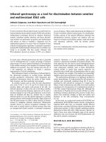
The electroneuromyograph at workFigure 1
The electroneuromyograph at work. A subject with
masseter, submental group, infrahyoid, and trapezius loca-
tions of SEMG electrodes.
Head & Face Medicine 2009, 5:9 />Page 6 of 11
(page number not for citation purposes)
(4) Two electrodes are to be placed on the left or right side
of the thyroid cartilage to record from the laryngeal strap
and infrahyoid muscles (INF-location).
Variation
when OO-location is uninformative, but some general
muscular-neurologic disease is suspected, the fourth elec-
trode can be attached above a muscle not involved in
deglutition, for example, above m. trapezius (Fig. 1). The
same variation is preferable in cases of psychogenic swal-
low or malingering, when a patient strains muscles not
involved in deglutition.
The exact electrode positions for each muscle group have
been known since the 19
th
century [54,55], and, in addi-
tion, can be clarified following anatomical correlates [56].
The proposed electrode locations cover all stages of a
swallow. The staging of normal deglutition can be clini-
cally important as an additional tool for establishing aeti-
ology and localization – oral, pharyngeal, or oesophageal
– of causes for dysphagia or odynophagia. Each stage has
its mean normal duration and its specific graphic pattern.
Each pair of electrodes has a third electrode as ground.
Electrical impedance at the sites of electrode contact
might be reduced since the target areas are lightly
scrubbed with alcohol gauze pads. Radiates skin, edema-
tous skin and status post neck dissection are possible
counter-indications for SEMG investigation.
Protocol tests
A set of four tests might be suggested: voluntary single
swallows of saliva ("dry" swallow), voluntary single water
swallows from an open cup ("normal"), voluntary single
swallows of an excessive amount of water (20 ml, "stress
test"), and continuous drinking of 100 cc of tap water
from an open cup [31-36,38-44]. Subjects are permitted
to move their chins slightly upwards while swallowing if
needed when it emerged that there is no changes of the
graphic and numerical baseline associated with this
movement. (This movement involves the mm. rectus
capitis posterior minor and minor, as well as some other
posterior neck muscles, and does not affect signals from
the above-mentioned electrode locations.) After electrode
placement, a patient is asked to perform the following
tasks:
1. Three trials of "dry" swallowing. Instruction given:
"Swallow your saliva".
2. Three trials of swallowing normal volume of tap water
for a particular person. These volumes, calculated for a
particular age group [31-36], are: Group 1 (age 18–40) –
16,5 cc; Group 2 (age 41–70) – 14,5 cc; Group 3 (age 70+)
– 12 cc. Instruction given: "Swallow this water in one
gulp".
3. Three trials of swallowing 20 cc of tap water to check
adaptation abilities of the patients ("stress test", larger
bolus volume accommodation). Instructions given:
"Swallow this water in one gulp".
4. One trial of continuous drinking of 100 ml of tap water.
Instruction given: "Drink this water as normal".
The main test is a single water swallow as normal. Saliva
swallow might be very important test in case of salivary
glands diseases like, for example, Sjögren syndrome.
Stress test with excessive amount of water swallowed in
one gulp might reveal lack of larger bolus volume accom-
modation abilities in cases of anatomical changes of the
pharynx, neurological problems or might provoke regur-
gitation in patients with Zenker's diverticulum. Testing of
continuous drinking is important not only in the evalua-
tion of dysphagia but also of odynophagia and in the dif-
ferential diagnosis in cases of compulsive water drinking,
excessive water drinking, the malingering of dysphagia
and psychogenic disorders expressed by symptoms of dys-
phagia. This is the test that can help in better evaluation of
slight dysphagia in fast-fatigable subjects. For fast-fatiga-
ble subjects continuous non-interrupted drinking is a
stress test [57]. The amount of water for continuous drink-
ing test was set at 100 cc, approximately one-half of a
standard glass because a smaller volume, e.g., 50 cc, can
be swallowed in two gulps and provide inadequate data
while we had observed that 200 cc of water involves sig-
nificant swallowing/ventilation interactions which can
confound the validity of the obtained data.
In addition to water swallow tests, there are tests with
food, involving mastication. They can be justified in cases
of diseases of temporomandibular joint and some other
disorders. For screening purposes, however, simple water
swallow tests are sufficient.
Protocol analysis
Protocol analysis includes assessment of duration (in sec),
amplitude of electric activity (mean, in μV), graphic pat-
terns and number of swallows (in continuous drinking).
To carry out the rapid assessment of patients, the SEMG
record should be clear and easily understandable. A com-
prehensive study was recently performed in which SEMG
was used for monitoring functionally distinct muscle acti-
vation during swallowing [58]. This study supports the
statement that raw SEMG records should be rectified and
filtered before evaluation.
A typical single water swallow of a healthy individual is
observed graphically at the rectified and low-pass filtered
SEMG as a normal wave with upward deflections and a
sharp apex when recorded from the MS, SUB and, to lesser
extent, from INF locations [31-36]. The reflex part of a
Head & Face Medicine 2009, 5:9 />Page 7 of 11
(page number not for citation purposes)
swallow is recorded from the MS, SUB and INF locations,
while the conscious part can be recorded from the OO
and MS locations. This upward stroke, as recorded from
the MS, SUB and much less from INF locations, can be
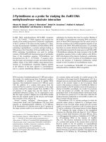
divided into three main parts (Fig. 2). In swallowing
water, the first part (A) is usually seen as a mild elevation
of the line and represents the final oral stage of a swallow
which occurs when the tongue is moved so as to squeeze
the liquid volume against the hard palate. Submental
muscles and the masseter muscle support the tongue-
induced pressure. At this stage, the automatic reflexive
motion of swallowing is triggered. When the reflex is ini-
tiated, the second stage, the pharyngeal, begins. It is seen
as a rapid voltage line elevation on the second part of the
wave (B). When the bolus is finally passed into the
oesophagus by the relaxation of the sphincter at the
cricopharyngeal juncture, the third part (C) of the stroke
is seen as a rapid descent of the SEMG line when the mus-
cles relax and their voltage decreases. This part indicates
the initiation of the oesophageal stage of swallowing. In
all healthy people during the tests, the mean electric activ-
ity at SUB location is 30–50% higher than the activity at
MS and INF locations. Amplitude of the OO line is usually
significantly higher than the SUB and especially with the
MS and INF graphic lines. Being under conscious control,
the oral phase of swallowing is very variable and should
be taken into consideration with caution during the eval-
uation of the recordings. By the same reason, the data
taken from the OO electrode location is the least inform-
ative and safely can be omitted if we deal with pharyngo-
laryngeal or oesophageal problems rather than with oro-
maxillo-facial.
There is no visible difference between the shapes of SEMG
recordings of swallows based on gender [31-36,38-44].
Elderly patients (age 70+) showed age-induced peculiari-
ties in the recorded swallows. In general, prevalence of
dysphagia increases with age and poses particular prob-
lems in the older patient, potentially compromising nutri-
tional status, complicating the administration of solid
medications, increasing the risk of aspiration pneumonia
and undermining the quality of life [59]. For them, the
muscle activity is usually longer in duration, and suggests
a lack of coordination between activities of different mus-
cles involved in deglutition both in single swallows and
continuous drinking. For children, the duration of muscle
activity during swallows and drinking in all tests showed
decrease with the age, and this tendency is statistically sig-
nificant. There is no statistically significant difference in
electric amplitude measurements between children and
adults [36].
Slow swallows and drinking are usually observed in cases
of various neurological disorders affecting deglutition
[60-63]. Analyzing timing one needs to remember that
the times indicated by an SEMG device represent the dura-
tion of SEMG activity which lasted longer than the actual
time required to pass a bolus from the oral cavity to the
oesophagus.
Electric amplitude is also considered in the SEMG analy-
sis. The range (amplitude, in μV) and mean of electric
activity are less important for stage-by-stage evaluation of
a SEMG recording [32-34]. These data might be useful,
however, when abnormal swallows are investigated. For
example, a person usually presents low electric activity at
the MS location after undergoing a tooth extraction [39].
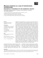
Patients with the Zenker's diverticulum present unusually
high electric activity at the LSM location. The main sEMG
patterns of Zenker's are: A) duration of swallowing and
drinking is longer than normal; B) electric amplitude of
laryngeal strap muscles during swallowing activity is
higher than normal; C) regurgitation peaks immediately
after swallow followed by secondary swallow of the regur-
gitated portion of a bolus as seen at the sEMG records are
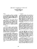
specific graphic patterns (Fig. 3) [44]. Patients with recur-
rent tonsillitis present abnormally high electric activity of
LSM and infrahyoid muscles. Acute tonsillitis and recur-
rent tonsillitis affect muscle activity significantly by
involving additional muscles (mainly infrahyoid) in swal-
lowing (Fig. 4). Acute tonsillitis triggers temporary electric
hyperactivity of LSM and infrahyoid muscles. Recurrent
tonsillitis affects MS and infrahyoid even during periods
of remission (fixed pathologic changes) [40-43]. There
were also numerous reports of authors who indicated
changes of the masseter electric activity in patients with
diseases of temporomandibular joint [64-66].
Stages of the normal swallow (reflex part)Figure 2
Stages of the normal swallow (reflex part). A – final
oral stage, B – pharyngeal stage, C – beginning of oesophageal
stage. In normal deglutition the INF record is the lowest and
less informative (eliminated here for clarity).
Head & Face Medicine 2009, 5:9 />Page 8 of 11
(page number not for citation purposes)
Published studies [67,68] on normal subjects show a very
wide range of normal electric amplitudes for SEMG stud-
ies. These variations are not only due to biologic causes
but are also greatly affected by such technical factors as
skin/electrode impedance, depth of the muscle from the
skin surface, location of the recording electrodes in rela-
tion to anatomic structures, variation in muscle size
among individuals, and temperature. It is because of the
wide variation in the normal values that an absolute value
of the amplitude is considered less clinically useful.
The SEMG amplitude, however, remains an important
aspect in the relationship between muscle force and the
associated electric activity. There is, however, no simple
relationship between an SEMG signal and muscle force.
When all the different types of neuromuscular disorders
are considered collectively, amplitudes are by far the most
informative features [69]. Some authors argue that ampli-
tudes are the only components that have a direct relation-
ship to clinical symptoms (muscle weakness) in
neurogenic lesions [70].
Also, during SEMG testing, there is a certain amount of
impedance noise that arises directly from the resistance of
the electrodes' connection to the skin. This makes skin
resistance a significant factor when working with the low-
level SEMG signals typical of small muscles involved in
swallowing. Wiping the skin with isopropyl alcohol in a
water solution has proven to be the best form of prepara-
tion for most situations. The alcohol removes the dead
skin and surface oils, and the water moistens the skin and
provides improved ion flow. The SEMG sensors we used
are designed so that the use of electrode gel is generally
not necessary.
Combined analysis of timing, amplitude and a shape of
the graphic record during the screening procedure is pre-
sented in the Table 3. The data for the Table is collected
from numerous studies and compiled [23,28,29,38-
44,57,60,61,64].
This study emphasizes that a surface SEMG analysis of all
the muscle groups involved in swallowing process, fol-
lowing a proper placement of the electrodes and selection
of tests, can give reliable indications of muscle activity and
provide data for screening evaluation of complaints a
patient came with. Further investigation might help to
develop a proper combination of flexible endoscopic eval-
uation of swallowing (FEES) with a nasopharyngoscope
(or flexible endoscopic evaluation of swallowing with
sensory testing, FEESST)[71] with SEMG to achieve com-
plete evaluation of swallowing without exposure to radia-
tion. Such screening might help a general practitioner to
direct a patient to a neurologist, ENT physician or other
proper specialist and to a specific radiographic or, per-
haps, manofluorographic investigation [72-74]. Other
topics for further investigation might be SEMG detecting
of sensory predeglutitive oropharyngeal swallowing disor-
ders, detecting dysphagia in patients after head and neck
tumor therapy.
A typical single swallow + regurgitation peak + secondary swallow of a person with Zenker's diverticulum (MS, SUB and INF locations)Figure 3
A typical single swallow + regurgitation peak + sec-
ondary swallow of a person with Zenker's diverticu-
lum (MS, SUB and INF locations). The complete
swallow (2.5–4.5 period) is slightly longer than the normal
swallow. The SUB peak is normal, MS peak is high and
appears in front of the SUB peak, LSM is normal. After a
short pause (4.5–5.5) regurgitation peaks appeared with high
INF line and low MS and SUB lines (5.5–9 period). Then the
secondary swallow of the regurgitated bolus followed with
high SUB peak (9.5–11).
Typical single swallows and drinking of a person with recur-rent tonsillitis, age 24 years, (MS, SUB and INF locations)Figure 4
Typical single swallows and drinking of a person with
recurrent tonsillitis, age 24 years, (MS, SUB and INF
locations). Trials 1–3 – saliva swallows, trials 4–6 – normal
swallows, trial 7 – excessive swallow, trial 8 – 100 ml drink-
ing. The SUB peaks are normal (blue line) except trial 1; MS
peaks are somewhat high, especially in single swallows being
almost similar to the SUB amplitude (green line), INF is very
high compare to normal database (red line).
Head & Face Medicine 2009, 5:9 />Page 9 of 11
(page number not for citation purposes)
Dysphagia due to neurologic problems, however, might
present difficulties when investigated by surface EMG. It
may be possible to collect data using surface electrodes,
sufficient to make conclusions with respect to patients
with odynophagia or non-neurological disorders such as
ENT disturbances. It is, however, difficult to nearly impos-
sible to collect proper knowledge about neurogenic dys-
phagia including important and urgent etiologies such as
stroke, ALS and so on. While surface kind of recording
picks up not only muscle activity related with swallowing
but also that of some other contiguous muscles with ran-
dom or associated activity during swallowing, direct and
precise neurologic assessment seems problematic. Despite
that, screening assessment might be successful even in
neurological cases if the record shows abnormal timing
(prolongation), abnormal voltage (decreased) and abnor-
mal shape of a signal (no peak).
In this respect it might be added that neddle EMG pro-
vides valuable data when neurologic problems are inves-
tigated. Numerous researches using needle EMG
contributed significantly to our knowledge of single mus-
cle actions during deglutition [75,76]. Specifically these
studies were important for neurologic diseases [77]. Tech-
nically, however, needle EMG is an invasive and time-con-
suming investigation requiring expensive devices and
being potentially dangerous in the neck area [76]. While
this type of EMG testing might be invaluable to establish
precise diagnosis, this method might be hardly recom-
mended as a screening technique.
Surface EMG assessment of odynophagia also requires
special attention. Pain is a subjective experience and one
may argue that it is impossible to be assessed objectively.
Partially this is correct. However, since Johann Bohn's
(1686) observations of reflex movements in decapitated
frogs, medical scientists are aware about muscular reac-
tions on irritation of pain receptors. While pain is subjec-
tive in general, these muscle reactions are objective and
EMG can provide us with qualitative and quantitative data
Table 3: Quick reference simplified set of SEMG data for screening purposes.
Disorder SEMG locations
MS SUB INF Additional peculiarity
T A S T A S T A S
Neurological ↑ ↓ Abn ↑ ↓ Abn ↑ N N Disorganized Peaks*
Ent N N N N ↓ Abn N ↑ N Multiple Peaks**
Maxillofacial (Oral)*** ↑ ↓ Abn ↑ N N ↑ N N
Gastroenterological N N N ↑ N Abn ↑ ↑ Abn Regurgitation Peaks
Psychogenic**** N N N N N N N N N Shoulder Tension
Abbreviations: MS – masseter electrode location, SUB – submental electrode location, INF – infrahyoid electrode location; T – timing, A – electric
amplitude, S – shape of a graphic record; N – normal, AbN – abnormal, ↑ – higher than normal, ↓ – lower than normal.
* Appearance of MS, SUB and INF peaks is not coordinated
** One bolus can be swallowed in several shares
*** Including salivary gland diseases
**** Excluding malingering
An example of a single swallow and normal drinking of 100 cc of water by a patient with severe throat problem (dysphagia and odynophagia due to tonsillectomy, second postoperative day, pain score 7)Figure 5
An example of a single swallow and normal drinking
of 100 cc of water by a patient with severe throat
problem (dysphagia and odynophagia due to tonsil-
lectomy, second postoperative day, pain score 7). MS
– masseter activity, SUB – submental activity, INF – infrahy-
oid activity. (In real EMG records these lines are of different
colors). INF line is very high (normally this line is the lowest
at the record), SUB line is lower than normal and almost sim-
ilar to MS line. The single swallow is prolonged and done in
two shares. Drinking is arrhythmic, swallows are small.
Head & Face Medicine 2009, 5:9 />Page 10 of 11
(page number not for citation purposes)
for their assessment (Fig. 5) [39,42]. In fact, since electric-
ity was introduced for diagnostic purposes by Duchenne
de Boulogne in 1850s–70s, these muscular reactions to
painful stimulation being studied before for neurophysi-
ological purposes, gained additional clinical importance
[78]. During this period of time and up to 1930s, how-
ever, Faradic current was used for muscle testing instead of
non-irritating modern EMG [79]. It helped, for example,
to improve the differential diagnosis between central and
peripheral paralyses and attempts were made to investi-
gate dysphagia after stroke [80]. But naturally, no screen-
ing methods were proposed, physicians' interest shifted to
electrotherapy and further EMG investigation of dys-
phagia/odynophagia was delayed.
It is estimated that 15 million patients suffered from a
swallowing disorder during each year in the United States
alone [81,82]. These data make us to believe that the
screening method for this disorder might be a very timely
addition to our evaluation techniques.
Summary
Surface SEMG of swallowing is a simple and reliable
method for screening and initial evaluation of dysphagia
and odynophagia complaints of various origins. This non-
invasive radiation-free examination has low level of dis-
comfort, simple, time-saving and inexpensive. With
proper standard technique and established normative
database surface SEMG can serve as a reliable screening
method for assessment of dysphasia of unknown origin in
order to refer a patient to a neurologist or some other
proper specialist and proper further investigation. The
findings are the impetus for further study regarding the
mechanisms of muscle activity changes in disorders affect-
ing swallowing.
Consent
Written informed consent was obtained from the patient
for publication of accompanying image. A copy of the
written consent is available for review by the Editor-in-
Chief of this journal.
Competing interests
The authors declare that they have no competing interests.
Authors' contributions
General concept, data collection, assessment, writing.
References
1. Logemann JA: Swallowing Physiology and Pathophysiology.
Otolaryngol Clin North Amer 1988, 4(21):613-23.
2. Wilson JD, eds, et al.: Harrison's Principles of Internal Medicine 12th edi-
tion. New York: McGraw-Hill; 1991:417.
3. Magendie F: Vomissement. Procès-verb Acad d Sc 1813, 5:152-9.
4. Magendie F: L'epiglotte et ses usages dans le deglutition.
Procès-verb Acad d Sc 1813, 5:192-9.
5. Logemann JA: Evaluation and Treatment of Swallowing Disorders San
Diego, CA: College Hill Press; 1983.
6. Sonies BC, Baum BJ: Evaluation of swallowing pathophysiology.
Otolaryngol Clin North Amer 1988, 21(4):637-48.
7. Bastian RW: Contemporary diagnosis of the dysphagic
patient. Otolaryngol Clin North Amer 1998, 31(3):489-506.
8. Halpert RD, Dubin L, Feczko PJ, Weitz J: Air contrast tube
esophagram: technique and clinical examples. Technical
note. J Can Assoc Radiol 1984, 1(35):58-60.
9. Balfe DM, Helken JP: Contrast evaluation of structural lesions
of the pharynx. Curr Probl Diagn Radiol 1986, 15:77-91.
10. Leonard R, Belafsky PC, Rees CJ: Relationship between fluoro-
scopic and manometric measures of pharyngeal constric-
tion: the pharyngeal constriction ratio. Ann Otol Rhinol Laryngol
2006, 115(12):897-901.
11. Hurvitz AL, Nelson JA, Haddad JK: Oropharyngeal dysphagia:
Manometric and cine esophagraphic findings. Dig Dis 1975,
20:313-324.
12. Sokol EM, Heitmann P, Wolf BS, Cohen BR: Simultaneous cinera-
diographic and manometric study of the pharynx, hypophar-
ynx, and cervical esophagus. Gastroenterology 1966, 51:960-973.
13. McConnell FMS, Cerenko D, Jackson RT: Clinical application of
the manofluorogram. Laryngoscope 1988, 98:705-711.
14. Perlman A, Van Deale D: Simultaneous videoendoscopic and
ultrasound measures of swallowing. J Med Speech Lang Pathol
1993,
1:223-232.
15. Aviv JE, Murry T: FEESST: Flexible Endoscopic Evaluation of Swallowing
with Sensory Testing San Diego – Oxford, Plural Publishing; 2005.
16. Bastian RW: The videoendoscopic swallow study: an alterna-
tive and partner to the videofluoroscopic swallowing study.
Dysphagia 1993, 8:359-367.
17. Logemann J: Upper digestive tract anatomy and physiology. In
Head and Neck Surgery-Otolaryngology Edited by: Bailey B. Philadelphia,
JB Lippincott; 1993:485-491.
18. Dray TG, Hillel AD, Miller RM: Dysphagia caused by neurologic
deficits. Otolaryngol Clin North Amer 1998, 31(3):507-524.
19. Darrow DH, Harley CM: Evaluation of swallowing disorders in
children. Otolaryngol Clin North Amer 1998, 31(3):405-418.
20. Arvedson JC: Management of pediatric dysphagia. Otolaryngol
Clin North Amer 1998, 31(3):453-476.
21. Trate D, Parkman H, Fisher R: Dysphagia: Evaluation, diagnosis,
and treatment. Gastroenterology 1996, 23:417-432.
22. Dellow PG: The general physiologic background of chewing
and swallowing. In Mastication and Swallowing Edited by: Sessle BJ,
Hannam AG. Toronto – Buffalo, Univ of Toronto Press; 1976:6-18.
23. Perlman AL: Electromyography and the study of oropharyn-
geal swallowing. Dysphagia 1993, 8:351-355.
24. Palmer JB: Electromyography of the muscles of oropharyngeal
swallowing: basic concepts. Dysphagia 1989, 3:192-198.
25. Logemann JA: Non-imaging techniques for the study of swal-
lowing. Acta Otorhino laryngol Belg 1994, 48:139-142.
26. Ertekin C, Palmer JB: Physiology and electromyography of swal-
lowing and its disorders. Clinical Neurophysiology at the Beginning of
the 21st Century; Suppl to Clin Neurophysiol 2000, 53:148-154.
27. Gupta V, Reddy NP, Canilang EP: Surface SEMG measurements
at the throat during dry and wet swallow. Dysphagia
1996,
11:173-179.
28. Crary MA, Baldwin BO: Surface electromyographic character-
istics of swallowing in dysphagia secondary to brainstem
stroke. Dysphagia 1997, 12:180-187.
29. Logemann JA: Screening, diagnosis and management of neuro-
genic dysphagia (review). Semin Neurol 1996, 16:319-327.
30. Schultz JL, Perlman AL, VanDaele DJ: Laryngeal movement,
oropharyngeal pressure, and submental muscle contraction
during swallowing. Arch Phys Med Rehabil 1994, 75:183-8.
31. Vaiman M, Eviatar E, Segal S: Evaluation of Stages of Normal
Deglutition with the help of Rectified Surface Electromyog-
raphy Records. Dysphagia 2004, 19, 2:125-132.
32. Vaiman M, Eviatar E, Segal S: Surface electromyographic studies
of swallowing in normal subjects: a review of 440 adults.
Report 1: Quantitative data – Timing measures. Otorhynolaryn-
gol Head Neck Surg 2004, 131, 4:548-555.
33. Vaiman M, Eviatar E, Segal S: Surface electromyographic studies
of swallowing in normal subjects: a review of 420 adults.
Report 2: Quantitative data – Amplitude measures. Oto-
rhynolaryngol Head Neck Surg 2004, 131, 5:773-80.
34. Vaiman M, Eviatar E, Segal S: Surface electromyographic studies
of swallowing in normal subjects: a review of 440 adults.
Head & Face Medicine 2009, 5:9 />Page 11 of 11
(page number not for citation purposes)
Report 3: Qualitative data. Otorhynolaryngol Head Neck Surg 2004,
131, 6:977-985.
35. Vaiman M, Eviatar E, Gabriel Ch, Segal S: Rectified and filtered sur-
face electromyography of continuous drinking in healthy
adults. Laryngoscope 2005, 115(1):68-73.
36. Vaiman M, Segal S, Eviatar E: Surface electromyographic studies
of swallowing in normal children, age 4–12. J Pediatric Otorhi-
nolaryngol 2004, 68:65-73.
37. Katirji B: The clinical electromyography examination. An
overview. Neurol Clin North Amer 2002, 20, 2:291-304.
38. Vaiman M, Nahlieli O, Eviatar E, Segal S: Electromyography mon-
itoring of patients with salivary gland diseases. Otolaryngol
Head Neck Surg 2005, 133(6):869-73.
39. Vaiman M, Nahlieli O, Eliav E: Odynophagia in Patients after
Dental Extraction: Surface Electromyography Study. Head
and Face 2007, 2:34-40.
40. Vaiman M, Krakovski D, Gavriel H: Swallowing before and after
tonsillectomy as evaluated by surface electromyography.
Otolaryngol Head Neck Surg 2007, 137(1):138-45.
41. Vaiman M: The influence of Tonsillitis on Oral and Throat
Muscles in Adults. Otolaryngol Head Neck Surg 2007, 136(5):832-7.
42. Vaiman M, Gavrieli H, Krakovski D: Electromyography in Evalu-
ation of Pain after Different Types of Tonsillectomy: pro-
spective randomized study. ORL 2007, 69:256-264.
43. Vaiman M, Krakovsky D, Eviatar E: The influence of Tonsillitis on
Oral and Throat Muscles in Children. Int J of Pediatric Otorhi-
nolaryngol 2006, 70(5):891-8.
44. Vaiman M: Surface electromyography in Preoperative Evalua-
tion and Postoperative Monitoring of Zenker's diverticulum.
Dysphagia 2006, 21:1-7.
45. Stalberg E, Fuglsang-Frederiksen A, Bischoff C: Quantitation and
standardization in SEMG and neurography. Suppl Clin Neuro-
physiol 2000, 53:
101-11.
46. Bolton CF, Benstead TJ, Grand'Maison F, Tardif GS, Weston LE: Min-
imum standards for electromyography in Canada: a state-
ment of the Canadian Society of Clinical Neurophysiologists.
Can J Neurol Sci 2000, 27(4):288-91.
47. Stalberg E, Falck B, Gilai A, Jabre J, Sonoo M, Todnem K: Standards
for quantification of SEMG and neurography. The Interna-
tional Federation of Clinical Neurophysiology. Electroencepha-
logr Clin Neurophysiol Suppl 1999, 52:213-20.
48. Fuglsang-Frederiksen A, Johnsen B, Vingtoft S, Carvalho M, Fawcett P,
Liguori R, Nix W, Schofield I, Veloso M, Vila A: Variation in per-
formance of the SEMG examination at six European labora-
tories. Electroencephalogr Clin Neurophysiol 1995, 97(6):444-50.
49. Farina D, Madeleine P, Graven-Nielsen T, Merletti R, Arendt-Nielsen
L: Standardising surface electromyogram recordings for
assessment of activity and fatigue in the human upper trape-
zius muscle. Eur J Appl Physiol 2002, 86(6):469-78.
50. Podnar S, Vodusek DB, Stalberg E: Standardization of anal
sphincter electromyography: normative data. Clin Neurophys-
iol 2000, 111(12):2200-7.
51. Hermans HJ, Boon KL, Zivold G: The clinical use of surface EMG.
Electromyography Clin Neurophysiol 1984, 24:243-265.
52. Giroux B, Lamontagne M: Comparisons between surface elec-
trodes and intermuscular wire electrodes in isometric and
dynamic conditions. Electromyography Clin Neurophysiol 1990,
30:397-405.
53. Viitasalo JH, Saukkonen S, Komi PV: Reproducability of measure-
ment of selected neuromuscular performance variables in
man. Electromyography Clin Neurophysiol 1980, 20:487-501.
54. Erb W: Handbuch der Electrotherapie 2 Aufl., Leipzig: Vogel;
1886:34-45.
55. Remak E: Grundriss der Electrodiagnostik und Electrotherapie 2 Aufl.,
Wien: Meyer; 1909:26-34.
56. Goodgold J: Anatomical Correlates of Clinical Electromyography 1st edi-
tion. Baltimore: The Williams & Wilkins Company; 1975:2-3.
57. Schalow G, Paasuke M, Ereline J, Gapeyeva H: Coordination
dynamics in Parkinson's disease patients and healthy sub-
jects quantified by the coordination dynamics recording
method and sEMG. Electromyogr Clin Neurophysiol 2003,
43(8):473-85.
58. McKeown MJ, Torpey DC, Gehm WC: Non-invasive monitoring
of functionally distinct muscle activations during swallowing.
Clin Neurophysiol 2002, 113:354-66.
59. Morris H: Dysphagia in the elderly – a management challenge
for nurses. Br J Nurs 2006, 15(10):558-62.
60. Gallas S, Moirot P, Debono G, Navarre I, Denis P, Marie JP, Verin E:
Mylohyoid motor-evoked potentials relate to swallowing
function after chronic stroke dysphagia. Neurogastroenterol
Motil 2007, 19(6):453-8.
61. Arai T: Post stroke swallowing dysfunction and aspiration
pneumonia. Nippon Rinsho 2006, 64(Suppl 7):463-6.
62. Logemann JA: Levels of evidence supporting dysphagia inter-
ventions: where are we going? Semin Speech Lang 2006,
27(4):219-26.
63. Dimond B: Mental capacity and professional advice in a
patient with dysphagia. Br J Nurs 2006, 15(10):574-5.
64. da Silva MA, Issa JP, Vitti M, da Silva AM, Semprini M, Regalo SC: Elec-
tromyographical analysis of the masseter muscle in dentu-
lous and partially toothless patients with
temporomandibular joint disorders. Electromyogr Clin Neuro-
physiol 2006, 46(5):263-8.
65. Klasser GD, Okeson JP: The clinical usefulness of surface elec-
tromyography in the diagnosis and treatment of temporo-
mandibular disorders. J Am Dent Assoc 2006, 137(6):763-71.
66. Sato S, Kawamura H: Natural course of non-reducing disc dis-
placement of the temporomandibular joint: changes in elec-
tromyographic activity during chewing movement. J Oral
Rehabil 2005, 32(3):159-65.
67. Ruark JL, McCullough GH, Peters RL, Moore SA: Bolus consistency
and swallowing in Children and Adults. Dysphagia 2002,
17:24-33.
68. Sorin S, McClean MD: Electroglottographic evaluation of the
swallow. Arch Phys Med Rehabil
1987, 68:232-5.
69. Wilbourn AJ, Ferrante MA: Clinical electromyography. In Baker's
clinical neurology [book on CD-ROM] Edited by: Joynt RJ, Griggs Rc. Phil-
adelphia: WB Saunders; 2000.
70. Wilbourn AJ: Nerve Conduction Studies: Types, Components,
Abnormalities, and value in Localization. In Clinical Electromyog-
raphy Volume 20, 2. Edited by: Katirji B. Neurologic Clinics;
2002:310-11.
71. Aviv JE, Murry T: FEESST: Flexible Endoscopic Evaluation of Swallowing
with Sensory Testing San Diego – Oxford, Plural Publishing Inc; 2005.
72. Kahrilas PJ, Shi G: First measurement standards, then catheter
standards, for manofluorography. Dysphagia 1998, 13(2):111-2.
73. Dejaeger E, Pelemans W, Bibau G, Ponette E: Manofluorographic
analysis of swallowing in the elderly. Dysphagia 1994,
9(3):156-61.
74. McConnel FM, Cerenko D, Hersh T, Weil LJ: Evaluation of pha-
ryngeal dysphagia with manofluorography. Dysphagia 1988,
2(4):187-95.
75. Palmer J: Electromyography of the muscles of oropharyngeal
swallowing: basic concepts. Dysphagia 1989, 3:192-8.
76. Palmer J, Tanaka E, Siebens AA: Electromyography of the pha-
ryngeal musculature: technical considerations. Arch Phys Med
Rehabil 1989, 70:283-7.
77. Logemann J: Evaluation and treatment of swallowing disorders Boston;
College Hill; 1983:19-26.
78. Duchene de Boulogne GBA: De lé lectrisation localisée, et de son appli-
cation a la thérapeutique Paris; JB Bailliere et Fils; 1855.
79. Meyer M: Electricity in its relation to practical medicine. Transl from ed 3
German edition. Edited by: Hammond WA. New York; D. Appleton
& Co; 1869:56-78.
80. Reynolds JR: Lectures on the Clinical Uses of Electricity London; J & A
Churchill; 1871:80-92.
81. Byles J: The epidemiology of communication and swallowing
disorders. Advances in Speech Language Pathology 2005, 7, 1:1-7.
82. Murry T, Carrau RL, Eibling DE: Epidemiology of swallowing dis-
orders (Chapter 1). In Comprehensive Management of Swallowing
Disorders Edited by: Carrau R, Murry T. San Diego, CA: Singular Pub-
lishing; 1998:3-7.