báo cáo khoa học: " Condylar growth after non-surgical advancement in adult subject: a case report" pptx
Bạn đang xem bản rút gọn của tài liệu. Xem và tải ngay bản đầy đủ của tài liệu tại đây (630.06 KB, 5 trang )
BioMed Central
Page 1 of 5
(page number not for citation purposes)
Head & Face Medicine
Open Access
Case report
Condylar growth after non-surgical advancement in adult subject: a
case report
Antonino Marco Cuccia* and Carola Caradonna
Address: Section of Orthodontics, Department of Dental Sciences "G. Messina", University of Palermo, Via del Vespro 129, 90127, Palermo, Italy
Email: Antonino Marco Cuccia* - ; Carola Caradonna -
* Corresponding author
Abstract
Background: A defect of condylar morphology can be caused by several sources.
Case report: A case of altered condylar morphology in adult male with temporomandibular
disorders was reported in 30-year-old male patient. Erosion and flattening of the left mandibular
condyle were observed by panoramic x-ray. The patient was treated with splint therapy that
determined mandibular advancement. Eight months after the therapy, reduction in joint pain and a
greater opening of the mouth was observed, although crepitation sounds during mastication were
still noticeable.
Conclusion: During the following months of gnatologic treatment, new bone growth in the left
condyle was observed by radiograph, with further improvement of the symptoms.
Background
The temporomandibular joint (TMJ) is a complex joint
essential for speech, mastication and swallowing.
The mandibular condyle is an ovoidal bony structure that
articulates with the temporal bone by means of a bicon-
cave disk.
Both articular surfaces are covered by a connective fibrous
tissue (condylar cartilage). On the articular surface of the
condyle, the collagen fibres are parallel to the condylar
surface, and are in continuity with the fibrous layer of the
periosteum.
The condylar cartilage covers very dense undifferentiated
mesenchyme, within which are multipotential cells, form-
ing either cartilage or bone, depending upon the environ-
mental circumstances [1]. The bony tissue forms the
deepest part.
The TMJ grows and functions in an environment of
mechanical forces that interact with cells and tissues.
These forces (muscular activity, mastication, swallowing)
influence the shape of mandibular condyle, through the
process of biological adaptation termed "remodeling" [2].
Condylar resorption (CR) is a specific condition that
affects TMJs. A number of local and systemic pathologies
may cause mandibular CR. Local factors include osteoar-
thritis, reactive arthritis, avascular necrosis, infection,
traumatic injuries and temporomandibular disorders
(TMD). CR may also be due to systemic connective tissue
or autoimmune diseases including rheumatoid arthritis,
psoriatic arthritis, scleroderma, systemic lupus erythema-
tosus, Sjögren syndrome, ankylosing spondylitis, and oth-
ers [3-5].
Changes in condylar morphology have also been
observed in experimental protrusion or retrusion of the
Published: 20 July 2009
Head & Face Medicine 2009, 5:15 doi:10.1186/1746-160X-5-15
Received: 27 December 2007
Accepted: 20 July 2009
This article is available from: />© 2009 Cuccia and Caradonna; licensee BioMed Central Ltd.
This is an Open Access article distributed under the terms of the Creative Commons Attribution License ( />),
which permits unrestricted use, distribution, and reproduction in any medium, provided the original work is properly cited.
Head & Face Medicine 2009, 5:15 />Page 2 of 5
(page number not for citation purposes)
jaw, in surgical induction of disc displacement and exper-
imental disc perforation [6-9].
In this paper, we report a case of an adult male with TMD
and left CR, in which, after occlusal modification, new
bone growth in the left condyle was observed.
Case presentation
A 30 year old male was referred to our department with a
4 years history of pain (pain scale VAS 80) and crepitus in
the left TMJ during mastication, increased left facial pain,
and limited functional mandibular movements.
Bruxism was reported by the patient for a period of about
18 months. He had natural molar contacts in each dental
quadrant, and no parodontal disorders.
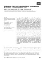
Intraoral examination revealed a bilateral Class II molar
relationship and a severe overjet [Figure 1]. The lower
dental midline deviated to the left of the upper by 4 mm.
Moderate crowding was observed in both arches.
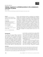
Clinical examination confirmed acute muscular pain, lat-
eral deviation of the mandible to the left during opening
and closing of the mouth, persistent pain and crepitus in
left TMJ, limited opening (interincisal distance 20 mm),
lateral movement to the right (3 mm), lateral movement
to the left (7 mm), and difficulty protruding the mandible
[Figure 2]. Crepitus and pain were determined by palpa-
tion of both joints during maximal protrusion and maxi-
mum mouth opening.
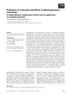
A panoramic radiograph of the patient's jaws prior to
removal of the mandibular left third molar, revealed left
CR [Figure 3]. This type of radiographic examination does
not offer as clear and reliable images as those of other
techniques such as computerized or linear tomography,
but does demonstrate the condyles with a degree of clarity
[10].
Routine haematological analysis did not reveal any evi-
dence of underlying systemic bone disease such as rheu-
matoid arthritis.
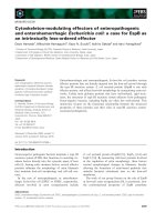
Treatment was anterior repositioning of the mandible
with a hard acrylic splint in the maxilla.
The splint was 3 mm thick, and it was constructed with an
inclined plane for mandibular advancement of 2,5 mm,
re-centring the lower deviated dental midline [Figure 4].
The splint surface was adjusted to obtain a balanced mus-
cular activity, and checked with conventional clinical con-
trol of the dental contacts.
The splint was used consistently, though due to work
commitments, not in the mornings. The patient was
reviewed every month and showed progressive sympto-
matic improvement on each occasion. After 8 months, a
new panoramic radiograph confirmed new bone forma-
tion on the condylar surface [Figure 5]. Clinical features
were improved, with reduced pain (pain scale VAS 20)
and an increase in mouth opening (30 mm), although
deviation of the mandible and crepitus were still evident
during mastication [Figure 6]. After 18 months there was
complete resolution of the symptoms, with no pain, and
similar morphology of both condyles [Figure 7, 8]. At
completion of treatment, there were no occlusal abnor-
malities.
Discussion
Mandibular condylar cartilage is characterised histologi-
cally as fibrocartilage containing a layer of pre-chondrob-
Occlusal relationship of the patient's dentition (top: right side, middle: front view, bottom: left side)Figure 1
Occlusal relationship of the patient's dentition (top:
right side, middle: front view, bottom: left side).
Pre-treatment maximal active mouth openingFigure 2
Pre-treatment maximal active mouth opening.
Pre-treatment panoramic radiograph showing normal mor-phology of right condyle and left condylar resorptionFigure 3
Pre-treatment panoramic radiograph showing nor-
mal morphology of right condyle and left condylar
resorption.
Head & Face Medicine 2009, 5:15 />Page 3 of 5
(page number not for citation purposes)
lastic mesenchymal stem cells which can undergo rapid
differentiation into chondrocytes [11,12].
Other forms of mature articular cartilage do not have such
progenitor cells and only poorly responsive chondrocytes
[13].
This structural difference between mandibular condylar
cartilage and hyaline articular cartilage may explain the
relative difference in their regenerative potential.
The growth of mandibular condylar cartilage may be
influenced by exogenic factors including mechanical fac-
tors.
These phenomena are also present in the adult, though to
a lesser extent [14], since subcondylar trabecular bone for-
mation is apparently not affected by age [15].
Animal exprerimentaion confirms that mandibular
advancement causes cellular changes in rats' condyles
with increased neo-vascularization and new bone forma-
tion significantly higher or equal to the levels towards the
end of growth spurt [16]. Recently, McNamara et al.
reported histological changes associated with mandibular
advancement in adult Rhesus monkeys. In these monkeys,
adaptive changes of the condylar cartilage were evident
after 3 weeks of advancement. Furthermore, the dimen-
sions of the condylar cartilage showed a gradual increase
throughout the experimental period, whereas an
untreated control group had a bony outer layer [17]. Fur-
thermore, Rabie et al. found that 60-day forward mandib-
ular positioning causes adaptive morphological changes
in the condylar head of adult rats [18].
In particular, bone deposition was differential, occurring
not on the anterior surface of the condyle but only on the
posterior and superior surfaces, with compensatory
resorption along the posterior surface of the post-glenoid
tubercle, and the insertion of the lateral pterygoid muscle
into the neck of the condyle [19].
Several authors have suggested that CR is possibly related
to orthodontic treatment [20,21], but no previous ortho-
dontic treatment was reported by this patient.
Splint used for occlusal rehabilitationFigure 4
Splint used for occlusal rehabilitation.
Panoramic radiograph revealing enhanced density of the cor-tical layer over the left condyle 8 months after commence-ment of treatmentFigure 5
Panoramic radiograph revealing enhanced density of
the cortical layer over the left condyle 8 months after
commencement of treatment.
Maximal active mouth opening after 8 monthsFigure 6
Maximal active mouth opening after 8 months.
Panoramic radiograph revealing new growth in the left con-dyle after 18 months of therapyFigure 7
Panoramic radiograph revealing new growth in the
left condyle after 18 months of therapy.
Head & Face Medicine 2009, 5:15 />Page 4 of 5
(page number not for citation purposes)
TMD or bruxism (that may cause TMD), may cause degen-
erative disease of the TMJ [22]. A rewiew of our patient's
clinical data revealed that he had suffered from TMD for
about 4 years and from bruxism for about 18 months.
No evidence of any bone-involving systemic diseases such
as rheumatoid factors and hyperparathyroidism were
found in this patient. However, it is not known how long
the changes of mandibular bone structure had existed,
since the condylar alteration was first noted in the
patient's x-ray prior to the extraction of the left wisdom
tooth. It is probable that the excessive loads produced by
the force of bruxism or TMD were the causes of CR in this
particular case.
Yamada et al. have found that the flattening of the condy-
lar head was the most frequent unilateral condylar
change. Furthermore, these authors noted that CR may be
related to a lateral mandibular shift and a retrognathic
mandible in patients who demonstrate TMD symptoms
[23].
The capacity of TMD to remodel after acute or chronic
trauma, can be used clinically not only in the correction of
skeletal malocclusion, but also in treating occlusal disor-
ders.
Splint therapy is one modality for the management of
TMD. In this case, the use of a full coverage occlusal splint
with mandibular advancement brought about an
improvement of the clinical symptoms and new bone
growth was evidenced radiographically after 18 months.
The occlusal splint can correct the effects of muscle micro-
trauma and associated symptoms of pain or discomfort of
TMJ, and also improve jaw support, as well as facilitating
the spatial re-orientation of the jaw into an optimal posi-
tion.
Mandibular advancement stimulates a differentiation of
proliferative zone cells into chondroblasts with significant
morphological changes in the TMJ [8].
Historically, treatment for CR included, apart from occlu-
sal splint to minimize joint loading (with or without
orthodontics and/or prosthetic therapy), arthroscopic
lysis and lavage, condylectomy and condylar replacement
with a costochondral graft, removal of hyperplastic syno-
vial and bilaminar tissue with disk repositioning and lig-
ament repair, and orthognathic surgery (to correct only
the functional and aesthetic facial deformity) [24-27].
Conclusion
TMJ rehabilitation of patients with CR requires careful
treatment planning.
Studies suggest that increasing age and altered loading
may diminish condylar growth capacity of the TMJ.
Although aging may diminish the capacity for condylar
growth, this case suggests that careful mandibular reposi-
tioning can positively influence the process of remodel-
ling of the condyle.
Consent
Written informed consent was obtained from the patient
for publication of this case report and accompanying
images. A copy of the written consent is available for
review by the Editor-in-Chief of this Journal.
Competing interests
The authors declare that they have no competing interests.
Authors' contributions
AMC and CC carried out the case study. AMC wrote the
article. Both authors read and approved the final manu-
script.
Acknowledgements
The authors would like to thank Prof. A F Markus for his assistance revising
the manuscript.
References
1. Meikle MC: In vivo transplantation of the mandibular joint of
the rat; an autoradiographic investigation into cellular
changes at the condyle. Arch Oral Biol 1973, 18:1011-1020.
2. Moffett BC: Alterations in craniofacial growth resulting from
unilateral fracture of the mandibular condyle in a young rhe-
sus monkey. J Dent Res 1971, 50:1486-1487.
3. Ferretti C, Bryant R, Becker P, Lawrence C: Temporomandibular
joint morphology following post-traumatic ankylosis in 26
patients. Int J Oral Maxillofac Surg 2005, 34:376-381.
4. Roberts WE, Hartsfield JK: Bone development and function:
genetic and environmental mechanisms. Semin Orthod 2004,
10:100-122.
5. Wolford ML: Idiopathic condylar resorption of the temporo-
mandibular joint in teenage girls (cheerleaders syndrome).
Bumc Proceedings 2001, 14:246-252.
6. Ali AM, Sharawy MM: Histopathological changes in rabbit tem-
poromandibular joint associated with experimentally
induced anterior disc displacement. J Oral Pathol Med 1994,
23:364-374.
7. McNamara JA, Hinton RJ, Hoffman DL: Histologic analysis of tem-
poromandibular adaption to protrusive function in young
adult rhesus monkeys. Am J Orthod 1982, 82:288-298.
Close-up view of the left condyleFigure 8
Close-up view of the left condyle.
Publish with Bio Med Central and every
scientist can read your work free of charge
"BioMed Central will be the most significant development for
disseminating the results of biomedical research in our lifetime."
Sir Paul Nurse, Cancer Research UK
Your research papers will be:
available free of charge to the entire biomedical community
peer reviewed and published immediately upon acceptance
cited in PubMed and archived on PubMed Central
yours — you keep the copyright
Submit your manuscript here:
/>BioMedcentral
Head & Face Medicine 2009, 5:15 />Page 5 of 5
(page number not for citation purposes)
8. Isberg AM, Isacsson G: Tissue reaction of the temporomandib-
ular joint flowing retrusive guidance of the mandible. Cranio
1986, 4:143-148.
9. Hinton RJ: Alteration in rat condylar cartilage following dis-
cectomy. J Dent Res 1992, 71:1292-1297.
10. Molina M: Concetti fondamentali di gnatologia moderna ILIC. Milano;
2001:498.
11. Milam SB, Klebe RJ, Triplett RG, Herbert D: Characterization of
the extra-cellular matrix of the primate temporomandibular
joint. J Oral Maxillofac Surg 1991, 49:381-39l.
12. Ellis E, Carlson DS: Histologic comparison of the costo-chon-
dral, sternoclavicular and temporomandibular joints during
growth in Macaca mulatta. J Oral Maxillofac Surg 1986,
44:312-321.
13. Robinson D, Halperin H, Nevo Z: Regenerating hyaline cartilage
in articular defects of old chickens using implants ofembryo-
nal chick chondrocytes embedded in a new natural delivery
substance. Calcýf Tissue Imt 1990, 46:246-253.
14. Bouvier M: Effects of age on the ability of the rat temporo-
mandibular joint to respond to changing functional
demands. J Dent Res 1988, 67:1206-1212.
15. Bouvier M, Zimny ML: Effects of mechanical loads on surface
morphology of the condylar cartilage of the mandible in rats.
Acta Anat 1987, 129:293-300.
16. Rabie ABM, Leung FYC, Chayanupatkul A, Hägg U: The Correlation
Between Neovascularization and Bone Formation in the
Condyle During Forward Mandibular Positioning. Angle
Orthod 2002, 72:431-438.
17. McNamara JA Jr, Peterson JE Jr, Pancherz H: Histologic changes
associated with the Herbst appliance in adult rhesus mon-
keys (Macaca mulatta). Semiars in Orthodontics 2003, 9:26-40.
18. Rabie AB, Xiong H, Hägg U: Forward mandibular positioning
enhances condylar adaptation in adult rats. Eur J Orthod 2004,
26:353-358.
19. Meikle MC: Remodeling the dentofacial skeleton: the biologi-
cal basis of orthodontics and dentofacial orthopedics. J Dent
Res 2007, 86:112-124.
20. Kato Y, Hiyama S, Kuroda T, Fujisaki T: Condylar resorption 2
years following active orthodontic treatment: a case report.
Int J Adult Orthodon Orthognath Surg 1999, 14:243-250.
21. De Meyer MD, De Boever JA: The role of bruxism in the appear-
anc of temporomandibular joint disorders. Rev Belge Med Dent
1997, 52:124-138.
22. Shen YH, Chen YK, Chuang SY: Condylar resorption during
active orthodontic treatment and subsequent therapy:
report of a special case dealing with iatrogenic TMD possibly
related to orthodontic treatment. J Oral Rehabil 2005,
32:332-336.
23. Yamada K, Hiruma Y, Hanada K, Hayashi T, Koyama J, Ito J: Condy-
lar bony change and craniofacial morphology in orthodontic
patients with temporomandibular disorders (TMD) symp-
toms: a pilot study using helical computed tomography and
magnetic resonance imaging. Clin Orthod Res 1999, 2:133-142.
24. Arnett GW, Tamborello JA: Progressive class II development:
female idiopathic condylar resorption. Oral Maxillofac Surg Clin
North Am 1990, 2:699-716.
25. Crawford JG, Stoelinga PJ, Blijdorp PA, Brouns JJ: Stability after
reoperation for progressive condylar resorption after
orthognathic surgery: report of seven cases. J Oral Maxillofac
Surg 1994, 52:460-466.
26. Huang YL, Pogrel MA, Kaban LB: Diagnosis and management of
condylar resorption. J Oral Maxillofac Surg 1997, 55:114-119.
27. Arnett GW, Milam SB, Gottesman L: Progressive mandibular
retrusion – idiopathic condylar resorption. Part I. Am J Orthod
Dentofacial Orthop 1996, 110:8-15.