báo cáo khoa học: " In vitro proliferation of human osteogenic cells in presence of different commercial bone substitute materials combined with enamel matrix derivatives" pdf
Bạn đang xem bản rút gọn của tài liệu. Xem và tải ngay bản đầy đủ của tài liệu tại đây (442.85 KB, 9 trang )
BioMed Central
Page 1 of 9
(page number not for citation purposes)
Head & Face Medicine
Open Access
Research
In vitro proliferation of human osteogenic cells in presence of
different commercial bone substitute materials combined with
enamel matrix derivatives
Christoph Reichert
1
, Bilal Al-Nawas
2
, Ralf Smeets*
3
, Adrian Kasaj
4
,
Werner Götz
1
and Marcus O Klein
2
Address:
1
Department of Orthodontics, Rheinische Friedrich-Wilhelms-University, Bonn, Germany,
2
Department of Oral and Maxillofacial
Surgery, Johannes Gutenberg-University, Mainz, Germany,
3
Department of Oral and Maxillofacial Surgery, University Hospital Aachen, Aachen,
Germany and
4
Department of Operative Dentistry and Periodontology, Johannes Gutenberg-University, Mainz, Germany
Email: Christoph Reichert - ; Bilal Al-Nawas - ; Ralf Smeets* - ;
Adrian Kasaj - ; Werner Götz - ; Marcus O Klein -
* Corresponding author
Abstract
Background: Cellular reactions to alloplastic bone substitute materials (BSM) are a subject of
interest in basic research. In regenerative dentistry, these bone grafting materials are routinely
combined with enamel matrix derivatives (EMD) in order to additionally enhance tissue
regeneration.
Materials and methods: The aim of this study was to evaluate the proliferative activity of human
osteogenic cells after incubation over a period of seven days with commercial BSM of various origin
and chemical composition. Special focus was placed on the potential additional benefit of EMD on
cellular proliferation.
Results: Except for PerioGlas
®
, osteogenic cell proliferation was significantly promoted by the
investigated BSM. The application of EMD alone also resulted in significantly increased cellular
proliferation. However, a combination of BSM and EMD resulted in only a moderate additional
enhancement of osteogenic cell proliferation.
Conclusion: The application of most BSM, as well as the exclusive application of EMD
demonstrated a positive impact on the proliferation of human osteogenic cells in vitro. In order to
increase the benefit from substrate combination (BSM + EMD), further studies on the interactions
between BSM and EMD are needed.
Background
The treatment of quantitative and qualitative defects of
supporting bone tissue is one major aspect of modern
dentoalveolar surgery and periodontology. In this con-
text, alloplastic bone substitute materials (BSM) are well
documented as alternatives to autogenous bone grafts for
certain indications in the management of hard tissue defi-
ciencies [1-5].
Various commercial BSM of different origin, chemical
composition, and micro or macro-structural properties
have been introduced and investigated in recent years [6-
Published: 12 November 2009
Head & Face Medicine 2009, 5:23 doi:10.1186/1746-160X-5-23
Received: 23 May 2009
Accepted: 12 November 2009
This article is available from: />© 2009 Reichert et al; licensee BioMed Central Ltd.
This is an Open Access article distributed under the terms of the Creative Commons Attribution License ( />),
which permits unrestricted use, distribution, and reproduction in any medium, provided the original work is properly cited.
Head & Face Medicine 2009, 5:23 />Page 2 of 9
(page number not for citation purposes)
8]. Today, a large percentage of these BSM are based on
calcium phosphate composites, such as hydroxyapatite
(HA) and tricalcium phosphate (TCP), as well as bioactive
glass (silicate: SiO
2
) [9]. In addition to the various well-
employed substitutes with rather homogenous chemical
compositions, such as Bio-Oss
®
(HA), Cerasorb
®
(β-TCP),
and PerioGlas
®
(SiO
2
), recent developments have focused
on "composites" with different chemical phases, such as
Straumann
®
BoneCeramic (HA + β-TCP) [10], NanoBone
®
(SiO
2
+ HA), and BONIT
®
matrix (SiO
2
+ HA + β-TCP)
[11]. The latter biomaterials have been designed to com-
bine the biological advantages of calcium phosphate and
bioactive glass. Hence, the various BSM feature different
biological behaviour in vitro and in vivo [12-14]. In a
recent in vitro comparison of five commercial bone substi-
tutes, Kuebler et al. [13] demonstrated significant differ-
ences among the investigated specimens with regard to
osteogenic cell proliferation, pointing out the need for
further research.
Emdogain
®
, a commercial mixture of porcine derived
enamel matrix derivatives (EMD), is an evidence-based
option for the treatment of bony defects in periodontal
therapy [15-17]. Biologically active EMD ingredients are
ligands, such as amelogenin, ameloblastin, enamelin, and
tuftelin, that play a crucial role in the development of
teeth and supporting structures [18,19]. A recent system-
atic review summarised the effect of EMD on relevant cell
populations in the periodontal region, such as epithelial
cells, gingival fibroblasts, periodontal ligament cells, oste-
ogenic cells, and cementoblasts as stimulatory rather than
inhibitory [18]. For osteogenic cells specifically, EMD
have been shown to support cell viability and prolifera-
tion in a dose dependent manner [20,21], as well as
encourage cell attachment [22], cell motility [23], and cell
differentiation [22,24,25]. Recent studies of periodontal
regeneration focused on the augmentation of BSM with
EMD [5,26]. However, up to now, no significant clinical
benefit could be measured, making further research on
this approach desirable.
The application of either BSM or EMD into the hard tissue
defect should ideally initiate and support tissue regenera-
tion. For osteogenic cells, cell recruitment and migration
into the defect (osteoconduction), and cell proliferation
precede osteogenic cell differentiation [27], while cell pro-
liferation plays a pivotal role for further successful regen-
eration. In cellular research, many biological assays focus
on cell proliferation. The toxic or radioactive properties of
assays like the H
3
-thymidin or BrDU assay are disadvanta-
geous. The Alamar Blue
®
assay is a well established, non-
toxic, and non-radioactive method for continuously
quantifying cellular proliferation over a long time interval
[28].
The aim of this study was to compare the impact of vari-
ous bone substitute materials on the proliferation of
human osteogenic cells in vitro, employing the Alamar
Blue
®
assay over 7 days. Furthermore, the impact of the
Table 1: Bone substitute materials investigated
Chemical composition and origin Abbr.Commercial name, manufacturer Investigated particle size,
manufacturer's data
tricalcium phosphate:
β-TCP
synthetic CBM Cerasorb
®
M, Curasan 500-1000 μm
BRE Bioresorb
®
Macro Pore,
Oraltronics
®
500-1000 μm
biological apatite: HA bovine BIO Bio-Oss
®
, Geistlich 250-1000 μm
silicate:
SiO
2
synthetic PGL PerioGlas
®
, Sunstar Butler 90-710 μm
biphasic:
β- TCP, HA
synthetic BOC Straumann
®
BoneCeramic,
Straumann
500-1000 μm
biphasic:
SiO
2
, HA
synthetic NBO NanoBone
®
, Artoss mean particle size: 600 μm
triphasic:
SiO
2
, gβ-TCP, HA
synthetic BIM Bonit
®
matrix, DOT 300 x 600 μm
(β-TCP: β-tricalcium phosphate, HA: hydroxyapatite, SiO
2
: silicon dioxide). Abbr.: Abbreviation. Manufacturers: Cursan AG (Kleinostheim,
Germany), Oraltronics
®
Dental Implant Technology GmbH (Bremen, Germany), Geistlich Biomaterials (Baden, Germany), John O. Butler GmbH
(Kriftel, Germany), Straumann GmbH (Freiburg, Germany), Artoss GmbH (Rostock, Germany), DOT GmbH (Rostock, Germany).
Head & Face Medicine 2009, 5:23 />Page 3 of 9
(page number not for citation purposes)
additional application of EMD on osteogenic cell prolifer-
ation activity was investigated.
Materials and methods
Cell Line
A commercial hip bone derived osteoblastic cell line
(HHOBc, PromoCell, Heidelberg, Germany) was utilised.
Cells were cultivated using a standard osteoblast cultiva-
tion medium, consisting of fetal calf serum (FCS, Gibco
Invitrogen, Karlsruhe, Germany), Dulbecco's modified
Eagle's medium (DMEM, Gibco Invitrogen), dexametha-
sone (100 nmol/l, Serva Bioproducts, Heidelberg, Ger-
many), L-glutamine (Gibco Invitrogen), and
streptomycin (100 mg/ml, Gibco Invitrogen). Cultivation
was carried out at 37°C in a constant, humidified atmos-
phere with 95% room air and 5% CO
2
.
Prior to our experiments, the cell line was qualitatively
characterised by the immunohistochemical expression of
alkaline phosphatase (AP) and osteocalcin (labelled
streptavidin-biotin/horseradish peroxidase). Cells were
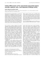
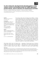
Table 2: Bonferroni's t-test for AB reduction of osteogenic cells
cultivated on the various BSM compared to the control after 7 d
Comparison Diff of Means t p
control vs. BRE 1473 26.8 <0.001**
control vs. NBO 1286 23.4 <0.001**
control vs. BOC 1061 19.3 <0.001**
control vs. CBM 1047 19.1 <0.001**
control vs. BIM 1002 18.2 <0.001**
control vs. BIO 267 4.8 0.001*
control vs. PGL 51 0.9 0.936
(BIO = Bio-Oss
®
, NBO = NanoBone
®
, BRE = Bioresorb
®
, CBM =
Cerasorb
®
M, PGL = PerioGlas
®
, BOC = Straumann
®
BoneCeramic,
BIM = BONIT
®
matrix; t = probability; p = p-value; *significant, **
highly significant).
figure illustrating Bonferroni's t-test for AB reduction of osteogenic cells cultivated on the various BSM compared to the con-trol after 7 dFigure 1
figure illustrating Bonferroni's t-test for AB reduction of osteogenic cells cultivated on the various BSM com-
pared to the control after 7 d.
Head & Face Medicine 2009, 5:23 />Page 4 of 9
(page number not for citation purposes)
passaged at regular intervals, depending on their growth
characteristics, using 0.25% trypsin (Seromed Biochrom
KG, Berlin, Germany).
All trials were carried out at the 4
th
cell passage. Osteo-
genic cells were detached and seeded on the different test
substrates.
Test Substrates and Incubation
Seven different commercial alloplastic BSM were investi-
gated. Except for the biological sample derived from
bovine bone (Bio-Oss
®
), all other samples were synthetic,
composed of pure β-tricalcium phosphate (Cerasorb
®
M,
Bioresorb
®
Macro Pore), pure bioactive glass (PerioGlas
®
),
biphasic BSM (β-tricalcium phosphate + hydroxyapatite:
Straumann
®
BoneCeramic; silicon dioxide + hydroxyapa-
tite: NanoBone
®
) or triphasic BSM (silicon dioxide + β-tri-
calcium phosphate + hydroxyapatite: Bonit
®
matrix). Table
1 provides a synopsis.
The porcine derived protein mixture Emdogain
®
(Strau-
mann, Freiburg, Germany) was utilised as a commercial
EMD.
In our investigation, 100 mg of the respective BSM were
loosely placed into black 24 well plates (Thermo Fisher
Scientific, Langenselbold, Germany), ensuring complete
coverage of the well surface. Wells without BSM served as
a control group. For those wells incubated additionally
with EMD, an emulsion of 100 μg Emdogain
®
/ml was pre-
pared and added to the respective wells. Osteogenic cells
were added to the respective compositions at a density of
1*10
4
cells per well, and further cultivated at 37°C in a
constant, humidified atmosphere of 95% room air and
5% CO
2
.
Alamar Blue
®
proliferation assay
The Alamar Blue
®
(AB) assay (Biozol, Echingen, Germany)
was performed according to manufacturer's guidelines for
the quantification of cellular proliferation. The AB assay is
based on the incorporation of a fluorogenic redox indica-
tor of cell growth in culture. The turnover of AB is a reflec-
tion of cell proliferation, and is quantified by measuring
the fluorescence in Relative Flourescence Units (RFU).
Fluorescence was detected using a fluorescence reader
(FLx800 Microplate Fluorescence Reader, BIO-TEK Instru-
ments, Vinooski, Vermont, USA) at 560/20 nm and 620/
40 nm at the following time points: immediately after the
addition of AB (0 h), then at 3 h, 6 h, 12 h, 24 h, 2 d, 3 d,
4 d and 7 d. Uncultured wells served as a reference. Assays
were run in triplicate for each BSM and BSM/EMD com-
position, and at each time point.
Statistics
Statistical analysis was performed using the statistical soft-
ware SigmaStat (Version 3.1.; Systat Software, Inc., Rich-
mond, USA). Means and standard deviations were
calculated for each group. Results are shown graphically
in a plot (abscissa: point of time, ordinate: RFU values). In
order to identify the BSM or BSM/EMD composition
showing the greatest proliferation after both 24 h and 7 d,
all groups were compared using Bonferroni's t-test. Fur-
thermore, the groups were compared against pure EMD.
To verify the differences between BSM without EMD and
BSM with EMD, a separate t-test was performed. The out-
come each statistical test was considered to be significant
with p < 0.05 and highly significant with p < 0.001.
Results
In general, all of the investigated BSM and BSM/EMD
compositions revealed continuous cell proliferation over
the observation period, with some significant differences.
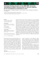
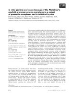
Table 3: Bonferroni's t-test for AB reduction of osteogenic cells
cultivated on the various BSM compared to the untreated
control or EMD after 7 d
Comparison Diff of Means t p
control vs. BRE + EMD 1544 14.0 <0.001**
control vs. NBO + EMD 1327 12.0 <0.001**
control vs. BIM + EMD 1206 10.9 <0.001**
control vs. BOC + EMD 1084 9.8 <0.001**
control vs. CBM + EMD 1002 9.1 <0.001**
control vs. EMD 758 6.9 <0.001**
control vs. BIO + EMD 400 3.6 0.015*
control vs. PGL + EMD 54 0.4 >1.0
EMD vs. PGL + EMD 812 7.0 <0.001**
EMD vs. BRE + EMD 786 6.8 <0.001**
EMD vs. NBO + EMD 569 4.9 0.001*
EMD vs. BIM + EMD 448 3.8 0.009*
EMD vs. BIO + EMD 358 3.1 0.048*
EMD vs. BOC + EMD 326 2.8 0.085
EMD vs. CBM + EMD 244 2.1 0.352
(BIO = Bio-Oss
®
, NBO = NanoBone
®
, BRE = Bioresorb
®
, CBM =
Cerasorb
®
M, PGL = PerioGlas
®
, BOC = Straumann
®
BoneCeramic,
BIM = BONIT
®
matrix; t = probability; p = p-value; *significant, **
highly significant).
Head & Face Medicine 2009, 5:23 />Page 5 of 9
(page number not for citation purposes)
After 24 h, the mean values (standard deviation in paren-
theses) for AB reduction of osteogenic cells cultivated on
the various BSM without EMD were: control 832 (± 25)
RFU, Cerasorb
®
M 963 (± 16) RFU, Bioresorb
®
1073 (± 19)
RFU, Bio-Oss
®
863 (± 18) RFU, PerioGlas
®
705 (± 8) RFU,
Straumann
®
BoneCeramic 963 (± 45) RFU, NanoBone
®
1088 (± 6) RFU, BONIT
®
matrix 1184 (± 32) RFU.
After seven days, the values for AB reduction of osteogenic
cells cultivated on the various BSM without EMD were:
control 1447 (± 20) RFU, BONIT
®
matrix 2450 (± 48)
RFU, Straumann
®
BoneCeramic 2508 (± 100) RFU, Peri-
oGlas
®
1396 (± 31) RFU, Cerasorb
®
M 2494 (± 61) RFU,
Bioresorb
®
2921 (± 69) RFU, NanoBone
®
2733 (± 34)
RFU, Bio-Oss
®
1714 (± 23) RFU. After 7 days, a significant
increase in AB reduction, compared to the negative con-
trol, was found in decreasing order for Bioresorb
®
>
NanoBone
®
> Straumann
®
BoneCeramic > Cerasorb
®
M >
BONIT
®
matrix > Bio-Oss
®
. Furthermore, a slight, but not
significant decrease in AB reduction was documented for
PerioGlas
®
(figure 1, table 2).
After 24 h, AB reduction values for osteogenic cells culti-
vated on the various BSM with EMD were: control 1055 (±
16) RFU, Cerasorb
®
M 1034 (± 40) RFU, Bioresorb
®
1166
(± 13) RFU, Bio-Oss
®
918 (± 24) RFU, PerioGlas
®
701 (±
12) RFU, Straumann
®
BoneCeramic 1045 (± 24) RFU,
NanoBone
®
1181 (± 37) RFU, BONIT
®
matrix 1182 (± 93)
RFU.
After 7 days, AB reduction values for osteogenic cells cul-
tivated on the various BSM with EMD were: control 1447
(± 80) RFU, EMD 2212 (± 80) RFU, BONIT
®
matrix 2660
(± 206) RFU, Straumann
®
BoneCeramic 2538 (± 105)
RFU, PerioGlas
®
1399 (± 30) RFU, Cerasorb
®
M 2456 (±
98) RFU, Bioresorb
®
2998 (± 83) RFU, NanoBone
®
2781
(± 162) RFU, Bio-Oss
®
1854 (± 54) RFU. Compared to the
untreated control group, the AB reduction showed a sig-
nificant increase in descending order for Bioresorb
®
>
NanoBone
®
> BONIT
®
matrix > Straumann
®
BoneCeramic
> Cerasorb
®
M > Emdogain
®
> Bio-Oss
®
. A slight, but not
significant decrease in AB reduction was documented for
PerioGlas
®
(figure 2, table 3). Table 3 also provides a com-
parison between EMD and BSM enriched with EMD.
figure illustrating Bonferroni's t-test for AB reduction of osteogenic cells cultivated on the various BSM compared to the untreated control or EMD after 7 dFigure 2
figure illustrating Bonferroni's t-test for AB reduction of osteogenic cells cultivated on the various BSM com-
pared to the untreated control or EMD after 7 d.
Head & Face Medicine 2009, 5:23 />Page 6 of 9
(page number not for citation purposes)
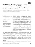
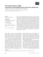
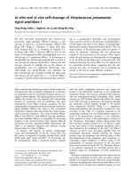
In a comparison of pure BSM and the BSM/EMD compo-
sition, all of the BSM, except for PerioGlas
®
and
BONIT
®
matrix, showed an increase in AB reduction values
at 24 h with the addition of EMD. For NanoBone
®
and
Bioresorb
®
, the addition of EMD resulted in significantly
increased AB reduction values. After 7 days, The only BSM
to show a decrease in the AB reduction value with EMD as
compared to without EMD was Cerasorb
®
M. For Bio-Oss
®
,
the addition of EMD resulted in a significantly increased
AB reduction value (figures 3 and 4, table 4).
Discussion
When employing alloplastic bone substitute materials
(BSM) for guided bone regeneration, the biocompatibility
and biological activity of the material used plays an essen-
tial role, alongside the distinct physical properties of the
graft, like stiffness and stability, for the overall therapeutic
success. In this context, the development of an "ideal"
synthetic bone graft that fulfils the attributes "biocompat-
ible", "degradable", "osteoconductive", and "osteoinduc-
tive" is the focus of recent research. A major issue for the
clinical practitioner is whether a bone graft acts as a plain
defect filler, or has additional osteoconductive or osteoin-
ductive capacities [29]. The pore size of the BSM plays a
crucial role in enhancing the osteoconductive potential of
the BSM. Current literature postulates a minimum pore
size of between 200-400 μm as necessary for osteoconduc-
tion, vascularisation, and formation of mineralised tissue
within a scaffold [30-32]. Furthermore, it is known that an
increasing number of interconnective pores raises the
internal surface area of a BSM, with promotion of the
growth of regenerative cells [33].
The assessment of cell proliferation in vitro provides valu-
able clues about substrate biocompatibility. Furthermore,
proliferating cells are a precondition for osteoconductivity
and osteoinductivity. The BSM investigated in our study
represent a cross-section of the currently commercially
available grafting materials, reflecting the most popular
and well-documented chemical compositions (HA, TCP,
bioactive glasses). The sample size of 100 mg of BSM was
chosen in order to completely cover the floor of a well in
a 24 well plate. This ensured that the majority of the cul-
tivated cells was in close contact with the BSM particles.
Our results suggest that none of the grafting materials
used in this study has a significantly negative influence on
cellular proliferation, as compared to the control. In fact,
all but one of the BSM tested led to an increased AB reduc-
tion over the observation period of 7d. Only PerioGlas
®
showed a slight, but not significant decrease in AB reduc-
tion, compared to the control. Our findings are, to a cer-
tain extent, contrary to former studies [13,34]. Possible
explanations might be dissimilarities in the experimental
set-up. Furthermore, it should be kept in mind that in vitro
studies only give a limited reflection of the complex in vivo
situation.
Although the biomaterial Bio-Oss
®
showed very good
results in various clinical trials [26,35], our in vitro inves-
tigation showed weaker results for cell proliferation as
compared to the other test materials, with the exception of
PerioGlas
®
. These findings for Bio-Oss
®
are in agreement
with other in vitro studies [13]. In our study, all of the
other investigated BSM clearly promoted osteogenic cell
proliferation, with the highest values after 24 h for
BONIT
®
matrix, and after 7 d for Bioresorb
®
Macro Pore.
Nanocrystalline HA (NanoBone
®
) has been shown to pro-
mote other cell lines with osteogenic potential, in a fash-
ion similar to that observed in our study [36].
Table 4: Comparison of BSM without EMD to BSM + EMD on
osteogenic cell proliferation after 24 h and 7 d using the t-test
24 h
Comparison Diff of Means t p
BIO vs. BIO + EMD -54 -2.5 0,06
NBO vs. NBO + EMD -93 -3.4 0,025 *
BRE vs. BRE + EMD -93 -5.7 0,004 *
CBM vs. CBM + EMD -71 -2.3 0,078
PGL vs. PGL + EMD +4 0.4 0,686
BOC vs. BOC + EMD -82 -2.2 0,086
BIM vs. BIM + EMD +2 -1.4 0,231
7d
Comparison Diff of Means T p
BIO vs. BIO + EMD -139 -3.3 0.028 *
NBO vs. NBO + EMD -48 -0.4 0.701
BRE vs. BRE + EMD -83 -0.3 0.400
CBM vs. CBM + EMD +38 -0.4 0.663
PGL vs. PGL + EMD -3 -0.1 0.911
BOC vs. BOC + EMD -29 -0.2 0.787
BIM vs. BIM + EMD -210 -1.4 0.231
(BIO = Bio-Oss
®
, NBO = NanoBone
®
, BRE = Bioresorb
®
, CBM =
Cerasorb
®
M, PGL = PerioGlas
®
, BOC = Straumann
®
BoneCeramic,
BIM = BONIT
®
matrix; t = probability; p = p-value; *significant).
Head & Face Medicine 2009, 5:23 />Page 7 of 9
(page number not for citation purposes)
figure illustrating Comparison of BSM without EMD to BSM + EMD on osteogenic cell proliferation after 24 h and 7 d using the t-testFigure 3
figure illustrating Comparison of BSM without EMD to BSM + EMD on osteogenic cell proliferation after 24 h
and 7 d using the t-test.
figure illustrating Comparison of BSM without EMD to BSM + EMD on osteogenic cell proliferation after 24 h and 7 d using the t-testFigure 4
figure illustrating Comparison of BSM without EMD to BSM + EMD on osteogenic cell proliferation after 24 h
and 7 d using the t-test.
Head & Face Medicine 2009, 5:23 />Page 8 of 9
(page number not for citation purposes)
Conclusion
In our study, the addition of EMD resulted in an increase
in AB reduction for almost all test groups, but significantly
for the control, NanoBone
®
, and Bioresorb
®
Macro Pore
after 24 h, as well as for the control, and Bio-Oss
®
after 7
d. We observed a minimal, EMD-dependent decrease in
AB reduction for PerioGlas
®
after 24 h, and for Cerasorb
®
M after 7 d. Schwarz et al. observed a benefit in the func-
tionalisation of titanium surfaces with EMD [21]. Alto-
gether, the addition of EMD seems to promote osteogenic
cell proliferation to a certain degree. In the routine clinical
situation, the benefit of combining BSM and EMD is well
established, and scientifically documented [15,16,26].
In our study, we found no clear correlation between the
BSM chemical composition or structural properties, and
osteogenic cell proliferation - regardless of the addition of
EMD. Further research must be conducted to understand
the exact modus of interaction between EMD and BSM,
e.g. studies of protein release kinetics from BSM with dif-
ferent chemical and structural properties. We could iden-
tify promising BSM candidates for enhancing osteogenic
cell activity.
Competing interests
The authors declare that they have no competing interests.
Authors' contributions
The study design was established by MOK, CR and BA. CR
and MOK carried out the in vitro experiments and wrote
the manuscript. RS performed the data management and
data analysis. AK and WG carried out the manuscript edit-
ing and manuscript review. All authors read and approved
the final version of the manuscript.
Acknowledgements
This project is supported by a grant (MAIFOR 135/2007) from the Univer-
sity Mainz, medical section, for the promotion of medical research, Ger-
many.
The authors thank the respective companies for providing the bone substi-
tute materials.
References
1. Eppley BL, Pietrzak WS, Blanton MW: Allograft and alloplastic
bone substitutes: a review of science and technology for the
craniomaxillofacial surgeon. J Craniofac Surg 2005, 16(6):981-9.
2. Esposito M, et al.: The efficiancy of various bone augmentation
procedures for dental implants: a Cochrane systematic
review of randomized controlled clinical trials. Int J Oral Max-
illofac Implants 2006, 21(5):696-710.
3. Orsini M, et al.: Long-term clinical results on the use of bone-
replacement grafts in the treatment of intrabony periodon-
tal defects. Comparison of the use of autogenous bone graft
plus calcium sulfate to autogenous bone graft covered with
a bioabsorbable membrane. J Peridontol 2008, 79(9):1630-1637.
4. Sculean A, et al.: Healing of human intrabony defects following
regenerative periodontal therapy with a bovine-derived
xenograft and guided tissue regeneration. Clin Oral Investig
2004, 8(2):70-74.
5. Sculean A, et al.: Clinical and histologic evaluation of an enamel
matrix derivative combined with a biphasic calcium phos-
phate for the treatment of human intrabony periodontal
defects. J Peridontol 2008, 79(10):1991-1999.
6. Klein MO, et al.: Pore characteristics of bone subsitute materi-
als assessed by microcomputed tomography. Clin Oral Implants
Res 2009, 20:67-74.
7. Tadic D, Epple M: A thorough physicochemical characterisa-
tion of 14 calcium phosphate-based bone substitution mate-
rials in comparison to natural bone. Biomaterials 2004,
25(6):987-94.
8. Weibrich G, et al.: [Determining the size of the specific surface
of bone substitutes with gas adsorption]. Mund Kiefer Gesich-
tschir
2000, 4(3):148-52.
9. Kao ST, Scott DD: A review of bone substitutes. Oral Maxillofac
Surg Clin North Am 2007, 19(4):513-21.
10. Dietze S, et al.: The ultrastructure and processing properties of
Straumann Bone Ceramic and NanoBone. Folia Morphol
(Warsz) 2006, 65(1):63-5.
11. Thimm BW, et al.: Biocompatibility studies of endothelial cells
on a novel calcium phosphate/SiO2-xerogel composite for
bone tissue engineering. Biomed Mater 2008, 3(1):15007.
12. Henkel KO, et al.: Macroscopical, histological, and morpho-
metric studies of porous bone-replacement materials in
minipigs 8 months after implantation. Oral Surg Oral Med Oral
Pathol Oral Radiol Endod 2006, 102(5):606-13.
13. Kübler A, et al.: Growth and proliferation of human osteoblasts
on different bone graft substitutes: an in vitro study. Implant
Dent 2004, 13(2):171-179.
14. Schwartz Z, et al.: Differential effects of bone graft substitutes
on regeneration of bone marrow. Clin Oral Implants Res 2008,
19(12):1233-1245.
15. Sculean A, et al.: Ten-year results following treatment of intra-
bony defects with enamel matrix proteins and guided tissue
regeneration. J Clin Periodontol 2008, 35(9):817-24.
16. Sculean A, et al.: The application of an enamel matrix protein
derivative (Emdogain) in regenerative periodontal therapy:
a review. Med Princ Pract 2007, 16(3):167-80.
17. Trombelli L, Farina R: Clinical outcomes with bioactive agents
alone or in combination with grafting or guided tissue regen-
eration.
J Clin Periodontol 2008, 35(s8):117-135.
18. Bosshardt DD: Biological mediators and periodontal regener-
ation: a review of enamel matrix proteins at the cellular and
molecular levels. J Clin Periodontol 2008, 35(s8):87-105.
19. Bosshardt DD, Nanci A: Hertwig's epithelial root sheath,
enamel matrix proteins, and initiation of cementogenesis in
porcine teeth. J Clin Periodontol 2004, 31(8):184-192.
20. Guida L, et al.: In vitro biologic response of human bone mar-
row stromal cells to enamel matrix derivative. J Periodontol
2007, 78:2190-2196.
21. Schwarz F, et al.: Effect of enamel matrix protein derivative on
the attachment, proliferation, and viability of human
SaOs(2) osteoblasts on titanium implants. Clin Oral Investig
2004, 8(3):165-71.
22. Rincon JC, et al.: Enhanced proliferation, attachment and oste-
opontin expression by porcine periodontal cells exposed to
Emdogain. Arch Oral Biol 2005, 50:1047-1054.
23. Klein MO, et al.: In vitro assessment of motility and prolifera-
tion of human osteogenic cells on different isolated extracel-
lular matrix components compared with enamel matrix
derivative by continuous single-cell observation. Clin Oral
Implants Res 2007, 18(1):40-5.
24. Schwartz Z, et al.: Porcine fetal enamel matrix derivative stim-
ulates proliferation but not differentiation of pre-osteoblas-
tic 2T9 cells, inhibits proliferation and stimulates
differentiation of osteoblast-like MG63 cells, and increases
proliferation and differentiation of normal human osteoblast
NHOst cells. J Periodontol 2000, 71:1287-1296.
25. He J, et al.:
Emdogain promotes osteoblast proliferation and
differentiation and stimulates osteoprotegerin expression.
Oral Surg Oral Med Oral Pathol Oral Radiol Endod 2004, 97(2):239-45.
26. Sculean A, et al.: Clinical evaluation of an enamel matrix pro-
tein derivative (Emdogain) combined with a bovine-derived
xenograft (Bio-Oss) for the treatment of intrabony perio-
dontal defects in humans. Int J Periodontics Restorative Dent 2002,
22(3):259-67.
Publish with Bio Med Central and every
scientist can read your work free of charge
"BioMed Central will be the most significant development for
disseminating the results of biomedical research in our lifetime."
Sir Paul Nurse, Cancer Research UK
Your research papers will be:
available free of charge to the entire biomedical community
peer reviewed and published immediately upon acceptance
cited in PubMed and archived on PubMed Central
yours — you keep the copyright
Submit your manuscript here:
/>BioMedcentral
Head & Face Medicine 2009, 5:23 />Page 9 of 9
(page number not for citation purposes)
27. Aubin JE, Heersche JM: Osteoprogenitor cell differentiation to
mature bone-forming osteoblasts. Drug Development Research
2000, 49(3):206-215.
28. Larson EM, et al.: A new, simple, nonradioactive, nontoxic in
vitro assay to monitor corneal endothelial cell viability. Invest
Ophthalmol Vis Sci 1997, 38(10):1929-33.
29. Albrektsson TJC: Osteoinduction, osteoconduction and
osseointegration. Eur Spine J 2001:96-101.
30. Tsuruga E, et al.: Pore size of porous hydroxyapatite as the cell-
substratum controls BMP-induced osteogenesis. J Biochem
1997, 121(2):317-24.
31. Karageorgiou V, Kaplan D: Porosity of 3D biomaterial scaffolds
and osteogenesis. Biomaterials 2005, 26(27):5474-91.
32. Dong J, et al.: Promotion of bone formation using highly pure
porous beta-TCP combined with bone marrow-derived
osteoprogenitor cells. Biomaterials 2002, 23(23):4493-502.
33. Webster TJ, Ahn ES: Nanostructured biomaterials for tissue
engineering bone. Adv Biochem Eng Biotechnol 2007, 103:275-308.
34. Xynos ID, et al.: Bioglass 45S5 stimulates osteoblast turnover
and enhances bone formation In vitro: implications and
applications for bone tissue engineering. Calcif Tissue Int 2000,
67(4):321-9.
35. Traini T, et al.: A histologic and histomorphometric evaluation
of anorganic bovine bone retrieved 9 years after a sinus aug-
mentation procedure. J Periodontol 2007, 78(5):955-61.
36. Kasaj A, et al.: Human periodontal fibroblast response to a
nanostructured hydroxyapatite bone replacement graft in
vitro. Arch Oral Biol 2008, 53(7):683-9.