báo cáo khoa học: " Management of venous thrombosis in fibular free osseomusculocutaneous flaps used for mandibular reconstruction: clinical techniques and treatment considerations" pdf
Bạn đang xem bản rút gọn của tài liệu. Xem và tải ngay bản đầy đủ của tài liệu tại đây (1.45 MB, 5 trang )
HEAD & FACE MEDICINE
Draenert et al. Head & Face Medicine 2010, 6:8
/>Open Access
SHORT REPORT
© 2010 Draenert et al; licensee BioMed Central Ltd. This is an Open Access article distributed under the terms of the Creative Commons
Attribution License ( which permits unrestricted use, distribution, and reproduction in
any medium, provided the original work is properly cited.
Short report
Management of venous thrombosis in fibular free
osseomusculocutaneous flaps used for mandibular
reconstruction: clinical techniques and treatment
considerations
Florian G Draenert*
1
, Martin Gosau
2
and Bilal Al Nawas
1
Abstract
Background: Mandibular reconstruction by means of fibula transplants is the standard therapy for severe bone loss
after subtotal mandibulectomy. Venous failure still represents the most common complication in free flap surgery. We
present the injection of heparine into the arterial pedicle as modification of the revising both anastomoses in these
cases and illustrate the application with a clinical case example.
Methods: Methods consist of immediate revision surgery with clot removal, heparin perfusion by direct injection in
the arterial vessel of the pedicle, subsequent high dose low-molecular weight heparin therapy, and leeches. After 6
hours postoperatively, images of early flap recovery show first sings of recovery by fading livid skin color.
Results: The application of this technique in a patient with venous thrombosis resulted in the complete recovery of
the flap 60 hours postoperatively. Other cases achieved similar success without additional lysis Therapy or revision of
the arterial anastomosis.
Conclusion: Rescue of fibular flaps is possible even in patients with massive thrombosis if surgical revision is done
quickly.
Background
Mandibular and maxillary reconstruction with fibular
osseomusculocutaneous free flaps represents a common
procedure that is often applied in primary and secondary
reconstructions of large bony defects in these areas [1,2].
A possible complication of free flap procedures is venous
failure of the anastomosis [2], which demands immediate
revision surgery involving clot removal and anticoagula-
tion therapy. We avoid the reopening of the arterial anas-
tomosis by injecting the necessary rinsing with heparin in
the arterial vessel with a small syringe.
Methods
We apply standard anti-thrombosis prophylaxis with low
molecular weight heparin, for instance, Fragmin P, but do
not preoperatively use any further anti-coagulatives, such
as ASS or high dose heparin. Signs of venous failure after
flap surgery, which becomes visible by livid skin color,
represent a peracute indication for revision surgery.
Therefore, nursing staff in the intensive care unit control
the flap every 2 hours within the first 72 hours after initial
surgery. This procedure includes visual control of the flap
color, refill control by mild compression, and palpation of
the flap consistence. The revision procedure includes
opening of the venous anastomosis, clot removal, and
flap perfusion with 3 ml heparin solution (5000 I.E./ml).
This solution is injected in the pedicle artery three times,
resulting in high frequency coagulation of the punctual
bleeding. In this technique, the arterial anastomosis is
not opened but anticoagulation is injected in the pedicle
artery. The venous vessel is re-anastomozed after several
minutes of continuous blood flow from the venous pedi-
cle vessel. Post-surgical treatment includes the use of
leeches applied three times a day (four to six leeches on
the skin island) until return of normal skin color.
* Correspondence:
1
Clinic for Maxillofacial Surgery, University of Mainz, Augustusplatz 2, 55131
Mainz, Germany
Full list of author information is available at the end of the article
Draenert et al. Head & Face Medicine 2010, 6:8
/>Page 2 of 5
Case report
We report the successful clinical management of a 55-
year old man with venous thrombosis of the pedicle after
mandibular reconstruction by means of a osseomusculo-
cutaneous fibular flap. Because of the diagnosis of a
squamous cell carcinoma in the mandibular region in
January 2007, the patient underwent hemimandibulec-
tomy and primary soft tissue reconstruction with a radial
forearm flap in combination with bilateral neck dissec-
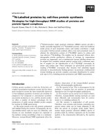
tions (see fig. 1a and 1b). The histopathological examina-
tion showed a TNM-classification of T4a, N2c, Mx, R0,
G2. After surgery, the patient underwent radiotherapy
with 60 Gy, which resulted in partial necrosis of the lower
lip and radiofibrosis of the surrounding soft tissue. In
June 2008, the reconstruction plate perforated the epider-
mis and was subsequently removed.
Two years after the first surgical intervention, the
patient received a second mandibular reconstruction
without recurrence on 4 May 2009. A fibular osseomus-
culocutaneous flap was harvested from the right lower
limb, transplanted in the mandibular defect site, and
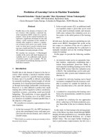
fixed with a reconstruction plate (see fig. 1c and fig. 2).
The artery was re-anastomosized to an appropriate vessel
in the area of the main branch of the arteria thyroidea.
Because of the lack of small vessels, venous anastomosis
was done at the internal jugular vein. No complications
occurred during the first 60 postoperative hours (see fig.
3a). The flap developed venous failure visible by livid skin
color 60 hours after surgery (see fig. 3b). The venous part
of the pedicle showed a massive thrombus at revision sur-
gery (see fig. 3c). The clot was removed and the flap was
perfused with 3 ml heparin solution (5000 I.E./ml), which
was injected in the pedicle artery three times, resulting in
high frequency coagulation of the punctual bleeding. The
flap showed recovery of the venous function 6 hours after
revision surgery detectable by the fading of the dissemi-
nated spots of livid color (see fig. 3d). The patient
received Fraxiparine 0.9 mg twice per day for 2 weeks,
and leeches were applied to the skin island of the flap
three times per day. In the following weeks, the flap
showed complete recovery with small areas of necrosis at
the margins of the flap (see fig. 3e).
Results and discussion
The described technique for treating venous thrombosis
in microvascular flap surgery avoids the opening of the
arterial anastomosis. This procedure has been success-
fully applied in several patients at the Departments for
Maxillofacial Surgery of the Universities of Mainz and
Regensburg as presented in this case example from Mainz
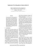
(see table 1).
Late bony reconstruction after radiotherapy is still
widely applied in Germany, even though early bony
reconstruction has promised some advantages [3].
Patients with osteoradionecrosis and large bony defects
require microvascular bony flaps, such as fibula or scap-
ula transplants [4,5]. After radiotherapy, the number of
venous vessels suitable for microsurgical re-anastomosis
of the flap is often limited to jugular veins [6-8]. Compro-
Figure 1 A: 3D-CT image before tumor resection. The infiltration of the bone is clearly visible. B: 3D-CT image after tumor resection with a man-
dibular continuity defect. C: 3D-CBCT after fibular osseomusculocutaneous flap reconstruction.
Figure 2 Fibular flap harvested from the right lower limb.
Draenert et al. Head & Face Medicine 2010, 6:8
/>Page 3 of 5
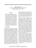
Figure 3 A: 24 h after re-anastomosis. A regular pink color of the skin island can be observed. B: Livid skin color after 60 hours indicates venous
thrombus. C: The situs during revision surgery (a: artery, v: vein, star: location of venous anastomosis). D: Signs of venous function visible by fading livid
skin color 6 hours after revision surgery. E: Regular wound healing and correct vessel function 5 weeks after fibular transplant surgery.
Draenert et al. Head & Face Medicine 2010, 6:8
/>Page 4 of 5
mised venous vessels in the donor region may lead to
venous failure [9]. A further risk of thrombus formation is
the higher prethrombotic activity in irradiated vessels
[10]. Imaging techniques, such as angiography, can be
applied to evaluate the vascular situation in advance [11].
We keep to the recommended practice of a minimum
surveillance time of 45 min after the anastomosis of flap
vessels [12].
Our described monitoring regiment includes visual
control, palpation, and a manual refill test that is also
described by other authors [13]. Further methods, such as
a Doppler probe, are not applied [13]. Intensive care unit
personnel densely control during the first 72 postopera-
tive hours. The surgeons of our clinic additionally check
the flap at least twice a day.
We immediately revise venous complications. This reg-
iment is also described by other authors [13-16]. Local
heparin injection is a well-known procedure in the man-
agement of venous thrombosis [13]. We avoid the open-
ing of the artery by injecting high dose heparin into the
pedicle artery.
Adjuvant therapy with leeches is also common practice
[13,17]. Flap survival after venous thrombosis in fibula
flaps is possible in most patients, but the survival rate of
flaps with a bony component is lower [14]. The presented
technique is one possible regiment in patients with
venous thrombosis after mandibular reconstruction by
means of fibular free osseomusculocutaneous flaps. We
did not apply lysis therapy and never did it. However this
is also a known practice with good results in several pub-
lications [18-21].
Consent
Written informed consent was obtained from the patient
for publication of this case report and accompanying
images. A copy of the written consent is available for
review by the Editor-in-Chief of this journal.
Competing interests
The authors declare that they have no competing interests.
Authors' contributions
FGD wrote the manuscript and operated the case report patient, MG docu-
mented the patients in Regensburg, BA was correcting senior author on the
manuscript. All authors read and approved the final manuscript.
Author Details
1
Clinic for Maxillofacial Surgery, University of Mainz, Augustusplatz 2, 55131
Mainz, Germany and
2
Clinic for Maxillofacial Surgery, University Hospital
Regensburg, Franz-Josef-Strauss-Allee 11, 93053 Regensburg, Germany
References
1. Wei FC, Celik N, Yang WG, Chen IH, Chang YM, Chen HC: Complications
after reconstruction by plate and soft-tissue free flap in composite
mandibular defects and secondary salvage reconstruction with
osteocutaneous flap. Plast Reconstr Surg 2003, 112:37-42.
2. Brown JS, Magennis P, Rogers SN, Cawood JI, Howell R, Vaughan ED:
Trends in head and neck microvascular reconstructive surgery in
Liverpool (1992-2001). Br J Oral Maxillofac Surg 2006, 44:364-370.
3. Thorwarth M, Eulzer C, Bader R, Wolf C, Schmidt M, Schultze-Mosgau S:
Free flap transfer in cranio-maxillofacial surgery: a review of the
current data. Oral Maxillofac Surg 2008, 12:113-124.
4. Curi MM, Oliveira dos Santos M, Feher O, Faria JC, Rodrigues ML, Kowalski
LP: Management of extensive osteoradionecrosis of the mandible with
radical resection and immediate microvascular reconstruction. J Oral
Maxillofac Surg 2007, 65:434-438.
5. Chang DW, Oh HK, Robb GL, Miller MJ: Management of advanced
mandibular osteoradionecrosis with free flap reconstruction. Head
Neck 2001, 23:830-835.
6. Chalian AA, Anderson TD, Weinstein GS, Weber RS: Internal jugular vein
versus external jugular vein anastamosis: implications for successful
free tissue transfer. Head Neck 2001, 23:475-478.
7. Hong P, Taylor SM, Trites JR, Maclean J, Hart RD: Use of the external
jugular vein as the sole recipient vein in head and neck free flap
reconstruction. J Otolaryngol 2006, 35:361-365.
8. Yazar S: Selection of recipient vessels in microsurgical free tissue
reconstruction of head and neck defects. Microsurgery 2007,
27:588-594.
9. Fukuiwa T, Nishimoto K, Hayashi T, Kurono Y: Venous thrombosis after
microvascular free-tissue transfer in head and neck cancer
reconstruction. Auris Nasus Larynx 2008, 35:390-396.
10. Halle M, Ekstrom M, Farnebo F, Tornvall P: Endothelial activation with
prothrombotic response in irradiated microvascular recipient veins. J
Plast Reconstr Aesthet Surg 2010 in press.
11. Whitley SP, Sandhu S, Cardozo A: Preoperative vascular assessment of
the lower limb for harvest of a fibular flap: the views of vascular
Received: 2 November 2009 Accepted: 7 June 2010
Published: 7 June 2010
This article is available from: 2010 Draenert et al; licensee BioMed Central Ltd. This is an Open Access article distributed under the terms of the Creative Commons Attribution License ( .0), which permits unrestricted use, distribution, and reproduction in any medium, provided the original work is properly cited.Head & Face Medicine 2010, 6:8
Table 1: Free fibula osseomusculocutaneous flaps with venous thrombosis treated following the described regiment.
gender age (years) diagnosis reconstruction type radiatiotherapy before flap
surgery
result of
revision
male 55 osteoradionecrosis late yes flap survived
male 48 squamous cell
carcinoma
immediate yes flap survived
male 46 squamous cell
carcinoma
immediate no flap survived
male 26 Ewing sarcoma late no, but chemotherapy flap lost
Draenert et al. Head & Face Medicine 2010, 6:8
/>Page 5 of 5
surgeons in the United Kingdom. Br J Oral Maxillofac Surg 2004,
42:307-310.
12. Wolff KD, Holzle F, Wysluch A, Mucke T, Kesting M: Incidence and time of
intraoperative vascular complications in head and neck microsurgery.
Microsurgery 2008, 28:143-146.
13. Kubo T, Yano K, Hosokawa K: Management of flaps with compromised
venous outflow in head and neck microsurgical reconstruction.
Microsurgery 2002, 22:391-395.
14. Arai K, Toh S, Tsubo K, Nishikawa S, Narita S, Miura H: Complications of
vascularized fibula graft for reconstruction of long bones. Plast
Reconstr Surg 2002, 109:2301-2306.
15. Hirsch DL, Bell RB, Dierks EJ, Potter JK, Potter BE: Analysis of microvascular
free flaps for reconstruction of advanced mandibular
osteoradionecrosis: a retrospective cohort study. J Oral Maxillofac Surg
2008, 66:2545-2556.
16. Suh JD, Sercarz JA, Abemayor E, Calcaterra TC, Rawnsley JD, Alam D,
Blackwell KE: Analysis of outcome and complications in 400 cases of
microvascular head and neck reconstruction. Arch Otolaryngol Head
Neck Surg 2004, 130:962-966.
17. Conforti ML, Connor NP, Heisey DM, Hartig GK: Evaluation of
performance characteristics of the medicinal leech (Hirudo
medicinalis) for the treatment of venous congestion. Plast Reconstr Surg
2002, 109:228-235.
18. Serletti JM, Moran SL, Orlando GS, O'Connor T, Herrera HR: Urokinase
protocol for free-flap salvage following prolonged venous thrombosis.
Plast Reconstr Surg 1998, 102:1947-1953.
19. Yii NW, Evans GR, Miller MJ, Reece GP, Langstein H, Chang D, Kroll SS,
Wang B, Robb GL: Thrombolytic therapy: what is its role in free flap
salvage? Ann Plast Surg 2001, 46:601-604.
20. Tran NV, Bishop AT, Convery PA, Yu AY: Venous congestive flap salvage
with subcutaneous rtPA. Microsurgery 2006, 26:370-372.
21. Panchapakesan V, Addison P, Beausang E, Lipa JE, Gilbert RW, Neligan PC:
Role of thrombolysis in free-flap salvage. J Reconstr Microsurg 2003,
19:523-530.
doi: 10.1186/1746-160X-6-8
Cite this article as: Draenert et al., Management of venous thrombosis in
fibular free osseomusculocutaneous flaps used for mandibular reconstruc-
tion: clinical techniques and treatment considerations Head & Face Medicine
2010, 6:8