báo cáo khoa học: " Surgical treatment of gingival overgrowth with 10 years of follow-up" potx
Bạn đang xem bản rút gọn của tài liệu. Xem và tải ngay bản đầy đủ của tài liệu tại đây (586.3 KB, 7 trang )
RESEARC H Open Access
Surgical treatment of gingival overgrowth with
10 years of follow-up
Andrea Ballini
1
, Adele Scattarella
1
, Vito Crincoli
1
, Roberto Gianfranco Carlaio
1
, Francesco Papa
1
, Letizia Perillo
3
,
Teodoro Romanazzo
1
, Maria Virginia Bux
1
, Gianna Maria Nardi
2
, Angela Dituri
1
, Stefania Cantore
1
,
Francesco Pettini
1
, Felice Roberto Grassi
1*
Abstract
Background: In some pathological conditions, gingivitis caused by plaque accumulation can be more sev ere, with
the result of an overgrowth. Nevertheless, the overgrowth involves the gingival margin with extension to the inter-
dental papilla. The lesion may involve the inter-proximal spaces, and become so extensive that the teeth are
displaced and their crowns covered. Severe overgrowth may lead to impairment in aesthetic and masticatory
functions, requiring surgical excision of the excessive tissue. Aim of this study is to describe an operative protocol
for the surgical treatment of localized gingival overgrowth analyzing the surgical technique, times and follow-up.
Methods: A total of 20 patients were enrolled and underwent initial, non surgical, periodontal treatment and
training sessions on home oral hygien e training. The treatment plan involved radical exeresis of the mass follo wed
by positioning of an autograft of connective tissue and keratinized gingiva.
Results: During 10 years of follow-u p, all the grafts appeared well vascularized, aesthetically satisfactory, and
without relapse.
Conclusions: Periodontal examinations, surgical procedures, and dental hygiene with follow-up are an essential
part of the treatment protocol. However, additional effort is needed from the patient. Hopefully, the final treatment
result makes it all worthwhile.
Background
The term gingival overgrowth (GO) only provides a
topographic description of the lesion but no histological
diagnosis.
Moreover, the histological classification is still unclear,
owing to the wide range of possible histological mor-
photypes [1,2].
In fact, elements of granulation tissue are frequently
observed, as are giant cells, mesenchymal cells combined
or not with fibroblasts, collagen, epithelial cells, calcifi-
cation zones and vessels [2].
From the epidemiologic point of view, GO most often
affects the female sex, at ages ranging from 6 to 80
years but with a prevalence between the second and
fifth decades of life [3,4].
The etiology is still unknown, although there is a con-
sensus from some Authors that chronic local trauma
(plaque, poor oral hygiene, defective restoration, foreign
bodies such as food impaction or toothbrush bristle)
can trigger chronic inflammation of the periodontal tis-
sue, together with an endocrine or metabolic imbalance,
which may determine the onset of the lesions [1,3,4].
Among the important systemic conditions in the etio-
pathogenesis of GO, hormonal factors must be borne in
mind, which have a fundamental role in amplifying the
tissue reaction to chronic inflammatory conditions [5].
In current clinical descriptive terminology, GO can be
classificated as [1,3,6]:
A-) According to etiologic factors an pathologic
changes, GO could be listed out as:
I-) Inflammatory overgrowth
a. Chronic
b. Acute
II-) Drug-induced overgrowth
* Correspondence:
1
Department of Dental Sciences and Surgery, University of Bari, Bari, Italy
Full list of author information is available at the end of the article
Ballini et al. Head & Face Medicine 2010, 6:19
/>HEAD & FACE MEDICINE
© 2010 Ballini et al; licensee BioMed Central Ltd. This is an Open Access article distributed under the terms of the Creative Commons
Attribution License ( which permits unrestricted use, distribut ion, and reproduction in
any medium, provided the original work is properly cited.
III-) Overgrowth associated with systemic disease
a. Conditioned overgrowth
1. Pregnancy
2. Puberty
3. Vitaminic C deficiency
4. Plasma Cell gingivitis
5. Non- specific conditioned overgrowth (granuloma
pyogenicum)
b. Systemic diseases causing gingival overgrowth:
1. Leukemia
2. Granulomatous diseases
IV-) neoplastic overgrowth (gingival tumors)
V-) False overgrowth
B-) According to location and distribution, gingival
overgrowth can be classified as:
Localized: gingival overgrowth limited to one or more
group of teeth
Generalized: Entire mouth
Papillary: Confined to the interdental papilla
Dif fuse: Involves all the parts of the gingival, i.e. mar-
ginal, attached and interdental gingival
Discrete: isolated sessile or peduncolated tumor-like
overgrowth.
Three different types of drugs are associated with GO,
namely anti-convulsant, calcium channel blockers and
the immunosuppressants like cyclosporine [6].
Cyclosporine A (CsA) has been the primary tool to
prevent the reje ction of organ transplants. CsA is still
the mostly used drug in renal transplant therapy [6].
However, there is evidence that use of Tacrolimus
causes fewer oral side-effects than CsA [7,8].
The histopathological classification of GO is as fol-
lows: gigant cell, fibromatous, peripheral ossification and
congenital [1,2,9].
There are various, controversial theories as to the ori-
gin of those cells, whereby some Authors believe that
they could derive from the osteoclasts, other Authors
attribute t hem a mesenchymal origin, or an endothelial
origin and yet other Authors consider that they derive
from pericapillary adventitial cells [9-11].
Finally, the epithelial lining of the giant cell form is of
multilayered type with signs of hyper- and para-keratosis
combined with ulcerative phenomena [4].
The peripheral ossification form shows a histological
drawn of layers of connective tissue with an irregular
appearance and a rich content of bone trabeculae and
calcified matter in the stroma [9,10].
Instead, in the third form of GO mature connective
tissue is present, lined by a hyper-para-keratosic
epithelium.
Ther e is often a modest degree of aspecific inflamma-
tory infiltrate[1-3].
In the past, treatment was obtained by complete exer-
esis of the mass and removal of the adjacent tooth or
teeth to avoid recurrence, thus resulting in a very poor
aesthetic and functional outcome [11].
Nowadays, classic treatment of GO is by surgical exci-
sion of the lesion with curettage of the dental and peri-
odontal structures in the involved area, and histological
analysis of the removed tissue [5,11,12].
Instead, some studies have proposed the use of laser
treatment as a valid alternative to conventional surgical
treatment [12-16].
According to these studies, traditional surgical
excision is not only extremely difficult but also causes
post-surgical pain, gingival deformity and a difficult
post-surgical course.
All this can complicate home oral hygiene procedures
and allow bacterial colonization, that can often delay
patients healing [17,18].
Aimofthisstudywastodescribeanoperativeproto-
col for localized GO (using free soft tissue grafts), the
surgical timing and follow-up.
In fact, as descri bed before, a number of surgical pro-
cedures have been proposed to treat GO.
In this study it is used free soft tissue grafts, because
this procedure increase the width of keratinised tissue
and improve aesthetics results.
Patients and Me thods
Case series presentation
We report on 20 patients (8 males and 12 females) with
a mean age of 29 ± 4 months, with different etiopatho-
genesis of localized GO present from 15 days to 12
month (Table 1).
Only two patients were in therapy with drugs that can
influence GO (phenylhydantoin, nifedipine).
Also in those cases of drug-induced hyperplasia the
GO were localized.
The study was performed in accordance with the
Dec laration of Helsinki [19] and t he guidel ine for Good
Clinical Practice [20].
All patients were able to give consent to participation
in the study after receiving oral and written information.
Each patie nt underwent an initial non surgical period-
ontal treatment, with full-mouth tooth polishing and
oral hygiene home instructions.
Home oral hygiene also included the use of a si ngle
tufted brush for the less accessible zones.
Patients were instructed to use a liqu id plaque indica-
tor (GC Plaque Indicator Kit®), to remove all visible pla-
queverymeticulouslywithtoothbrushandusinga1%
chlorhexidine gel (Cor sodyl dental gel®-GlaxoSmithKl ine
-Brantford, Middlesex, UK).
Root debridement was carried out with manual and
ultrasonic instruments to complete the baseline therapy.
This prot ocol was able to eliminate the local aggrava-
tion factors and thus guarantee a good surgical result.
Ballini et al. Head & Face Medicine 2010, 6:19
/>Page 2 of 7
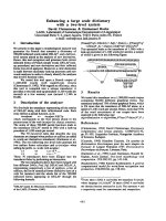
In 12 patients the GO was localized in the upper
jaw, between the central and the lateral (fig. 1) incisor
(7 subjects), between the lateral incisor and the canine
(5 subjects), while in the remaining 8 it was localized in
the mandible between the canines.
Before non-surgical therapy, three different indexes of
periodontal health were analyzed (for full mouth): prob-
ing depth (PD), plaque index according to 0’Leary (PI)
[21] and Gingival index according to Löe-Silness (GI)
[22,23]. (Table 2)
Surgical treatment
After local anesthesia, intrasulcular incisions were made
at the buccal and lingual sides with Bard-Parker surgical
blade n° 15, at least one tooth away from the mesial
portion, distally to the graft site, to create access for t he
tools and facilitate the direct clinical view of the defect.
A full-thickness flap was elevated and the granulation
tissue was removed showing the true extension and
depth of the periodontal defect.
On the palatal aspect, t he size of the grafts was mea-
sured using a periodontal probe (XP 23/UNC15, Hu-
Friedy MFG-Co, Inc., Chicago, IL, USA).
The autograft, obtained from the donor site (in our
case, the palatine mucosa of the maxillary pre-molars)
Table 1 Time of beginning, etiologic factor and associated disease distribution
PATIENTS AGE SEX DAYS/MONTHS SINCE ONSET ETIOLOGIC FACTORS OTHER DISEASES
AND DRUGS
THERAPY
1 25 M 15 days Calculus None
2 43 F 10 months Unsuitable prosthesis Diabetes mellitus
(insulin)
3 25 F 5 months Calculus Psoriasis
(betamethasone)
4 26 F 1 year Calculus, None
(oral contraceptives)
5 35 M 7 months Food Impaction Sjogren’s Syndrome
(pilocarpine, carboxymethyl cellulose collirium)
6 42 M 10 months Calculus Epilepsy
(phenylhydantoin)
7 68 F 1 year Plaque, calculus Hypertension (nifedipine)
8 50 F 8 months Plaque, calculus None
9 21 F 2 months Plaque None
10 33 M 2 months Plaque None
11 54 M 6 months Unsuitable prosthesis Rheumatoid arthritis
12 34 F 9 months Calculus Systemic lupus erythematosus
(betamethasone)
13 65 M 1 year Unsuitable prosthesis osteoarthrosis
14 24 F 4 months Calculus None
15 39 F 1 year Calculus None
(oral contraceptives)
16 43 F 2 years Calculus,
deciduous roots
Crohn’s disease
(prednisolone, azathioprine)
17 52 M 8 months Unsuitable prosthesis None
18 63 M 1 year Calculus Behcet ‘s disease
(prednisolone)
19 56 F 10 months Food Impaction Osteoarthrosis
20 34 F 6 months Calculus None
Figure 1 Intraoral view of gingival overgrowth.
Ballini et al. Head & Face Medicine 2010, 6:19
/>Page 3 of 7
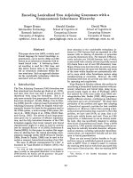
and free from keratinized tissue, were positioned in the
host site consisting of bone and periosteum (fig. 2)
The graft were preserved in sterile physiological solu-
tion and then cleaned from the adipose tissue; it was
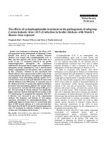
stabilized with stabilizing periosteal silk suture (fig. 2; 3).
Finally, the fla p were r e-positioned and sutured with
single stitches; in our protocol, the donor area was
closed by three interrupted sutures in 3-0 silk (two at
the borders and one in the centre) before grafting the
recipient site.
Firm pressure were exerted with fingers for 2 - 3 min-
utes using a gauze dipped in physiological solution, to
reduce the blood clot and promote healing.
All patients were placed on the following medication:
azithromycin 500 mg once a day, for 3 days.
Sutures were removed from the donor site after 1
week.
During sutures removal, no important tissues inflam-
mations were observed.
All bioptical samples were analyzed by the pathologist.
Maintenance and follow-up
After surgical procedures, patients were instructed to
rinse their mouths twice daily with 10 ml of and 0,12%
chlorexidine (Corsodyl mothwash® - GlaxoSmithKline
-Brantford, Middlesex, UK) rinse for 1 min, 3 times a
day, for 6 weeks.
Discomfort was assessed as the level of pain experi-
enced by the patients during the postoperative first week
due to the palatal wound by Visual analogue scale (VAS).
Table 2 Initial and final distribution of probe depth (P.
D.), plaque index (P.I.) and gingival index (G.I.), before
(T
0
) and after (T
1
) non surgical therapy in 20 Patients
(Pt) with different type of Epulides
Pt. P.D.
(T
0
)
P.D.
(T
1
)
P.I.
(T
0
)
P.I.
(T
1
)
G.I.
(T
0
)
G.I.
(T
1
)
Epulides
1 9 2 28% 19% 1 0 Fibromatous
2 8,5 3 23% 20% 1 0 Fissured
3 6 3 31% 22% 2 0 Gravidic
4 10 4 19% 9% 1 0 Peripheral ossification
5 11 2,5 25% 14% 2 0 Gigant-cell
6 7 2,5 21% 18% 1 0 Peripheral ossification
7 12 3 27% 25% 1 0 Fissured
8 10 4 40% 33% 3 1 Gigant-cell
9 7 2,5 51% 38% 3 1 Gravidic
10 8 3 45% 23% 3 1 Fibromatous
11 6 2 47% 27% 3 1 Fissured
12 9 3,5 55% 35% 3 1 Gigant-cell
13 9 3 60% 46% 3 0 Fissured
14 10 3 73% 45% 3 1 Gravidic
15
12 4 65% 31% 3 1 Fibromatous
16 8 4 68% 29% 3 1 Fibromatous
17 12 2,5 78% 34% 3 0 Fissured
18 12 3 71% 38% 3 1 Fibromatous
19 10 3 62% 29% 3 1 Peripheral ossification
20 9 3 64% 32% 3 1 Fibromatous
Figure 2 The graft was positioned in the host site with
stabilizing periosteal silk suture.
Figure 3 Palatal view of the donor site.
Ballini et al. Head & Face Medicine 2010, 6:19
/>Page 4 of 7
Three-point VAS (’ none’, ‘mild or moderate’, ‘severe’)
was used to record discomfort levels reported by the
participants.
Thefirstdayfromsurgeryanumberof13patients
referred for mild discomfort and 7 for a severe
discomfort.
At five days from surgery 15 patients referred for none
discomfort and 5 for mild discomfort.
At a 10-day follow-up, post-operative clinical assess-
ment demonstrated a G.I. grade of 0 or 1.
The participants in the study did not receive any di et-
ary guidance except for the day of the surgery itself,
when a diet based on soft and cold foods was suggested,
taking care to chew on the opposite side of the mouth
with respect to the donor site for the first week.
The patients then underwent a rigorous follow-up
sche dule at 30 days to assess the PI and GI, an d to per-
form periodontal debridement. Complete epithelialisa-
tion of the palatal wound occurred in all patients only 4
weeks after surgery. At the 6 months follow-up visit the
assessments of all the i ndexes were repeated. The PI
and GI notably improved in most patients (Table 2).
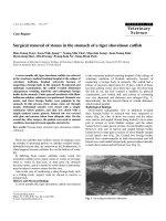
The follow-up schedule involved visits at 1 year,
2,3,4,5,6,7,8,9 and10 years from the procedure (fig. 4; 5; 6).
Results and Disc ussion
All patients referred that the post-operative course was
free from any complication either at the donor or the
host site.
After 10 years from the procedure, all patients had an
aesthetically satisfactory gingival appearance and no sign
of recurrence.
All the grafts were well vascularized and aesthetically
satisfactory.
Unlike the classic approach,thesurgicaltechnique
here described involved the use of a free gingival graft
obtained from the palatine m ucosa to cover the tissue
gap in the host site.
The palatal mucosa in the premolar region is the ideal
area for obtaining a graft for anatomic reasons [24], as
an adequate thickness of the graft is ensured [25] with-
out causing any damage to the greater palatine artery.
The main disadvantages of free soft tissue techniques
are the double surgical woun d and the relative discom-
fort suffered by the patient.
An other Author proposed the trap-door technique
with the aim of keeping the epithelial layer intact
to achieve healing by primary intention in the donor
area [26].
This method was described as more conservative and
less t raumatic for the patient with localized G O, ensur-
ing healing by primary intention and reducing palatal
discomfort as reported in VAS table (Table 3).
Not only did this markedly improve the patients com-
fort but it also yield an aesthetically satisfying result
thanks to the width and thickness of the keratinized tis-
sue used, as well as safeguarding the site from the risk
of recurrence [5].
The muco-gingival complex showed no functional or
aesthetic damage and no bone reabsorption occurred,
Figure 4 Clinical aspect two years after surgery.
Figure 5 Follow-up at six years later.
Figure 6 Follow-up at 10 years: an aesthetically satisfactory
gingival appearance and no signs of recurrence.
Ballini et al. Head & Face Medicine 2010, 6:19
/>Page 5 of 7
for exposure of the root surfaces in the involved
area [10].
In the twenty treated cases, not only did no recur-
rence develop, but no further surgical correction was
required.
Althoughtheroleofplaquehasnotbeenclearly
def ined consist ently with other several authors [17], the
hyper plastic tissue tends to aid plaque accumulation
and to inhibit plaque removal, increases the gingival
inflammation.
A treatment protocol including careful training in oral
hygiene, combined with a valid surgical technique is
therefore essential to resolve the problem of localized
gingival overgrowth.
The additional use of chlorhexidine both in the initial
and the maintenance therapy to ensure clinical control
of plaque was also highly beneficial.
All patients need to be instructed in the correct use of
oral hygiene measures and above all, to underg o regular
professional prophylactic treatment.
The role of the dental hygienist was fundamental for
co-adjuvant support therapy, and in ensuring good
patient compliance [17].
Conclusions
There are many reasons for GO.
Mostly, proper oral hygiene is sufficient to achieve
normal healthy gum
In some situations, however, gingival hyperplasia is
drug-induced or can be a manifestation of a genetic dis-
order. In the latter, it may exist as an isolated abnormal-
ity or as part of a syndrome.
In our study, We suggest an alternative surgical proto-
col that seems to yield good aesthetic results and a
stable muco-gingival complex in localized GO.
This technique is not app ropriate in generalized GO,
for the discomfort due to the multiple surgical sites
necessary for the procedure.
The patient overgrowth, generalized or localized,
should always be considered when choosing a course of
treatment.
Follow-up at 10 years demonstrated excellent gingival
health, satisfactory aesthetic results a nd no recurrence
of the lesions.
Competing interests
The authors declare that they have no competing interests.
Authors’ contributions
AB, AS and FRG made substantial contributions to conception and design
and drafted the manuscript. SC, LP and FP revised it critically for important
intellectual content and gave final approval of the version to be published.
VC, TR and FP help in the patients follow-up. AD and MVB documented this
study with digital pictures. GMN and RGC assisted the clinical procedures
and selected the cases reported. All authors read and approved the final
manuscript.
Consent Statement
Written informed consent was obtained from the patients for publication of
this study and accompanying images. A copy of the written consent is
available for review by the Editor-in-Chief of this Journal.
Author details
1
Department of Dental Sciences and Surgery, University of Bari, Bari, Italy.
2
Department of Dental Sciences, University of Rome, “Sapienza”, Rome, Italy.
3
Department of Orthodontics, University of Naples (Second University),
Naples, Italy.
Received: 23 November 2009 Accepted: 12 August 2010
Published: 12 August 2010
References
1. Trackman PC, Kantarci A: Connective tissue metabolism and gingival
overgrowth. Crit Rev Oral Biol Med 2004, 15(3):165-75.
2. Yuan K, Jin YT, Lin MT: The detection and comparison of angiogenesis-
associated factors in pathogenic granuloma by immunohistochemistry. J
Periodontol 2000, 71:701-709.
3. Doufexi A, Mina M, Ioannidou E: Gingival overgrowth in children:
epidemiology, pathogenesis, and complications. A literature review. J
Periodontol 2005, 76(1):3-10.
4. Kfir Y, Buchner A, Hansen LS: Reactive lesions of the gingival. A
clinicopathological study of 741 cases. J Periodontol 1980, 51:655-661.
5. Seymour RA: Effects of medications on the periodontal tissues in health
and disease. Periodontol 2000 2006, 40:120-9.
6. Hassell TM, Hefti AF: Drug-induced gingival overgrowth: old problem,
new problem. Crit Rev Oral Biol Med 1991, 2:103-37.
7. Budde K, Fritsche L: Clinical pharmacokinetics of tacrolimus in rescue
therapy after renal transplantation. Int J Clin Pharmacol Ther 1996,
34:493-7.
Table 3 Visual analogue scale (VAS) table
PATIENTS D1 D5
1 Mild None
2 Mild None
3 Mild None
4 Severe Mild
5 Mild None
6 Severe Mild
7 Mild None
8 Mild None
9 Mild None
10 Mild None
11 Severe Mild
12 Severe Mild
13 Mild None
14 Severe None
15 Severe None
16 Mild None
17 Severe Mild
18 Mild None
19 Mild None
20 Mild None
Three-point VAS (’none’, ‘mild or moderate’, ‘severe’) were used to record
discomfort (D) levels reported by the 20 patients a t 1 day from surgery (D1)
and at 5 days (D5) from surgery.
Ballini et al. Head & Face Medicine 2010, 6:19
/>Page 6 of 7
8. Hernández G, Arriba L: Reduction of severe gingival overgrowth in a
kidney transplant patient by replacing cyclosporin A with tacrolimus. J
Periodontol 2000, 71:1630-6.
9. Feller L, Buskin A, Raubenheimer EJ: Cemento-ossifying fibroma: case
report and review of the literature. J Int Acad Periodontol 2004, 6(4):131-5.
10. Korol UB, Schoor R, Nanda V, Almas K, Phelan JA: Gingival enlargement as
a manifestation of tuberous sclerosis: case report and periodontal
management. J Periodontol 2008, 79(4):759-63.
11. Wood NH, Anagnostopoulos C, Meyerov R, Lemmer J, Feller L: Idiopathic
gingival fibromatosis: a review of the literature and a case report. SADJ
2008, 63(5):298-300.
12. Carlson-Mann LD: Surgical intervention for hyperplastic gingival tissue.
Probe 1994, 28:233-234.
13. Adams TC, Pang PK: Lasers in aesthetic dentistry. Dent Clin North Am 2004,
48(4):833-60.
14. Mattson JS, Blankenau R, Keene JJ: Case report. Use of an argon laser to
treat drug-induced gingival overgrowth. J Am Dent Assoc 1998, 129:78-83.
15. Russo J: Periodontal laser surgery. Dent Today 1997, 16:80-81.
16. Research, Science and Therapy Commitee of American Academy of
Periodontology. Lasers in periodontics. J Periodontol 2002, 73(10):1231-9.
17. Research, Science and Therapy Commitee of American Academy of
Periodontology: Treatment of plaque-induced gingivitis, chronic
periodontitis, and other clinical conditions. J Periodontol 2001,
72:1790-1800, Erratum in: J Periodontol. 2003 Oct;74(10):1568.
18. Seymour RA: Dentistry and the medically compromised patient. Surgeon
2003, 1(4):207-14.
19. World Medical Association (WMA): WMA Declaration of Helsinki - Ethical
Principles for Medical Research Involving Human Subjects.[http://www.
wma.net/en/30publications/10policies/b3/index.html].
20. European Medicines Agency: ICHTopic E 6 (R1) Guideline for Good
Clinical Practice.[ />21. O’Leary TJ, Drake RB, Naylor JE: The plaque control record. J Periodontol
1972, 43:38.
22. Löe H, Silness J: Periodontal disease in pregnancy. Prevalence and
severity. Acta odontologica Scandinavica 1963, 21:533-551.
23. Löe H: The gingival index, the plaque index, and the retention index
systems. J Periodontol 1967, 38:610-616.
24. Reiser GM, Bruno JF, Mahan PE, Larkin LH: The subephitelial connective
tissue graft palatal donor site: anatomic consideration for surgeons. Int J
Periodontics Restorative Dent 1996, 16:131-137.
25. Studer SP, Allen EP, Rees TC, Kouba A: The thickness of masticatory
mucosa in the human hard palate and tuberosity as potential donor
sites for ridge augmentation procedure. J Periodontol 1997, 68:145-151.
26. Edel A: Clinical evaluation of free connective tissue grafts used to
increase the width of keratinised gingiva. J Clin Periodontol 1974,
1:185-196.
doi:10.1186/1746-160X-6-19
Cite this article as: Ballini et al.: Surgical treatment of gingival
overgrowth with 10 years of follow-up. Head & Face Medicine 2010 6:19.
Submit your next manuscript to BioMed Central
and take full advantage of:
• Convenient online submission
• Thorough peer review
• No space constraints or color figure charges
• Immediate publication on acceptance
• Inclusion in PubMed, CAS, Scopus and Google Scholar
• Research which is freely available for redistribution
Submit your manuscript at
www.biomedcentral.com/submit
Ballini et al. Head & Face Medicine 2010, 6:19
/>Page 7 of 7