báo cáo khoa học: " Early diagnosis of Gorlin-Goltz syndrome: case report" doc
Bạn đang xem bản rút gọn của tài liệu. Xem và tải ngay bản đầy đủ của tài liệu tại đây (409.33 KB, 5 trang )
CAS E REP O R T Open Access
Early diagnosis of Gorlin-Goltz syndrome: case
report
Ana R Casaroto
1*
, Daniela CN Rocha Loures
2
, Eduardo Moreschi
2
, Vanessa C Veltrini
2
, Cleverson L Trento
3
,
Vilmar D Gottardo
2
, Vanessa S Lara
1
Abstract
The Gorlin-Goltz syndrome, also known as nevoid basal cell carcinoma syndrome (NBCCS), is an infrequent
multisystemic disease inherited in a dominant autosomal way, which shows a high level of penetrance and
variable expressiveness. It is characterized by keratocystic odontogenic tumors (KCOT) in the jaw, multiple basal cell
nevi carcinomas and skeletal abnormities. This syndrome may be diagnosed early by a dentist by routine
radiographic exams in the first decade of life, since the KCOTs are usually one of the first manifestations of the
syndrome. This article paper reports the case of a patient, a 10-year-old boy with NBCCS, emphasizing its clinical
and radiographic manifestations. This study highlights the importance of health professionals in the early diagnos is
of NBCCS and in a preventive multidisciplinary approach to provide a better prognosis for the patient.
Introduction
Nevoid basal cell carcinoma syndrome (NBCCS), also
known as Gorlin-Goltz syndrome, is an aut osomal
dominant disorder characterized by a predisposition to
neoplasms and other developmental abnormalities [1].
Gorlin & Goltz [2] described the classical triad com-
posed of multiple basal cell carcinoma, keratocystic
odontogenic tumors (KCOTs) in the jaws and bifid ribs
that characterized the diagnosis of this syndrome. In
addition to this triad, calcification of the falx cerebri,
palmar and plantar epidermal pits, spine and rib anoma-
lies, relative macrocephaly, facial milia, frontal bossing,
ocular malformation, medulloblastomas, cleft lip and/or
palate, and developmental malformations were also
established as features of the syndrome [1,3].
This syndro me existed during Dynastic Eg yptian
times, as shown b y findings compatible with the syn-
drome in mummies dating back to 1,000 b.c. [4]. The
prevalence of NBCCS has been estimated from 1 in
57,000 [5] to 1 in 164,000 [6], but there is now general
agreement that the prevalence is about 1 per 60,000 [7].
This syndrome probab ly presents itself in all ethnic
groups, although a few cases have been published in
certain human races, and affects both men and women
in the same way [8].
During the last few years very important advances
have taken place in the knowledge about the genetic
characteristics of this syndrome [8]. The tumor suppres-
sor gene called Patched (PTCH), located in the 9q22.3
chromosome, has been identified as cause of NBCCS
[7,9]. However, mutations in others genes such as
Patched 2 (PTCH2), Smmothened (SMO) and Sonic
hedgehog (SHH) have been reported in isolated cases of
basal cell carcinoma and medulloblastoma [3].
In the case of NBCCS it is of great importance to
make an early diagnosis since the severity of complica-
tions, such as malignant skin and brain tumors can be
reduced, and maxillofacial deformities related to the jaw
cysts can be avoided [8]. The treatment of NBCCS
involves a therapeutic approach to its clinical findings.
The present report describes a patient with some typical
features of NBCCS, which were diagnosed for the first
time by preliminary orthodontic radiographic exams.
Furthermore, the case emphasizes the importance of the
dentist in recognizing these features in order to offer
early diagnosis and a multidisciplinary approach to
treatment of the syndromic patient.
Case report
The patient, a 10 year-old white boy was the first child
of non-consanguineous parents of normal stature
* Correspondence:
1
Department of Oral Pathology, Bauru School of Dentistry, University of São
Paulo, Bauru, Brazil
Full list of author information is available at the end of the article
Casaroto et al. Head & Face Medicine 2011, 7:2
/>HEAD & FACE MEDICINE
© 2011 Casaroto et al; licensee BioMed Central Ltd. This is an Open Access article distributed under the terms of the Creative
Commons Attribution License ( whic h permits unrestricted use, distribution, and
reproduction in any medium, provided the original work is properly cited.
(father’s height, 180 cm; mother’s height 165 cm). At the
time of patient’s birth, the father was 28 years old and
the mother 25. The patient was born at 40 weeks of
gestation after an uncomplicated pregn ancy. In the pre-
sent case, the syndrome did not affect the patient’s par-
ents and there were no familial antecedents.
Initially, panoramic radiography for orthodontic pur-
poses showed radiolucid images suggestive of multiple
KCOTs in the jaws. Patient was then examined by a n
oral and maxillofacial surgery team for removal of the
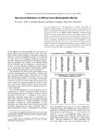
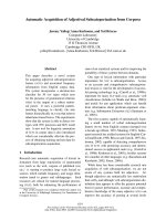
tumors. In the physical examination, however
,thepre-
sence of dysmorphic facial features was observed,
including relative macrocephaly (figure. 1a) and ocular
hypertelorism (figure. 1b); pectum excavatum (figure. 1c
and 1d), vertebral anomaly characterized by cyphosco-
liosis (figure. 1e) and polydactyly of both hands. Other
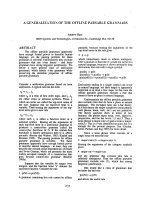
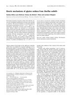
examinations were also perform ed which included, pos-
tero-anterior radiography of the skull and jaw, chest
radiographs and computed tomography. In addition to
images suggestive of K COTs in maxilla and mandible
(figure. 2a), imaging examinations revealed calcification
of the falx cerebri (figure. 2b), rib anomalies (figure. 2c)
and spine bifida (figure. 2d).
The tumors were surgically removed in consecutive
sessions, through the enucleation and marsupializat ion
technique. The specimens were fixed in 10% formalin
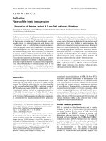
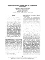
and submitted to histopathological examination. The
microscopic analysis showed epithelium with palisade
basalcelllayerwithdark-staining nuclei and a corru-
gated surface with parakerati nization (figure. 3a and 3b).
Prominent daughter-cysts containing keratin whorls
were found in the thin capsular connective tissue
(figure. 3d). In add ition, tumors presented inflammatory
changes, with consequent partial loss of epithelium lin-
ing features (figure. 3c). Based on clinical, radiographic
and microscopic data, the hypothesis of KCOTs was
confirmed and the diagnosis of NBCCS was established.
The patient was referred to a dermatologist for appro-
priate dermatological care including investigation and
early diagnosi s of future skin lesions ( basocellular
carcinomas).
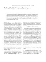
New bone formation sites were identified in the three-
month radiological follow-up (figure. 4). The patient
and his parents are aware of the importance of regular
examination.
Figure 1 Clinical features of NBCCS. (a and b) F acial appearance
of patient showed dysmorphic facial features, including relative
macrocephaly (a) and ocular hypertelorism (b). (c and d) Lateral and
frontal view showing pectum excavatum. (e) Vertebral anomaly
characterized by cyphoscoliosis.
Figure 2 Imaging f indings of NBCCS. (a) Orthopantographic
examination suggesting the presence of multiple KCOTs in the
maxilla and mandible (arrow). (b) Tomographic showing calcification
of the cerebral falx. (c) Thorax film showing anomalies of the ribs
characterized by flattening. (d) 3D tomographic reconstruction,
showing spina bifida.
Figure 3 Histopathology findings of KCOT. (a and b) Prominent
palisade basal cell layer with dark-staining nuclei and a corrugated
surface with parakeratinization (H-E staining, original magnification ×
40). (c) Presence of hyaline bodies and inflammatory changes that
have destroyed parts of the lining ephitelium (H-E staining, original
magnification × 4). (d) Prominent daughter cysts containing keratin
whorls (H-E staining, original magnification × 10).
Casaroto et al. Head & Face Medicine 2011, 7:2
/>Page 2 of 5
Discussion
Several studies have presented KCOTs, basal cell naevi
and skeletal anomalies as the principal clinical features
of NBCCS [3,10,11]. However, according to Manfredi
et al. [10], the diagnostic criteria of NBCCS requires the
presence of two major, or one major and two minor cri-
teria. Major criteria included the presence of more than
two basal cell carcinomas or one under the age of
20 years, histologically-proven KCOT of the jaw, cuta-
neous palmar or plantar pits, and bifid, fused or mark-
edly splayed ribs. Any one of the following features is
consider ed a minor criterion, such as orofacial congeni-
tal malformations (cleft lip or palate, frontal bossing or
mode rate or severe hypertelorism), skeletal and radiolo-
gical abnormalities (bridging of the sella turcica and ver-
tebral anomalies), ovarian fibroma and medulloblastoma.
The present case report showed a child patien t pre-
senting, among others, some of these features, such as
multiple KCOTs in the maxilla and mandibl e, r ib
anomalies, spine bifida, calcification of the falx cerebri,
ocular hypertelorism and vertebral anomaly character-
ized by kyphoscoliosis, which confirmed the diagnosis of
NBCCS or Gorlin-Goltz syndrome.
One of the features found in this syndrome and
emphatically mentioned in literature is the development
of multiple basal cell carcinomas, especially in the head
and neck region [1]. In this case it has not been possible
to identify the presence of basal cell carcinomas. This
fact can be explaine d by the patient’ s age (ten years
old). Possibly, these carcinomas may develop in the
future (second and third decade of life).
NBCCS is caused by mutations in a tumor suppressor
gene PTCH (human homologue of a Drosophila segment
polarity gene Ptch) located in chromosome 9q22.3
[1,3,12]. This protein can be found in the Hedgehog signal-
ing pathway [8]. PTCH in the absence of its ligand, it acts
as a cell-cycle regulator, normally inhibiting expression of
downstream genes that control cell fate, patterning and
growth [11]. Generally, for a tumor suppressor gene to be
inactivated, two mutagenic hits (two distinct episodes of
DNA damage) are required. The first hit involves a muta-
tion in one allele, which can be dominantly inherited if
present in a germ cell, but which is classically considered
to have no phenotypic effect. The second hit involves loss
of the other allele, known as loss of heterozygosity. When
both alleles are inactivated, tumor growth occurs [3]. Loss
of heterozygosity has been demonstrated in basal cell car-
cinomas, KCOTs and medulloblastoma, three features of
NBCCS [3,8]. Various physical anomalies of the brain,
ribs, vertebrae and limbs apparently need only one hit
[11]. The single germ cell hit may account for the malfor-
mations and their variability in NBCCS patients [3].
According to Marotto et al. [13], some of the most
common clinical findings of the syndrome are disc ov-
ered through radiography commonly used in orthodon-
tic treatment. In the case described in this study, a
panoramic radiograph for orthodontic purposes showed
radiolucent areas in the maxilla and mandible, suggest-
ing the presence of KCOTs. Chest radiography indicated
the presence of rib anomalies, post anterior of the skull,
computed tomography scan of the head and neck,
which showed calcification of the cerebral falx and spina
bifida which, according to Amezaga et al. [8], are char-
acteristic of the syndrome.
KCOTs are among the most consistent and common
features of NBCCS. They are found in 65 to 100% of
affected individuals [14]. Clinically, the lesions are char-
acterized by aggressive growth and a tendency to recur
after surgical treatment. The epithelial cells of the basal
layer show increased mitotic activity, together with a
potential for budding and the presence of daughter cysts
in the wall [14,15]. It has been reported that the pre-
sence of daughter cysts [16] was related to the recur-
renceofKCOT.Themandibleisinvolvedmore
frequently than the maxilla and the posterior regions are
the most commonly affected sites [17].
There are two methods for the treatment of KCOT, a
conservative and an aggr essive. In the conse rvative
method, simple enucleation with or without curettage
and marsupialization are suggested. Aggressive metho ds
include peripheral ostectomy, chemical curettage with
Carnoy’s solution, and resection [18].
Radical interventions as enucleation with shaving of
surrounding bone or sometime r esection might contri-
bute to preventing recurrences and to improve the prog-
nosis [16,18]. However serious c onsideration should be
given to en bloc resection in the following cases:
1) when KCOT recurs despite previous enucleation with
an adjunctive procedure; 2) when KCOT recurs despite
previous marsupialization followed by enucleation with
an adjunctive procedure; 3) in cases of multilocular
(multilobular) aggressive intraosseous KCOT; 4) in cases
Figure 4 Orthopantographic examination profile at three
months follow-up after the surgery to remove the cystic
lesions.
Casaroto et al. Head & Face Medicine 2011, 7:2
/>Page 3 of 5
of multiple nonsyndromic and syndromic KCOTs of
NBCCS; or 5) in a diagnosed KCOT exhibiting particu-
larly aggressive clinical behavior (eg, growth, destruction
of adjacent tissues) that sho uld require resection as the
initial surgical treatment [19].
If the patient is in the first decade and has still uner-
upted permanent teeth involving KCOTs, it would be
difficult to make a decision of aggressive surgery over
conservative management. In children who have yet to
be erupted, conservative management should be consid-
ered first because an aggressive operation can have an
adverse effect on teeth development, the eruption pr o-
cess, and the development of the involved jaw [20].
Thus, younger patients usually receive more conserva-
tive than aggressive treatment [20,21].
Although some authors believe that simple enuclea-
tion might be the most appropriate conservative method
for the treatment of KCOT [19,22], others have shown
the successful treatment of large or multiple KCOTs
using the marsupialization followed by enucleation
[23-27]. Furthermore, it has been repo rted that marsu-
pialization followed by enucleation results in the lowest
recurrence rate among the c onservative treatment
[21,28]. Moreover, considering the complication of radi-
cal surgery, marsupialization followed by enucleation
has been suggested as the conservative option for treat-
ment of KCOT in younger patients [20,21,28].
Histopathological examina tion of the removed tumors
should be performed to provide definitive diagnosis [8].
In this case, the microscopic an alysis confirmed the
diagnosis of KCOT and indicated the need for monitor-
ing the disease. Long follow-up periods are suggested
for this tumor [17]. In o rder to minimize secondary
morbidities after the treatment, patients with KCOT
should be observed carefully by radiographic imaging
particularly during the first year [16].
This case reinforces the idea that the dentist, spe-
cially the pediatric and orthodontic specialties, has an
important responsibility in early diagnosis and referral
to other specialists for further evaluation. A definitive
diagnosis of NBCCS should be made by a multidisci-
plinary team comprising medical specialists and den-
tists. Life expectancy in NBCCS is not significantl y
altered but there can be substantial morbidity as a
result of complications [8]. Regular follow-up by a
multi-specialists team should be offered. Moreover,
early diagnosis is important for counseling of patients
to prevent harmful exposure to ultraviolet and ionizing
radiations that increase the risk of d eveloping basal
cell carcinoma [1,11]. The patient in this case study
was sent to dermatologist for monitoring of possible
skin lesions.
In summary, it can be said that Gorlin-Goltz syn-
drome is a dominant autosomal genetic process, which
is of particular interest to the o ral and maxillofacial
health experts. Proper evaluation and characterization of
the clinical features are of the utmost importance for
the correct diagnosis and treatment of affected patients.
In order to be able to establish early diagnosis of
NBCCS, specialists should carry out clinical and imaging
examinations in early ages of life. Physicians and den-
tists must know the features of the syndrome well.
Acknowledgements
The authors are grateful to Patrícia Freitas-Faria (Bauru School of Dentistry,
University of São Paulo) and thank her for the valuable technical support
with images. The authors also wish to thank the patient and their family for
their contribution to this article. Written consent for publication was
obtained from the patient’s parent.
Author details
1
Department of Oral Pathology, Bauru School of Dentistry, University of São
Paulo, Bauru, Brazil.
2
Department of Dentistry, University Center of Maringá,
Maringá, Brazil.
3
Department of Dentistry, Federal University of Sergipe,
Aracaju, Brazil.
Authors’ contributions
CAR and LDCNR drafted the manuscript. LVS and VVC carried out the
histological analysis, wrote the histological part of the paper and
contributed to the writing of the final version. ME, TCL and GVD analysed
the patient’s history, reviewed the patient data and surgically removed the
tumors. Each author reviewed the paper for content and contributed to the
writing of the manuscript. All authors approved the final report.
Competing interests
The authors declare that they have no competing interests.
Received: 12 December 2009 Accepted: 25 Jan uary 2011
Published: 25 January 2011
References
1. Yamamoto K, Yoshihashi H, Furuya N, Adachi M, Ito S, Tanaka Y, Masuno M,
Chiyo H, Kurosawa K: Further delineation of 9q22 deletion syndrome
associated with basal cell nevus (Gorlin) syndrome: Report of two cases
and review of the literature. Congenit Anom 2009, 49:8-14.
2. Gorlin RJ, Goltz RW: Multiple nevoid basal cell epithelioma, jaw cysts and
bifid rib: A syndrome. New Engl J Med 1960, 262:908-12.
3. Cohen MM: Nevoid basal cell carcinoma syndrome: molecular biology
and new hypotheses. Int J Oral Maxillofac Surg 1999, 28:216-23.
4. Satinoff MI, Wells C: Multiple basal cell naevus syndrome in ancient. Egypt
Med Hist 1969, 13:294-7.
5. Farndon PA, Del Mastro RG, Evans DG, Kilpatrick MW: Location of gene for
Gorlin syndrome. Lancet 1992, 339:581-2.
6. Shanley S, Ratcliffe J, Hockey A, Haan E, Oley C, Ravine D, Martin N,
Wicking C, Chenevix-Trench G: Nevoid basal cell carcinoma syndrome:
review of 118 affected individuals. Am J Med Genet 1994, 50:282-90.
7. Gorlin RJ: Nevoid basal cell carcinoma (Gorlin) syndrome: Unanswered
issues. J Lab Clin Med 1999, 134:551-2.
8. Amezaga AOG, Arregui OG, Nuño SZ, Sagredo AA, Urizar JMA: Gorlin-Goltz
syndrome: Clinicopathologic aspects. Med Oral Patol Oral Cir Bucal 2008,
13:338-43.
9. Yang X, Pfeiffer RM, Goldstein AM: Influence of glutathione-S-transferase
(GSTM1, GSTP1, GSTT1) and cytochrome p450 (CYP1A1, CYP2D6)
polymorphism on numbers of basal cell carcinomas (BCCs) in families
with the naevoid basal cell carcinoma syndrome. J Med Genet 2006, 43:
e1-e16.
10. Manfredi M, Vescovi P, Bonanini M, Porter S: Nevoid basal cell carcinoma
syndrome: a review of the literature. Int J Oral Maxillofac Surg 2004,
33:117-24.
11. Lo Muzio L: Nevoid basal cell carcinoma syndrome (Gorlin syndrome).
Orphanet J Rare Dis 2008, 3:32.
Casaroto et al. Head & Face Medicine 2011, 7:2
/>Page 4 of 5
12. Ravel TJL, Ameye L, Ballon K, Borghgraef M, Vermeesch JR, Devriendt K:
Early detection of chromosome 9q22.32q31.1 microdeletion and the
nevoid basal cell carcinoma syndrome. Eur J Med Genet 2009, 52:145-7.
13. Marotto MR, Porras JLB, Saez RS, Rios MH, Gonzales LB: The role of the
orthodontist in the diagnosis of Gorlin’s syndrome. Am J Orthod
Dentofacial Orthop 1999, 115:89-98.
14. Sun LS, Li XF, Li TJ: PTCH1 and SMO gene alterations in keratocystic
odontogenic tumors. J Dent Res 2008, 87:575-9.
15. Daley TD, Multari J, Darling MR: A case report of a solid keratocystic
odontogenic tumor: is it the missing link? Oral Surg Oral Med Oral Pathol
Oral Radiol Endod 2007, 103:512-5.
16. Kuroyanagi N, Sakuma H, Miyabe S, Machida J, Kaetsu A, Yokoi M, Maeda H,
Warnakulasuriya S, Nagao T, Shimozato K: Prognostic factors for
keratocystic odontogenic tumor (odontogenic keratocyst): analysis of
clinico-pathologic and immunohistochemical findings in cysts treated by
enucleation. J Oral Pathol Med 2009, 38:386-92.
17. González-Alva P, Tanaka A, Oku Y, Yoshizawa D, Itoh S, Sakashita H, Ide F,
Tajima Y, Kusama K: Keratocystic odontogenic tumor: a retrospective
study of 183 cases. J Oral Sci 2008, 50:205-12.
18. Kolokythas A, Fernandes RP, Pazoki A, Ord RA: Odontogenic keratocyst: To
decompress or not to decompress? A comparative study of
decompression and enucleation versus resection/peripheral ostectomy. J
Oral Maxillofac Surg 2007, 65:640-44.
19. Tolstunov L, Treasure T: Surgical treatment algorithm for
odontogenic keratocyst: combined treatment of odontogenic
keratocyst and mandibular defect with marsupialization,
enucleation, il iac cr est bone graft, and dental implants. JOral
Maxillofac Surg 2008, 66:1025-36.
20. Hyun HK, Hong SD, Kim JW: Recurrent keratocystic odontogenic tumor in
the mandible: A case report and literature review. Oral Surg Oral Med
Oral Pathol Oral Radiol Endod 2009, 108:e7-e10.
21. Habibi A, Saghravanian N, Habibi M, Mellati E, Habibi M: Keratocystic
odontogenic tumor: a 10-year retrospective study of 83 cases in an
Iranian population. J Oral Sci 2007, 49:229-35.
22. Shmidt BL, Pogrel MA: The use of enucleation and liquid nitrogen
cryotherapy in management of odontogenic keratocysts. J Oral Maxillofac
Surg 2001, 59:720-25.
23. Maurette PE, Jorge J, De Moraes M: Conservative treatment protocolo of
odontogenic keratocyst: a preliminary study. J Oral Maxillofac Surg 2006,
64:379-83.
24. August M, Faquin WC, Troulis MJ: Diferentiation of odontogenic
keratocyst epithelium after cyst decompression. J Oral Maxillofac Surg
2003, 61:678.
25. Enislidis G, Fock N, Sulzbacher I, Ewers R: Conservative treatment of large
cystic lesions of the mandible: a prospective study of the effect of
decompression. Brit J Oral Maxillofac Surg 2004, 42:546-50.
26. Pogrel MA, Jordan RCK: Marsupialization as a definitive treatment for the
odontogenic keratocyst. J Oral Maxillofac Surg 2004, 62:655-56.
27. Pogrel MA: Treatment of Keratocysts: the case for descompression and
marsupialization. J Oral Maxillofac Surg 2005, 63:667-73.
28. Nakamura N, Mitsuyasu T, Mitsuyasu Y, Taketomi T, Higuchi Y, Ohishi M:
Marsupialization for odontogenic keratocysts: Long-term follow-up
analysis of the effects and changes in growth characteristics. Oral Surg
Oral Med Oral Pathol Oral Radiol Endod 2002, 94:543-53.
doi:10.1186/1746-160X-7-2
Cite this article as: Casaroto et al.: Early diagnosis of Gorlin-Goltz
syndrome: case report. Head & Face Medicine 2011 7:2.
Submit your next manuscript to BioMed Central
and take full advantage of:
• Convenient online submission
• Thorough peer review
• No space constraints or color figure charges
• Immediate publication on acceptance
• Inclusion in PubMed, CAS, Scopus and Google Scholar
• Research which is freely available for redistribution
Submit your manuscript at
www.biomedcentral.com/submit
Casaroto et al. Head & Face Medicine 2011, 7:2
/>Page 5 of 5