Báo cáo y học: " Prime immunization with rotavirus VLP 2/6 followed by boosting with an adenovirus expressing VP6 induces protective immunization against rotavirus in mice" pps
Bạn đang xem bản rút gọn của tài liệu. Xem và tải ngay bản đầy đủ của tài liệu tại đây (379.18 KB, 8 trang )
RESEARCH Open Access
Prime immunization with rotavirus VLP 2/6
followed by boosting with an adenovirus
expressing VP6 induces protective immunization
against rotavirus in mice
Hongli Zhou
1,2
, Li Guo
1,2
, Min Wang
1
, Jianguo Qu
1
, Zhendong Zhao
2*
, Jianwei Wang
2*
, Tao Hung
1,2
Abstract
Background: Rotavirus (RV) is the main cause of severe gastroenteritis in children. An effective vaccination regime
against RV can substantially reduce morbidity and mortality. Previous studies have demonstrated the efficacy of
virus-like particles formed by RV VP2 and VP6 (VLP2/6), as well as that of recombinant adenovirus expressing RV
VP6 (rAd), in eliciting protective immunities against RV. However, the efficacy of such prime-boost strategy, which
incorporates VLP and rAd in inducing protective immunities against RV, has not been addressed. We assessed the
immune effects of different regimens in mice, including rAd prime-VLP2/6 boost (rAd+VLP), VLP2/6 prime-rAd
boost (VLP+rAd), rAd alone, and VLP alone.
Results: Mice immunized with the VLP+rAd regimen elicit stronger humoral, mucosal, and cellular immune
responses than those immunized with other regimens. RV challenging experiments showed that the highest
reduction (92.9%) in viral shedding was achieved in the VLP+rAd group when compared with rAd+VLP (25%), VLP
alone (75%), or rAd alone (40%) treatment groups. The reduction in RV shedding in mice correlated with fecal IgG
(r = 0.95773, P = 0.04227) and IgA (r = 0.96137, P = 0.038663).
Conclusions: A VLP2/6 prime-rAd boost regimen is effective in conferring immunoprotection against RV challenge
in mice. This finding may lay the groundwork for an alternative strategy in novel RV vaccine development.
Background
Rotavirus (RV) infection is the most common cause of
severe gastroenteritis in children. RV-induced gastroen-
teritis is responsible for over 600, 000 deaths of children
every year; 85% of these deaths occur in developing
countries where nearly two million children are hospita-
lized annually due to RV infection [1,2].
The US Food and Drug Administration (FDA)
licensed the first RV vac cine (Rotashield™ ) in 1998.
However, this vaccine was withdrawn only one year
later due to a common side effect, intussusception [3].
In recent years, two more live RV vaccines, Rotarix™
(an attenuated human RV strain developed by
GlaxoSmithKline) and Rotateq™ (a pentavalent human-
bovine reassortant developed by Merck) were licensed
in several c ountries [4-6]. Yet the protective mechan-
isms of these RV vaccines have not been fully under-
stood [7].
Previous studies have shown that RV VP6 can interact
with a large fraction of human naive B cells [8] and that
the immunization using VP6 p rotein or DNA can
induce protective immunities in mice, gnotobiotic pigs,
and other animal models [9-14]. It has also been shown
that the double layered virus-like particles (VLPs)
formed by VP2 and VP6 (VLP2/6) of RV [15], together
with mucosal adjuvant, are able to induce protective
immunities [16-19]. These studies strongly suggest that
VP6 plays a key role in RV protective immunity.
Recombinant adenoviruses (rAds) have been widely
used in the development of viral vaccines due to their
safety and effectiveness in gene transfer and expression
* Correspondence: ;
2
State Key Laboratory for Molecular Virology and Genetic Engineering,
Institute of Pathogen Biology, Chinese Academy Medical Sciences & Peking
Union Medical College, Dong Dan San Tiao, Beijing 100730, PR China
Full list of author information is available at the end of the article
Zhou et al. Virology Journal 2011, 8:3
/>© 2011 Zhou et al; licensee BioMed Central Ltd. This is an Open Access article distributed under the terms of the Creative Commons
Attribution License ( which permits unrestricted use, distribution, and re production in
any medium, provid ed the original work is properly cited.
[20-24]. Administration of rAd expressing human RV
VP6 orally or intranasally stimulates effective specific
humoral, mucosal, and cellular immune responses and
confers protection against RV infection in mice [25].
Studies have also shown that combining rAds with
DNA or protein in prime-boost strategies effectively
enhance the immune response against target antigens.
Such methods have been applied to the development of
vaccines against HIV and many other viruses [26-29].
In the present study, we investigated the efficacy o f
prime-boost regimens in eliciting specific p rotective
immunities against RV infection in mice. We found that
mice immunized with VLP2/6 prime-rAd boost regimen
elicit stronger humoral, mucosal and cellular immune
responses and confer stronger protection against RV
challenge than those immunized with other regimens.
OurdatasuggesttheuseofaVLPprime-rAdboost
strategy for the development effective RV vaccines.
Results
Humoral immune responses
To asses the effectiveness of different vaccination regi-
mens in eliciting specific h umoral responses in mice
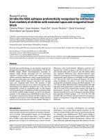
(Figure 1), serum IgG and IgA targeted to RV were ana-
lyzed by indirect ELISAs. We found tha t after the first
immunization (14 days post-inoculation), anti-VP6 IgG
were present in all mice subjected to VLP+rAd and VLP
treatment. Moreover, after the third immunization (35
dpi), the anti-VP6 IgG antibody levels of the VLP+rAd
group (GMT = 160948) and the VLP grou p (GMT =
1377449) were significantly higher than those of the
other two groups [VLP+rAd group vs. rAd+VLP group
(GMT = 11771), P = 0.02033; VLP +rAd group vs. rAd
group (GMT = 852), P = 0.00747; VLP group vs. rAd
+VLP group, P = 0.00126; VLP group vs. rAd group,
P = 0.00246]. Anti-VP6 IgG were present in all of the
mice in the rAd+VLP group until after the third immu-
nization. In the rAd group seroconversion was observed
in only 3 out of 5 mice (Figure 2A).
Anti-VP6 IgA were not detected at dpi14 in any
groups. However, these antibodies appeared at dpi 28
and dpi 35 only in mice immunized w ith VLP+rAd and
VLP (Figure 2B). The IgA level of the VLP +rAd group
was the highest, and at dpi 28, all mice in this group
were positive for anti-VP6 IgA. At dpi 35, the serum
IgA of the VLP+rAd group (GMT = 3482) was signifi-
cantly higher than that of the VLP group (GMT = 283,
P = 0.00425). In the VLP group, only 3/4 of the mice
showed that IgA were p ositive at dpi 35. The serum
anti-VP6 IgA in the rAd+VLP group and rAd alone
group remained negative in the duration of the study
(Figure 2B).
These results demonstrate that, among the four strate-
gies tested, the VLP2/6 prime-rAdVP6 boost strategy
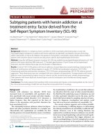
Figure 1 Schemes for animal experiments and sample
collection. BALB/c mice were randomized into five groups and
were immunized and sampled as described in the Materials and
Methods section. Mice were sacrificed at 35 days post-inoculation
(dpi) and the cellular immune responses were determined. At dpi
42, the remaining mice were challenged with the murine RV EDIM
strain, and stool samples were collected daily from dpi 42 to dpi 53.
Serum IgG
1
10
100
1000
10000
100000
1000000
10000000
14 28 35
Days Post Inoculation
GMT
PBS
VLP
VLP+rAd
rAd+VLP
rAd
1/5
0/5
0/5
5/5
4/4
4/4
5/5
5/5
5/5
2/5
3/4
4/4
1/5
3/5
3/5
A
Serum IgA
1
10
100
1000
10000
14 28 35
Days Post Inoculation
GMT
PBS
VLP
VLP+rAd
rAd+VLP
rAd
0/5
0/5
0/5
0/5
0/5
0/5
0/4
0/5
0/5
0/4
0/5
1/4
5/5
3/4
5/5
B
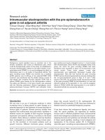
Figure 2 Serum RV VP6 specific antibody response following
immunization. Serum samples were collected from each mouse at
14, 28, and 35 days post-inoculation (dpi). Serum RV specific IgG (A)
and IgA (B) antibodies from individual mice were determined by
ELISA and used to calculate the GMTs for each group of mice. Days
post inoculation are shown on the X-axis. Error bars represent
standard errors of the means. Above each column is the number of
responders over the total number of mice tested.
Zhou et al. Virology Journal 2011, 8:3
/>Page 2 of 8
was the most effective in inducing the humoral immune
response against RV VP6 in mice.
Mucosal immune responses
We assessed the ability of various immunization regimen
in inducing specific mucosal antibody responses by deter-
mining the level of RV VP6 specific IgG (Figure 3A) and
IgA (Figure 3B) in fecal matter. Fecal suspensions were
measured after the third immunizations. Our results
showed that at dpi 14, IgA and IgG were both negative in
all experimental and control groups. After the second
immunization, the A450 of IgA in the VLP+rAd group
and in the VLP group increased to 0.663 ± 0.267 and
0.524 ± 0.200, with an increasing of IgG to 0.513 ± 0.184
and 0.639 ± 0.064, respectively, at A450. At dpi 35, the
A450 of IgA in the VLP+rAd group and in the VLP
group increased to 0.73 ± 0.14 and 0.46 ± 0.23, while the
A450 of IgG increased to 0.82 ± 0.05 and 0.87 ± 0.13,
respectively. But there was no significant differences
between the fecal IgA (P = 0.17412) and IgG (P =
0.34917) level of the two groups. Notably, the anti-VP6
IgA and IgG in the PBS, rAd+VLP, and rAd groups were
negative after each inoculation.
In the VLP+rAd group, 4 of 5 mice tested were posi-
tive for anti-VP6 IgA at dpi 28 and all mice were posi-
tive at dpi 35. This is in contrast to the VLP treated
group for which only 2 of 4 mice tested I gA positive at
dpi 35. Furthermore, all the VLP treated mice tested
positive for the presence of anti-VP6 IgG in fecal matter
at dpi 28, whereas 4 out of 5 mice in the VLP+rAd
group were positive at dpi 28 and dpi 35. These results
indicate that the VLP+rAd regimen is more effective
than the other regimens tested in eliciting mucosal
immune response.
Cellular immune responses
Secreted cytokines (TNF-a,IFN-g, IL-5, IL-4 and IL-2)
were analyzed by CBA technology to profi le the cellular
immune responses to the different vaccination regimens
(Figure 4). We found that the levels of both Th1 cyto-
kines (TNF-a,IFN-g, and IL-2) and Th2 cytokines (IL-4
and IL-5) increased following all immunization schemes.
Although we did not detect statistical differences in the
level of these specific cytokines, mice in the VLP+rAd
and the rAd+VLP group exhibited higher cytokine levels
overall. The TNF, IL-4, and IL-5 secretion in the VLP
group (TNF 70.5 pg/ml; IFN-g 40.3 pg/ml; IL-2 101.0
pg/ml; IL-4 1.2 pg/ml; IL-5 1.3 pg/ml) were nearly the
Fecal IgG
0
0.2
0.4
0.6
0.8
1
1.2
14 28 35
Days Post Inoculation
A450
PBS
VLP
VLP+rAd
rAd+VLP
rAd
0/5
0/5
4/4
4/4
4/5
4/5
0/5
0/5
0/5
0/5
0/4
0/4
0/5
0/5
0/5
A
Fecal IgA
0
0.2
0.4
0.6
0.8
1
14 28 35
Days Post Inoculation
A450
PBS
VLP
VLP+rAd
rAd+VLP
rAd
0/5
0/4
0/4
2/4
2/4
4/5
5/5
0/5
0/5
0/5
0/5
0/5
0/5
0/5
0/5
B
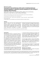
Figure 3 Fecal RV VP6 specific antibody response following
immunization. Fecal samples were collected from each mouse at
14, 28, and 35 days post-inoculation (dpi). Levels of specific IgG (A)
and IgA (B) antibodies in the feces were examined by indirect
ELISAs. Days post inoculation are shown on the X-axis. Error bars
show the standard errors of the mean. Above each column is the
number of responders over the total number of mice tested.
TNF-α
0.0
20.0
40.0
60.0
80.0
100.0
120.0
140.0
160.0
PBS VLP VLP+rAd rAd+VLP rAd
pg/ml
A
IFN-γ
0.0
200.0
400.0
600.0
800.0
1000.0
1200.0
1400.0
PBS VLP VLP+rAd rAd+VLP rAd
pg/ml
B
IL-5
0
5
10
15
20
25
PBS VLP VLP+rAd rAd+VLP rAd
pg/ml
C
IL-2
0.0
50.0
100.0
150.0
200.0
250.0
PBS VLP VLP+rAd rAd+VLP rAd
pg/ml
E
IL-4
0
5
10
15
20
25
30
PBS VLP VLP+rAd rAd+VLP rAd
pg/ml
D
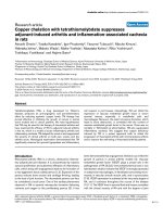
Figure 4 Cytokine production by splenocytes from immunize d
mice. Mice were sacrificed seven days after three immunizations.
The splenocytes were isolated and stimulated with RV VP6 peptide.
The concentrations of TNF-a (A), IFN-g (B), IL-5 (C), IL-4 (D) and IL-2
(E) in the culture supernatant were measured. Error bars represent
standard errors of the mean.
Zhou et al. Virology Journal 2011, 8:3
/>Page 3 of 8
same as that of the PBS group (TNF 39.1 pg/ml; IFN-g
1.2 pg/ml; I L-2 2.6 pg/ml; IL-4 2.3 pg/ml; IL-5 3.1 pg/
ml). Only IFN-g and IL-2 levels were higher than those
of the PBS group. All cytokines in the r Ad group (TNF
16.3 pg/ml; IFN-g 4.5pg/ml;IL-26.2pg/ml;IL-41.2
pg/ml; IL-5 1.4 pg/ml) were essentially the same as
those in the PBS group.
Protective efficacy against RV challenge
To determine the protection conferred by VLP2/6
prime-rAdVP6 boost, rAdVP6 prime-VLP2/6 boost, as
well as VLP2/6 and rAdVP6 alone, five mice from each
group were challenged with 10×DD50 of murine RV
EDIM at dpi 42. Vir al shedding curves (Figure 5A) indi-
cated that the mice in the PBS group shed virus as early
as 2 days after challenge. The viral shedding in each
experimental group decreased to various degrees after
challenge. Reduction in shedding (Figure 5B) of the
VLP+rAd group was the highest (92.9%), with more than
50% of reduction in each mouse. Reductions in shedding
of the VLP group, the rAd+VLP group, and the rAd
group were 76.7%, 36.1%, and 31.1%, respectively. These
numbers were lower than those of the VLP+rAd group,
and varied largely among individuals in each group. Our
results suggest that the VLP2/6 prime-rAdVP6 boost
regimen is more effective than other regimen in confer-
ring immunoprotection against RV challenge in mice.
Discussion
In the present study, we compared the effectiveness of
VLP prime-rAd boost and rAd prime-VLP boost regi-
mens in eliciting anti-RV protective immunities. Our
results demonstrate that the VLP2/6 prime-rAdVP6
boost regimen is more effective in stimulating VP6 spe-
cific immunities and conferred a higher protection than
the other regimens tested.
We administered mice with VLP2/6 via an intranasal
route to elicit vigorous mucosal immunity [18,30,31]. In
contrast, rAdVP6 was administered via an oral route to
ensure the safety of using adenovirus as a component of
a vaccine [32]. Studies have shown that immune
response elicited by oral rAd administration are poor
even in large doses [25,33]. We used a relatively small
dosage of adenovirus in each immunization (10
6
ifu/
dosage, approximately 1/100-1/10 of the documented
doses [ 34]) and found that the immune responses
induced by rAd alone were similar to those of the PBS
group, indicating that rAd alone was unable to protect
the mice against RV challenge.
Repeated immunization of VLP2/6 can effectively
induce humoral and mucosal immunity, but the induc-
tion of cellular immunity was not as effective as the
prime-boost regimens (VLP+rAd or rAd+VLP). After
the RV challenge, the mice immunized wi th VLP alone
still showed obvious virus shedding, with a large varia-
tion of shedding amount betwe en indiv iduals within the
group. In contrast, the VLP+rAd group not only elicited
high level h umoral, mucosal, and cellular immunities,
but also protected a gainst RV challenge and effectively
reduced the amount of virus shedding. After VLP prim-
ing, boosting twice with rAd at a small dosage was an
effective and economical immunization sche me. Our
results indicate that a prime-boost regimen may have
synergetic immune effects.
In our study, the mice immunized with the VLP+rAd
regimen elicited stronger humoral, mucosal, and cellular
immune responses than those immunized with o ther
regimens. The reasons for this disparity are unclear.
Onepossibleexplanationmaybethedifferencein
inducing innate immunity between rAd and VLP, which
leads to a difference in type and strength of the adaptive
immune responses [29]. VLP and rAd are recognized by
diff erent pattern recognition receptors, such as Toll-like
receptors [35,36], which may lead to differences in
cytokine activation. The sequence of prime-boost
0
0.5
1
1.5
2
2.5
01234567891011
Days Post Challenge
A450
VLP
VLP+rAd
rAd+VLP
rAd
PBS
A
-40%
-20%
0%
20%
40%
60%
80%
100%
VLP VLP+rAd rAd+VLP rAd
% Reduction of Virus Shedding
B
Figure 5 Protection from RV shedding in mice following
immunization. Five mice from each group were challenged with
the murine RV EDIM strain at dpi 42, and stool samples were
collected daily from dpi 42 to dpi 53. The presence of RV antigen in
fecal samples (A) was determined by a sandwich-ELISA. Reduction
in shedding (B) was calculated for each animal by comparing the
area under the curve for each individual animal to the mean of the
areas under the curves of the control group.
Zhou et al. Virology Journal 2011, 8:3
/>Page 4 of 8
immunization may also affect the cytokine milieu. This
milieu may determine the final direction, strength, and
breadth of various adaptive immunities, including the
balance between Th1 and Th2 immune responses
through different mechanisms [37]. However, these
mechanisms cannot be unravelled by our data alone. A
systems biology approach to analyze the markers of the
immune responses by different prime-boost regimens
may be needed [38].
Although the molecular mechanisms regulating immu-
noprotection against RV are still unclear and the immu-
nological indicators that can accurately reflect
protection against RV infection remain to be established,
mucosal immunity appears to be i mportant in anti-RV
protective immunities [11,13,30,39-44]. Our correlation
analysis between various immune indicators and reduc-
tion in RV shedding in mice indicate that a reduction in
shedding depends on the levels of fecal IgG antibody
(r = 0.95773, P = 0.04227) and IgA antibody (r = 0.96137,
P = 0.038663) (see Table 1). This finding suggests that
protection against RV is correlated with local intestinal
mucosal immunities. The observation is consistent with
the finding that immunities evoked by VP6 are mainly
present in intestines [45].
Several studies have suggested that cellular immunity
plays an important role in the clearance of RV infection
[14,46-48]. However, although the rAd+VLP regimen
induced a strong T cell response, w e did not observe a
correlation between this reaction and protective efficacy.
Future studies with multiple methods and epitopes
may be necessary to determine the cellular immune
responses more precisely and to assess their significance
in anti-RV immunities.
Conclusions
Our study has shown that a VLP2/6 prime-rAdVP6
boost regimen elicits p rotective immunities from RV
infection a nd is a superior regimen to those of VLP2/6
prime-rAdVP6 boost, VLP2/6 alone, or rAdVP6 alone.
Thus, the VLP2/6 prime-rAdVP6 boost regimen may
provide an alternative strategy for novel RV vaccine
development.
Methods
Preparation of recombinant adenovirus and VLP2/6
The recombin ant replication defective adenovirus sero-
type 5 (Ad5) expressing RV VP6, termed rAdVP6, was
generated with the AdEasy system (Stratagen, Cedar
Creek, TX) following the manufacturer ’s instructions.
Expression of VP6 was confirmed by Western blot ana-
lysis using an antibody against RV (Biodesign, Cat:
B65110G). The virus was titered with an Adeno-X
Rapid Titer Kit (BD Bioscienc es Clontech, Mountain
View, CA) and stored at -70°C prior to use.
VLP2/6 was produced by expression of RV VP2 and
VP6 simultaneously in Spodoptera frugiperda (Sf9) cells
through recombinant baculovirus. The recombinant
baculovirus was generated by the Bac-to-Bac
®
Baculo-
virus Expression System (Invitrogen, Carlsbad, CA)
according to the manufacturer’sprotocol.RVVLP2/6
was purified by ultracentrifugation as described pre-
viously [49,50]. Briefly, the supernatants of Sf9 cells
infected by the recombinant baculovirus were collected
at day 5 post infection and cellular debris was removed
by centrifugation (20 min at 10,000 rpm). VLP2/6 was
precipitated with PEG6000 (final concentration, 6%)
from the clarified supernatant. Precipitated pellets were
sonicated briefly followed by ultracentrifugation at
35,000 rpm for 3 hours thro ugh a 40% sucrose cushion.
The presence of the purified VLP2/6 was confirmed by
Western blot using an anti-RV antibody. Concentrated
VLP2/6 were verified by electron microscopy. The c on-
centration of purified VLP2/6 protein was determined
using the BCA Protein Assay Reagent Kit (Pierce, Rock-
ford, IL), and proteins were stored at -70°C prior to use.
Prime-boost regimens and animal experiments
Six- to eight-week old female BALB/c mice were
obtained fro m the Institute of Laboratory Animal
Science, Chinese Academy of Medical Scien ces, and
maintained in Animal Biosafety Level-2 facilities. Mice
were confirmed to be RV and Ad5 antibody-free by
ELISA prior to immunization and were randomized into
one of the five treatment groups as shown in Figure 1.
For the VLP group, mice were intranasally (i.n.) inocu-
lated with 10 μg RV VLP 2/6 at days 0, 14 , and 28,
respectively. For the VLP+rAd group, mice were i.n.
primed with 10 μg RV VLP2/6 at day 0, followed by
twice oral boosting of 1 × 10
6
ifu (infectious units)
rAdVP6 (in 0.1 ml each dose) at days 14 and 28, respec-
tively. For the rAd+VLP group, mice were orally primed
with 1 × 10
6
ifu of rAdVP6 (in 0.1 ml each dose) at day
0, followed by twice i.n. boosting with 10 μg RV VLP2/6
at da ys 14 and 28. For the rAd group, mice were orally
Table 1 Correlation analysis between all measurement
indicators and reduction in rotavirus shedding in mice
Indicators r P value
Serum IgA 0.94839 0.051611
Serum IgG 0.84071 0.15929
Fecal IgA 0.96137 0.038633
Fecal IgG 0.95773 0.04227
TNF-a 0.37996 0.62004
IFN-g -0.0793 0.92072
IL-5 -0.2375 0.76253
IL-4 0.01328 0.98672
IL-2 0.48413 0.515787
Zhou et al. Virology Journal 2011, 8:3
/>Page 5 of 8
inoculated with 1 × 10
6
ifu rAdVP6 (in 0.1 ml each
dose) at days 0, 14, and 28. In all the cases of VLP2/6
administration, 10 μgofCpGODN1826(5’ >TCC
ATG ACG TTC CTG ACG TT < 3’, synthesized by
Shanghai Sangon Biological Engineering Technology &
Services Co., Ltd., Shanghai, China), and 1 μgpolyI:C
(Sigma, St. Louis, MO) p er dose were used as adjuvant.
Control mice (PBS group) received intranasal immuniza-
tion of 0.1 ml PBS at days 0, 14, and 28.
At 0, 14, 28, and 35 days post-inoculation (dpi), serum
and stool samples were collected from each mouse
before each immunization. Sera were stored at -20°C
until analysis. Five mice from each group were eutha-
nized at dpi 35 and splenocytes were isolated for the
cytokine measurements. The remaining five mice from
each group were challenged with a 10 × 50% diarrhea-
inducing dose (DD50) of murine EDIM RV at 42 dpi
and stool samples were collected daily from dpi 42 to
53. Feces were weighed and resuspended i n PBS (pH
7.2; 1:10, wt/vol). Debris was removed by centrifugation
and supernatants were stored at -20°C until analysis.
Measurement of RV-specific antibodies by ELISA
Ninety-six-well polystyrene microtiter plates (Costar,
Bethesda, MD) were coated overnight at 4°C with 0.1
μg/well VP6 antigen diluted in carbonate buffer after
optimization of the experiments. Wells were washed
three times with 0.05% (vol/vol) Tween 20 in PBS (PBS-
T) and blocked with 200 μl of 1% BSA (Sigma, St.
Louis, MO) in PBS (PBS-BSA) for 2 hours at 37°C.
After washing, 100 μl/well of serum or stool homoge-
nates diluted in PBS-BSA were added, and plates were
incubated for 1 h our at 37°C to prevent non-specific
binding. Subsequently, plates were washed and incu-
bated for 1 hour at 37°C with 100 μl/well of horseradish
peroxidase (HRP)-labeled anti-mouse immunoglobulin
G (IgG) or IgA (Sigma, St. Louis, MO) at a dilution of
1:5000 in PBS-BSA. Color was developed by adding 100
μl/well of Sure Blue TMB (Sigma, St. Louis, MO) perox-
idase substrate, and absorbance was read at 450 nm
(A450) using an BioRad 550 ELISA plate reader (BioRad,
Hercules, CA). Serums were two-fold serially diluted to
determine antibody titers.
Detection of RV antigen in stools
The presence of RV antigen in fe cal samples was deter-
mined by a sandwich-ELISA using a Rotavirus Assay Kit
(Lanzhou Institute of Biological Products, Lanzhou,
China) according to the manufacturer’s protocol. Indivi-
dual stool samples were tested–10% (wt/vol)–and speci-
mens’ A450 was determined using an E LISA plate
reader (BioRad 550, Hercules, CA). Viral shedding
curves for each animal were plotted, and the areas
under the curves for each animal were calculated.
Reduction in shedding was calculated for each immu-
nizedanimalbycomparingtheareaunderthecurveto
the mean of the areas under the curves of the control
group. Reduction in shedding was then calculated for
each vaccination group by determining the mean reduc-
tion of each vaccinating group. A >50% reduction in
virus shedding for an individual animal or for a group
was considered significant protection from virus
challenge.
Multiple-cytokine assays
Freshly isolated murine splenocytes were cultured on
96-well round-bottom tissue culture plates at 5 × 10
5
cells/well in complete RPMI 1640 medium (Invitrogen,
Carlsbad, CA). C ells were stimulated with VP6 peptide
[9,51] (RLSFQLMRPPNMTP, synthesized by the Chi-
nese Academy of Military Medical Sciences) for 48
hours. Supernatants were collected and IL-2, IL-4, IL-5,
TNF-a,andIFN-g secretion were quantified using the
Mouse Th1/Th2 Cytokine Cytometric Array Bead
(CBA) Kit (BD PharMingen, San Diego, CA) according
to the manufacturer’s protocol. The IL-2, IL-4, IL-5,
TNF-a,andIFN-g secretion were detected with F ACS-
Calibur
®
Flow Cytometer (BD Biosciences, San Jose,
CA) using two-color detection and analyzed using CBA
software (BD PharMingen).
Statistical analysis
Antibody titers were log10-transformed and expressed
as geometric mean titers (GMTs). When RV-specific
antibodie s were not detected, a value of 50 (one-half the
lowest detectable level) was assigned to that sample, and
used in the calculation of the mean and standard error.
When the value of the sample was two times that of the
background, it was considered positive. Differences
between groups were compared by Student’s t-test. Cor-
relation analysis was performed by Pearson correlation.
All tests were two-tailed, and a P value of <0.05 was
considered significant.
Acknowledgements
The authors thank Drs. Li Ruan and Xiangrong Qi for their assistance in
ELISPOT assay, and Ms. Shan Mei and Li Li for their assistance in CBA assays.
This research was supported in part by the National 863 High-tech project
(2003AA215070).
Author details
1
National Institute for Viral Disease Control and Prevention, Chinese Center
for Disease Control and Prevention, Beijing 100052, PR China.
2
State Key
Laboratory for Molecular Virology and Genetic Engineering , Institute of
Pathogen Biology, Chinese Academy Medical Sciences & Peking Union
Medical College, Dong Dan San Tiao, Beijing 100730, PR China.
Authors’ contributions
HZ, LG and MW: constructed and characterized VLP2/6 and rAdVP6,
immunized mice and evaluated the immune response. JQ: characterized
VLP2/6 with electron microscopy. HZ and ZZ, JW: wrote the manuscript. ZZ,
Zhou et al. Virology Journal 2011, 8:3
/>Page 6 of 8
JW and TH: participated in the interpretation of data and critically revised
the manuscript. All authors read and approved the final manuscript.
Competing interests
The authors declare that they have no competing interests.
Received: 18 September 2010 Accepted: 5 January 2011
Published: 5 January 2011
References
1. Parashar UD, Gibson CJ, Bresee JS, Glass RI: Rotavirus and severe
childhood diarrhea. Emerg Infect Dis 2006, 12:304-306.
2. Parashar UD, Hummelman EG, Bresee JS, Miller MA, Glass RI: Global illness
and deaths caused by rotavirus disease in children. Emerg Infect Dis 2003,
9:565-572.
3. Withdrawal of rotavirus vaccine recommendations. MMWR 1999,
48:1007.
4. Glass RI, Parashar UD: The promise of new rotavirus vaccines. N Engl J
Med 2006, 354:75-77.
5. Ward R, Bernstein D: Rotarix: a rotavirus vaccine for the world. Clin Infect
Dis 2009, 48:222-228.
6. D. U, Alexander JP, Glass RI: Prevention of rotavirus gastroenteritis among
infants and children. Recommendations of the Advisory Committee on
Immunization Practices (ACIP). MMWR Recomm Rep 2006, 55:1-13.
7. Ward RL, Clark HF, Offit PA: Influence of potential protective mechanisms
on the development of live rotavirus vaccines. J Infect Dis 2010, 202:
s72-s79.
8. Parez N, Garbarg-Chenon A, Fourgeux C, Deist FL, Servant-Delmas A,
Charpilienne A, Cohen J, Schwartz-Cornil I: The VP6 protein of rotavirus
interacts with a large fraction of human naive B cells via surface
immunoglobulins. J Virol 2004, 78:12489-12496.
9. Choi AHC, Basu M, Mcneal MM, Flint J, Vancott JL, Clements JD, Ward RL:
Functional mapping of protective domains and epitopes in the rotavirus
VP6 protein. J Virol 2000, 74:11574-11580.
10. McNeal MM, VanCott JL, Choi AHC, Basu M, Flint JA, Stone SC, Clements JD,
Ward RL: CD4 T cells are the only lymphocytes needed to protect mice
against rotavirus shedding after intranasal immunization with a chimeric
VP6 protein and the adjuvant LT(R192G). J Virol 2002, 76:560-568.
11. García-Díaz A, López-Andújar P, Díaz JR, Montava R, Barceló CT, Ribes JM,
Buesa J: Nasal immunization of mice with a rotavirus DNA vaccine
that induces protective intestinal IgA antibodies. Vaccine 2004,
23:489-498.
12. Yuan L, Azevedo MS, Gonzalez AM, Jeong KI, Van Nguyen T, Lewis P,
Iosef C, Herrmann JE, Saif LJ: Mucosal and systemic antibody responses
and protection induced by a prime/boost rotavirus-DNA vaccine in a
gnotobiotic pig model. Vaccine 2005, 23:3925-3936.
13. Azevedo MS, Yuan L, Iosef C, Chang KO, Kim Y, Nguyen TV, Saif LJ:
Magnitude of serum and intestinal antibody responses induced by
sequential replicating and nonreplicating rotavirus vaccines in
gnotobiotic pigs and correlation with protection. Clin Diagn Lab Immun
2004, 11:12-20.
14. Ward RL, Monica MM: VP6: a candidate rotavirus vaccine. J Infect Dis 2010,
202:s101-s107.
15. Crawford SE, Labbe M, Cohen J, Burroughs MH, Zhou Y-J, Estes MK:
Characterization of virus-Like particles produced by the expression of
rotavirus
capsid proteins in insect cells. J Virol 1994, 68:5945-5952.
16. Fromantin C, Jamot B, Cohen J, Piroth L: Rotavirus 2/6 virus-like particles
administered intranasally in mice, with or without the mucosal
adjuvants cholera toxin and Escherichia coli heat-labile toxin, induce a
Th1/Th2-like immune pesponse. J Virol 2001, 75:11010-11016.
17. Gonzalez AM, Nguyen TV, Azevedo MS, Jeong K: Antibody responses to
human rotavirus (HRV) in gnotobiotic pigs following a new prime/boost
vaccine strategy using oral attenuated HRV priming and intranasal VP2/
6 rotavirus-like particle (VLP) boosting with ISCOM. Clin Exp Immunol
2004, 135:361-372.
18. Coste A, Sirard JC, Johansen K, Cohen J, Kraehenbuhl JP: Nasal
immunization of mice with virus-like particles protects offspring against
rotavirus diarrhea. J Virol 2000, 74:8966-8971.
19. Shuttleworth G, Eckery D, Awram P: Oral and intraperitoneal
immunization with rotavirus 2/6 virus-like particles stimulates a systemic
and mucosal immune response in mice. Arch Virol 2005, 150:341-349.
20. Mercier GT, Nehete PN, Passeri MF, Nehete BN, Weaver EA, Templeton NS,
Schluns K, Buchl SS, Sastry KJ, Barry MA: Oral immunization of rhesus
macaques with adenoviral HIV vaccines using enteric-coated capsules.
Vaccine 2007, 25:8687-8701.
21. Roy S, Kobinger GP, Lin J, Figueredo J, Calcedo R, Kobasa D, Wilson JM:
Partial protection against H5N1 influenza in mice with a single dose of a
chimpanzee adenovirus vector expressing nucleoprotein. Vaccine 2007,
25:6845-6851.
22. See RH, Petric M, Lawrence DJ, Mok CP, Rowe T, Zitzow LA, Karunakaran KP,
Voss TG, Brunham RC, Gauldie J, et al: Severe acute respiratory syndrome
vaccine efficacy in ferrets: whole killed virus and adenovirus-vectored
vaccines. J Gen Virol 2008, 89:2136-2146.
23. Richardson JS, Yao MK, Tran KN, Croyle MA, Strong JE, Feldmann H,
Kobinger GP: Enhanced protection against Ebola virus mediated by an
improved adenovirus-based vaccine. PLoS One 2009, 4:e5308.
24. Patel A, Zhang Y, Croyle M, Tran K, Gray M, Strong J, Feldmann H,
Wilson JM, Kobinger GP: Mucosal delivery of adenovirus-based vaccine
protects against Ebola virus infection in mice. J Infect Dis 2007, 196:
S413-420.
25. He J, Wang J, Jiang X, Wang D, Wen L, Dong J, Qu J, Hong T: Expression
of the main structural antigen VP6 of human rotavirus by recombinant
adenovirus and immune responses induced in vivo. Zhonghua Shi Yan
He Lin Chuang Bing Du Xue Za Zhi 2002, 16:109-113.
26. Shinoda K, Xin K-Q, Kojima Y, Saha S, Okuda K, Okuda K: Robust HIV-
specific immune responses were induced by DNA vaccine prime
followed by attenuated recombinant vaccinia virus (LC16m8 strain)
boost. Clin Immunol 2006, 119:32-37.
27. Rasmussen RA, Ong H, Kittel C, Ruprecht CR, Ferrantelli F, Hu S-L,
Policano P, McKenna J, Moon J, Travis B, Ruprecht RM: DNA prime/protein
boost immunization against HIV clade C: safety and immunogenicity in
mice. Vaccine
2006, 24:2324-2332.
28.
Cebere I, Dorrell L, McShane H, Simmons A, McCormack S, Schmidt C,
Smith C, Brooks M, Roberts JE, Darwin SC, et al: Phase I clinical trial safety
of DNA- and modified virus Ankara-vectored human immunodeficiency
virus type 1 (HIV-1) vaccines administered alone and in a prime-boost
regime to healthy HIV-1-uninfected volunteers. Vaccine 2006, 24:417-425.
29. Guo L, Zhou H, Wang M, Song J, Han B, Shu Y, Ren L, Si H, Qu J, Zhao Z,
et al: A recombinant adenovirus prime-virus-like particle boost regimen
elicits effective and specific immunities against norovirus in mice.
Vaccine 2009, 27:5233-5238.
30. O’Neal CM, Crawford SE, Estes MK, Conner ME: Rotavirus virus-like particles
administered mucosally induce protective immunity. J Virol 1997,
71:8707-8717.
31. Bertolotti-Ciarlet A, Ciarlet M, Crawford SE, Conner ME, Estes MK:
Immunogenicity and protective efficacy of rotavirus 2/6-virus-like
particles produced by a dual baculovirus expression vector and
administered intramuscularly, intranasally, or orally to mice. Vaccine 2003,
21:3885-3990.
32. Lemiale F, Kong W-p, Akyürek LM, Ling X, Huang Y, Chakrabarti BK,
Eckhaus M, Nabel GJ: Enhanced mucosal immunoglobulin A response of
intranasal adenoviral vector human immunodeficiency virus vaccine and
localization in the central nervous system. J Virol 2003, 77:10078-10087.
33. Vos A, Neubert A, Pommerening E, Müller T, Döhner L, Neubert L,
Hughes K: Immunogenicity of an E1-deleted recombinant human
adenovirus against rabies by different routes of administration. J Gen
Virol 2001, 82:2191-2197.
34. Lin SW, Cun AS, Harris-McCoy K, Ertl HC: Intramuscular rather than oral
administration of replication-defective adenoviral vaccine vector induces
specific CD8+ T cell responses in the gut. Vaccine 2007, 25:2187-2193.
35. Keller SA, Schwarz K, Manolova V, von Allmen CE, Kinzler MG, Bauer M,
Muntwiler S, Saudan P, Bachmann MF: Innate signaling regulates cross-
priming at the level of DC licensing and not antigen presentation. Eur J
Immunol 2010, 40:103-112.
36. Nayak S, Herzog RW: Progress and prospects: immune responses to viral
vectors. Gene Ther 2010, 17:295-304.
37. Liu MA: Immunologic basis of vaccine vectors. Immunity 2010, 33:504-515.
38. Pulendran B, Li S, Nakaya HI: Systems vaccinology. Immunity 2010,
33:516-529.
39. Ciarlet M, Crawford SE, Barone C, Bertolotti-Ciarlet A, Ramig RF, Estes MK,
Conner ME: Subunit rotavirus vaccine administered parenterally to
rabbits induces active protective immunity. J Virol 1998, 72:9233-9246.
Zhou et al. Virology Journal 2011, 8:3
/>Page 7 of 8
40. Chang KO, Vandal OH, Yuan L, Hodgins DC, Saif LJ: Antibody-secreting cell
responses to rotavirus proteins in gnotobiotic pigs inoculated with
attenuated or virulent human rotavirus. J Clin Microbiol 2001,
39:2807-2813.
41. Yuan L, Iosef C, Azevedo MSP, Kim Y, Qian Y, Geyer A, Nguyen TV, Chang K-
O, Saif LJ: Protective immunity and antibody-secreting cell responses
eicited by combined oral attenuated Wa human rotavirus and intranasal
Wa 2/6-VLPs with mutant Escherichia coli heat-labile toxin in gnotobiotic
pigs. J Virol 2001, 75:9229-9238.
42. Franco MA, Angel J, Greenberg HB: Immunity and correlates of protection
for rotavirus vaccines. Vaccine 2006, 24:2718-2731.
43. Liu X, Yang T, Sun Q, Sun M: Efficient intranasal immunization of
newborn mice with recombinant adenovirus expressing rotavirus
protein VP4 against oral rotavirus infection. Acta Virol 2005, 49:17-22.
44. Gonzalez R, Franco M, Sarmiento L, Romero M, Schael IP: Serum IgA levels
induced by rotavirus natural infection, but not following immunization
with the RRV-TV vaccine (Rotashield), correlate with protection. J Med
Virol 2005, 76:608-612.
45. Jaimes MC, Feng N, Greenberg HB: Characterization of homologous and
heterologous rotavirus-specific T-cell responses in infant and adult mice.
J Virol 2005, 79:4568-4579.
46. Ward RL: Possible mechanisms of protection elicited by candidate
rotavirus vaccines as determined with the adult mouse model. Viral
Immunol 2003, 16:17-24.
47. Narváez CF, Angel J, Franco MA: Interaction of rotavirus with human
myeloid dendritic cells. J Virol 2005, 79:14526-14535.
48. Ward RL: Rotavirus vaccines: how they work or don’t work. Expert Rev Mol
Med 2008, 10:e5.
49. Guo L, Wang J, Zhou H, Si H, Wang M, Song J: Intranasal administration of
a recombinant adenovirus expressing the norovirus capsid protein
stimulates specific humoral, mucosal, and cellular immune responses in
mice. Vaccine 2008, 26:460-468.
50. Guo L, Zhou H, Qu J, Wang J, Xu X, Hung T: Codon optimization and
expression of norovirus capsid proteins in insect cells. Virologica Sinica
2006, 21:121-125.
51. Baños DM, Lopez S, Arias CF, Esquivel FR: Identification of a T-helper cell
epitope on the rotavirus VP6 protein. J Virol 1997, 71:419-426.
doi:10.1186/1743-422X-8-3
Cite this article as: Zhou et al.: Prime immunization with rotavirus VLP
2/6 followed by boosting with an adenovirus expressing VP6 induces
protective immunization against rotavirus in mice. Virology Journal 2011
8:3.
Submit your next manuscript to BioMed Central
and take full advantage of:
• Convenient online submission
• Thorough peer review
• No space constraints or color figure charges
• Immediate publication on acceptance
• Inclusion in PubMed, CAS, Scopus and Google Scholar
• Research which is freely available for redistribution
Submit your manuscript at
www.biomedcentral.com/submit
Zhou et al. Virology Journal 2011, 8:3
/>Page 8 of 8