Báo cáo y học: "Japanese Encephalitis Virus wild strain infection suppresses dendritic cells maturation and function, and causes the expansion of regulatory T cells" doc
Bạn đang xem bản rút gọn của tài liệu. Xem và tải ngay bản đầy đủ của tài liệu tại đây (925.97 KB, 11 trang )
RESEARC H Open Access
Japanese Encephalitis Virus wild strain infection
suppresses dendritic cells maturation and function,
and causes the expansion of regulatory T cells
Shengbo Cao
1,2†
, Yaoming Li
1,2†
, Jing Ye
1,2
, Xiaohong Yang
1,2
, Long Chen
1,2
, Xueqin Liu
1,3
, Huanchun Chen
1,2*
Abstract
Background: Japanese encephalitis (JE) caused by Japanese encephalitis virus (JEV) accounts for acute illness and
death. However, few studies have been conducted to unveil the potential pathogenesis mechanism of JEV.
Dendritic cells (DCs) are the most prominent antigen-presenting cells (APCs) which induce dual humoral and
cellular responses. Thus, the investigation of the interaction betw een JEV and DCs may be helpful for resolving the
mechanism of viral escape from immune surveillance and JE pathogenes is.
Results: We examined the alterations of phenotype and function of DCs including bone marrow-derived DCs
(bmDCs) in vitro and spleen-derived DCs (spDCs) in vivo due to JEV P3 wild strain infection. Our results showed
that JEV P3 infected DCs in vitro and in vivo. The viral infection inhibited the expression of cell maturation surface
markers (CD40, CD80 and CD83) and MHCⅠ, and impaired the ability of P3-infected DCs for activating allogeneic
naïve T cells. In addition, P3 infection suppressed the expression of interferon (IFN)-a and tumor necrosis factor
(TNF)-a but enhanced the production of chemokine (C-C motif) ligand 2 (CCL2) and interleukin (IL)-10 of DCs. The
infected DCs expanded the population of CD4+ Foxp3+ regulatory T cell (Treg).
Conclusion: JEV P3 infection of DCs impaired cell maturation and T cell activation, modulated cytokine
productions and expanded regulatory T cells, suggesting a possible mechanism of JE development.
Background
JEVisacausativeagentofJEwhichcausesatleast
50,000 clinical cases and about 10,000 deaths each year.
It is a member of the mosquito-bor ne encephalitis com-
plex of the Flavivi ridae family and has recently been
discovered in previously non-affected areas like Australia
[1] and Pakistan [2]. The neurons in the central nervous
system (CNS) are target cells of JEV. Studies show that
a direct viral cytopathic response and both d irect and
indirect immunological responses can contribute to
CNS degeneration through JEV-infected cell exclusion
by macrophages and CTLs, secretion of cytokines and
chemokines and activation of microglia [3-6]. However,
few studies have investigated the mechanisms by which
JEVevadestheimmunesurveillanceofthehostand
passes through the blood-brain barrier (BBB) to the
CNS.
Dendritic cells (DCs) are the most prominent antigen-
presenting cells (APCs) which induce dual humora l and
cellular responses. While DCs also play unique role i n
inducing immune tolerance, avoiding immune surveil-
lance and causing persistent infecti on. T here are studies
about the interaction between virus and DCs which
showed that viral infection of DCs inhibited the cell
maturation and impaired the cell function [7-9]. Human
cytomegalovirus (HCMV) infection de-regulated the
expression of surface MHC classⅠ,CD40,CD80and
CD86 molecules on DCs. Furthermore, both T cell pro-
liferation and cytotoxicity of T cells specific to an anti-
gen presented by DCs were reduced via the release of
soluble CD83 when DCs were in fected with HCMV
[8,10,11]. Likewise, human immunodeficiency virus
(HIV) affected maturation of DCs within the thymus,
which contributed to the loss of the naive T cell and
* Correspondence:
† Contributed equally
1
State Key Laboratory of Agricultural Microbiology, Huazhong Agricultural
University, Wuhan, Hubei 430070, PR China
Full list of author information is available at the end of the article
Cao et al. Virology Journal 2011, 8:39
/>© 2011 Cao et al; licensee BioMed Central Ltd. This is an Open Access article distributed under the terms of the Creative Commons
Attribution License ( .0), which permits unrestricted use, distribution, and reproduction in
any medium, provided the original work is properly cited.
memory T cell population and even facilitated the disse-
mination of HIV [12].
Additionally, recent studies revealed that several
viruses belonging to the Flaviviridae family, such as
classical swine fever virus (CSFV), Dengue virus (DV)
and Yellow fever virus (YFV), infec ted DCs and altered
the cell phenotype and function [13-15]. Furthermore,
Aleyas et a l. [2009] rec ently reported that JEV Beijing
strain replicated both in bmDCs and macrophages, and
induced functional impairment of DCs through MyD88-
dependent and independent pathways w hich subse-
quently led to poor CD4+ and CD8+ T cell responses
[16]. Thus, the investigation of the interaction between
virusandDCsisimperativeforresolvingtheviral
escape from immune surveillance and JE pathogenesis.
Since there is no evidence for JEV infection of DCs
in vivo, we investigated the alteration of phenotype and
function of the JEV P3-infected DCs bo th in vitro and
in vivo. Our results indicated that JEV P3 severely
infected DCs in vitro an d in vivo, and the infection with
JEV impaired cell maturat ion and the capacity for T cell
activation. In addition, our study also showed that the
infection of DCs with P3 expanded the population of
CD4+ Foxp3+ regulatory T cell (Treg) with immuno-
suppressive potential, suggesting that the virus-induced
alteration of DCs is a likely cause of the immunosup-
pression found in JEV infection.
Results
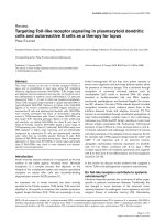
JEV P3 infection of DCs in vitro and in vivo
The purity of the bmDCs fraction from cel l culture or
infected mouse splenocytes was higher than 90% as
determined by FACS analysis with surface molecules
expression (CD11c). After JEV infection, a 467-bp speci-
fic RNA fragment of JEV was detected by RT-PCR
(Figure 1 A) and the E protein of the JEV was detected
by Western blotting in DCs (Figure 1B). FACS results
showed over 80% bmDCs and 90% spDCs were infected
by JEV P3 (Figure 1C). Analysis by real-time P CR
showed that DCs suppo rted JEV replication and yielded
infectious virus ( Figure 1D). These results suggest that
JEV infected DCs both in vitro and in vivo.
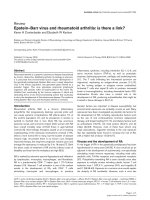
P3 infection suppressed the maturation of DCs
DCs present antigen to and activate T lymphocytes
through up-regulating the expression of costimulator y
and antigen presentation-associtated molecules at the
mature stage [17]. To examine whether the characteris-
tics of immature DCs were altered by P3 infection, we
tested the surface molecules of the infected DCs in vitro
and in vivo. The expression of maturation surface mar-
kers, including CD40, CD80, CD83 and MHCⅠwas up-
regulated in UV-P3-stimulated, but not in P3-infected
bmDCs and spDCs or mock-treated DCs (Figure 2),
indicating that UV-P3 stimulation accelerated the
maturity of DCs whereas P3 infection dramatically
inhibited the cell maturation process.
P3 infection modulated cytokine production of DCs
In many cases, virus does not directly result in the
destruction of host organism but i nstead causes indirect
damage through the disordered release of cytokines [18].
In additio n, imbalanced levels of cytokines may contri-
bute to viral persistence and irreversible immunsuppres-
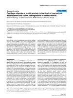
sion. Therefore, we examined the profiles of pro- and
anti-inflammatory cytokines produced by P3-infected
DCs in vitro and in vivo. Our results showed that P3
infection enhanced the releases of IL-10 and CCL2 of
DCs but suppressed the production of IFN-a and
TNF-a (Figure 3). And it was interesting to show that JEV
which was inactivated by UV irradiation failed to induce
the production of IL-10 and CCL2 but succeeded in indu-
cing the ex pression of IFN-a and TNF-a. This indicates
that the release of CCL2 and IL-10 from DCs was depen-
dent on viral replication, while the production of IFN-a
and T N F-a was independent on viral replication.
DCs infected with P3 attenuated allostimulatory activities
to T cells
To test whether P3 infection will impair the ability of
DCs to activate allogeneic naïve T cells, the direct effect
of P3-infected DCs in activation of naïve T cells was
analyzed by mixed lymphocyte reaction (MLR) and ELI-
SPOT assay. In MLR, the allo-stimulative capability of
DCs was significantly suppressed by P3 infection com-
pared to the UV-P3-stimulated group (P < 0.05). In
addition, the viral infection blocked the LPS-induced
allostimulatory activity of DCs (Figure 4A, B).
In ELI SPOT assay detecting IFN-g producing T cells,
the number of spot forming units/10
6
purified T cells
was counted after twenty four hour incubation with dif-
ferently treated bmDCs or spDCs. The results in vitro
showed that P3-infected bmDCs activa ted 25 ± 9 spots/
10
6
, while the UV-P3-stimulated bmDCs activated 68 ±
21 naïve T cells/10
6
. In v ivo, P3-infected spDCs pro-
duced 52 ±12 spots/10
6
whereas UV-P3-stimulated
spDCs produced 107 ± 34 spots/10
6
. This was consis-
tent with the result of MLR assay. P3 infection, in vivo
or in vitro, significantly suppressed the ability of DCs to
activate allogeneic naïve T cells in response to LPS
treatment (Figure 5A, B and 5C). It implied that P3
infection played an important role in the dysfunction of
DCs in activating allogeneic T cells.
P3-infected DCs expanded Treg
The immune response may be limited in magnitude and
efficacy when the host with normal Treg function is
infected with virus. We examined whether P3-infected
Cao et al. Virology Journal 2011, 8:39
/>Page 2 of 11
(A) (B)
(C)
(D)
Figure 1 P3 infects DCs in vitro and in vivo. (A) The in vitro infected bmDCs and the spDCs from P3-challenged mice were harvested and
analyzed with RT-PCR. Bands shown are 467-bp PCR products specific for JEV. (B) The bmDCs and spDCs were analyzed for E protein (JEV
envelope protein) by separation of the proteins on a 10% SDS-PAGE gel followed by electrotransfer to NC membranes and incubation with
monoclonal antibodies against E protein. (C) The bmDCs were harvested after 3 days infection and the spDCs were isolated from mice which
had been challenged for 5 days. 1 × 10
5
bmDCs or spDCs were doubly stained with FITC-anti-E and PE-anti-CD11c and analyzed by FACS
respectively. (D) The infected bmDCs and the spDCs from challenged mice were collected 3 times at day 1, 3 and 5, and a real-time PCR was
performed to quantitatively detect RNA copies of JEV. Each point represents the mean ± SD determinants in triplicate.
Cao et al. Virology Journal 2011, 8:39
/>Page 3 of 11
DCs would modulate Treg differentiation. The test
revealed that P3-infe cted bmDCs significantly enhanced
the differentiation of Foxp3+ Treg in vitro which was con-
sistent with the results in vivo (Figure 6A, B and 6C).
However, the UV-P3-stimulated DCs did not alter the
expansion of the Treg, as well as the mock-treated DCs.
Discussion
Most st udies conducted to evaluate the pathogenesis of
JEV infection have noted the interaction of the virus
with macrophages, microglia and astrocytes, which are
major contributors to the production of inflammatory
cytokines and CNS degeneration [3,4,6]. In the present
(
A) (B)
(C) (D)
Figure 2 Effects of P3 infection on DCs maturation.1×10
5
freshly purified bmDCs were left mock-treated or treated with 1 MOI of P3 or
UV-P3 with or without LPS (lipopolysacchide, Sigma-Aldrich, MO) for 3 days. The spDCs from mice, which have been challenged or immunized
for 5 days, were obtained and treated with or without LPS. Expressions of CD40, CD80, CD83 and MHCⅠ of the bmDCs (A,B) or spDCs (C,D) were
evaluated by FACS. Relative fluorescence intensity to mock group (fold induction) was expressed as the means ± SD of triplicates. *, P < 0.05; **,
P < 0.01.
Cao et al. Virology Journal 2011, 8:39
/>Page 4 of 11
study, we attempted to address the possible pathogen-
esis of JEV wild strain infection by testing the interac-
tion of JEV and DCs in vivo and in vitro.
Carrascoetal.,[2004]discoveredthatCSFVcould
infect and replicate in monocyte and myeloid-derived
DCs [14]. Therefore, we hypothesized that JEV, which
also belongs to the Flaviviridae family, may affect DCs
to facilitate viral spread by escaping immune surveil-
lance. Although Aleyas et al . [2009] recently r eported
JEV i nfection of DCs in vitro , whether JEV infects DCs
in vivo remained unknown until now. Our research not
only verified the results of Aleyas [16], but also investi-
gated the JEV infection of DCs in vivo. Additionally, one
of our preliminary experiments showed that when
BALB/c mice were inoculated with C6FeK4N6-labeled
P3-infected bmDCs or spDCs via intraperitoneal (i.p.),
JEV and C6FeK4N6-labeled DCs w ere detected simulta-
neouly in the brain of mice with severe symptoms of
immunohistochemistry (unpublished data). It is likely
that JEV could use DCs as a virus delivery vehicle as it
moves through the CNS.
The impaired surface molecule expression of APCs
maydirectlyaffecttheprocessofantigenpresentation
and T cell activation. Thus, we analyzed the alter ation of
the surface-molecule expression of infected DCs in vitro
and in vivo. The FACS analyses revealed an suppressed
expression of surface molecules, such as CD40, CD80,
CD83 and MHCI, on P3-infected DCs in vitro and
in vivo, which is in accordance with Aleyas’s results [16].
While we also discovered that the antigen presenting-
associated molecules on bmDCs were significantly
enhanced after JEV SA14-14-2 strain (a successful JEV
live vaccine strain) i nfection [19]. This suggests the
potential molecular mechanism of the immune escape of
P3 and the high immunopotency of SA14-14-2.
Since we have verified that JEV infection impaired the
expression of antigen p resenting-molecules and co-sti-
mulator molecules, whether this impairment of the cru-
cial components on DCs would affect their capa city to
activate CD4+ and CD8+ T cell directly i s needed to be
investigated[20,21]. Thus, we analyzed the capacity of
the infected DCs for activating allogene ic T cells by
MLR and ELISPOT assay. It was observed that the T
cell activating ability of was dramatically impaired by
P3-infection, but boosted by UV-P3 stimulation and
SA14-14-2 infect ion. It has been reported that Hepatitis
C virus (HCV), Ebola virus es and HIV escaped immune
surveillance during acute or chronic infection because of
the defect of APCs function for activating T cell [21-23].
Therefore it suggested that the impairment of activating
of all ogeneic naïve T cells of P3 infected DCs could be
involved in the JE development.
Treg is a subset of CD4+ T-cell with regulatory prop-
erties. Previous studies on the role of Tregs in viral
infections suggest that they suppresses antiviral effect or
T cell responses or local immune activation at the sites
of viral replication [24,25], which may subsequently
result in viral immune evasion and the establishment of
chronic infections [26-28]. Our FACS results showed
that P3 infection contributed to the differentiation of
Figure 3 Cytokine profiles of P3-infected DCs (IFN-a,TNF-a,CCL2andIL-10).1×10
5
freshly purified bmDCs were left mock-treated or
treated with 1 MOI of P3 or UV-P3 for 3 days. The spDCs from mice, which were challenged or immunized for 5 days, were obtained and
cultured for 3 days. The cell supernatants harvested at 3 days of post infection were analyzed with ELISA to measure the concentrations of
cytokines (IFN-a, TNF-a, CCL2 and IL-10). Cytokine concentrations were expressed as the means ± SD of triplicates. *, P < 0.05; **, P < 0.01.
Cao et al. Virology Journal 2011, 8:39
/>Page 5 of 11
Treg in vivo. The results also demonstrated the expan-
sion of Treg population after the co-culture of P3-
infected DCs and T cells. It suggested that JEV infection
of DCs might influence the mode of T-cell differentia-
tion. Thus, we assumed that induction and expansion of
Treg cells by JEV-infected DCs may be associated with
immunosuppression in JEV infection. It has pre viously
been shown that immature DCs induced Treg cells are
able to suppress other T-cell respon ses [29-33]. Further-
more, it has been demonstrated that the increased pro-
duction of IL-10 played an important role in Treg
responses which appeare d to contribut e to immune dys-
function, accountin g for viral persistence and acute tis-
sue damage. Therefore, the up-regulation of IL-10 in
P3-infected DCs may partly contribute to the expansion
of Treg. Based on these results, we suggest that P3
infection may have led to the expansion of Treg cell
population in vivo, which could have been involved in
the suppression of anti-JEV immune responses. In addi-
tion, it is essential to note that although CD25 is
expressed on most regulatory T cells, it is not specific
since it can also be expressed on activated CD4+ T cells
(
A
)
(B)
Figure 4 Effects of P3 infection on DCs activation of naïve T cells by MLR. Mock-treated, P3-infected or UV-P3 -stimulated DCs as well as
differently treated spDCs were added in grade dose to 1 × 10
5
allogeneic T cells at the indicated stimulator-responder ratios in triplicate, with
(B) or without (A) LPS treatment for 20 h before the addition of 50 μl of CellTiter 96
®
AQ
ueous
One Solution Cell Proliferation Assay. The bmDCs,
spDCs as well as T cells were served as spontaneous NADH/NADPH releases controls respectively. The presentation activities of differently treated
bmDCs were measured as 100% (OD490
DC+T exp.
-OD490
DC spont.
-OD490
T spont.
)/(OD490
T spont.
). Results were expressed as the means ± SD of
triplicates. *, P < 0.05.
Cao et al. Virology Journal 2011, 8:39
/>Page 6 of 11
(A)
(B)
(C)
Figure 5 IFN-g producing T cells were detected by ELISPOT
assay. P3-infected, UV-P3-stimulated or mock-treated DCs as well as
differently treated spDCs were harvested and treated with Mitomycin
C (Sigma-Aldrich, MO) at final concentration of 10 μg/ml for 1 h. The
differently treated or mock DCs were seeded (1 × 10
4
per well)
together with 1 × 10
5
per well T cells in triplicates for 20 h. LPS-
stimulated DC/T cell co-cultures served as positive controls. One
representative for IFN-g spot forming unit (SFU) by ELISPOT assay was
shown (A). The figure was representative of three independent
experiments. Corrected data (SFU)/well were shown for bmDCs and
spDCs activations for naïve T cells to expand and produce IFN- g by
ELISPOT assay (B, in vitro;C,in vivo). Results were expressed as the
means ± SD of triplicates. *, P < 0.05.
(
A
)
(B)
(C)
Figure 6 Effects of P3 infection on DCs-induced differentiation
of regulatory T cells.1×10
5
mock-, P3-, UV-P3- or LPS-treated
bmDCs were incubated with 1 × 10
6
allogeneic naïve T cells for
5 days. T cells were purified and doubly labeled for CD4 and Foxp3,
and assessed by FACS. The in vivo Treg in splenocytes were purified
and examined by FACS from mice inoculated with 1 × 10
5
PFU P3
or identical UV-P3 i.p. for 5 days. Representative result was shown
from three independent experiments (A). The percentage
represented the ratio of CD4+ Foxp3+ cells in CD4+ T cells. P3-
infected bmDCs elicited the Treg differentiation in vitro (B). After P3
infection or UV-P3 stimulation of mice i.p., Treg differentiation
in vivo was analyzed immediately (C). Results were expressed as the
means ± SD of triplicates. *, P < 0.05.
Cao et al. Virology Journal 2011, 8:39
/>Page 7 of 11
[34,35]. Foxp3 has been shown to be a better marker for
CD4+ CD25+ T regulatory cells.
The key cytokines secreted by DCs, including typeⅠIFN
(IFN-a/b), TNF-a, IL-10 and CCL2, restrict the prolif-
eration of invading pathogens and determine the polari-
zation of Th1 and Th2 [36-38]. In particular, secretion of
type I IFN is a key step in the innate immune response to
viral infection and TNF-a released by DCs can further
recruit DC precursors and sustain the antigen presenta-
tion [22]. The impaired expression of IFN-a and TNF-a
of DCs following the JEV P3 infection when compared
with UV-P3 was observed in the present study may con-
tribute to the attenuated generation of antiviral immune
response of the host. However, the report of Chang et al.,
[2005] revealed JEV infectio n induced IFN-b participated
in fighting the invading pathogens by using cell types of
A549 and SK-N-SH cells through IRF-3- and NF-B-
mediated pathway [39]. Similar results were also obtained
in the studies of West nile virus (WNV) infection which
induced the IFN-a production of pDCs and mDCs [40],
while inhibited the IFN-b expression of Hela cell [41].
Therefore, we hypothesize d that the different cell types
from different tissues may present distinct immune
response against viral infection. It is known that different
cell types usually exert different functions. For instance,
pDCs, which generate the crucial signal adaptor IRF7,
constitutively express IFN-I. On the contrary, the expres-
sion of IFN-I is extremely inhibited in those cell types in
absence of the receptor TLR7/TLR9 and IRF-7 [42,43].
Furthermore, different types of cytokines are usually used
to discriminate the patterns of immune responses. There-
fore, when only considering the individual cell type, dif-
ferent cell types may present distinct immune responses.
TNF-a level in serum and cerebrospinal fluid (CSF) of
the fatal case in significantly correlated with prognostic
outcome in wild type JEV infection [44]. Therefore,
TNF-a may play an important role in immunopathogi-
cal responses of the infected host. However, JEV infec-
tion of DCs reduced the expression of TNF-a in the
current study. On one hand, it usually appears of appro-
priate expressi on of TNF-a from the innate response of
the host when external pathogen invading. On the other
hand, the excess TNF-a induced cell degeneration could
be harmful to the survival of virus itself. Therefo re, we
speculate that the wild type virus may evolve a mechan-
ism by which to restrict the ex cess inflammatory factors
expression at the beginning of the infection, which may
facilitate the persistence of the virus survival. Moreover,
P3 infection significantly enhanced the release of CCL2
and IL-10. The IL-10 is considered as an anti-inflamma-
tory factor and plays an important role in the differen-
tiation of Treg cells [31,45,46]. The suppressed TNF-a
production in P3-infected DCs may be part ially regu-
lated by high-expressed IL-10. Our results indicated that
the release of CCL2 and IL-10 from DCs was positively
related to viral infection while the production of IFN-a
and TNF-a was negatively related to vi ral replication.
We specu late that the temporary presence of some non-
structure proteins or dsRNA of JEV during the viral
replication may play an important role in decelerating
or accelerating certain signaling pathway.
Additionally, most data obtained in our experiments
are consis tent with Aleyas’s results except for decreased
production of TNF-a . This contradicted finding about
decreased production of TNF-a might be due to various
factors, such as the DCs purity (>90% vs >75%), JEV
strain (P3 and Beijing) and MOI values. All together,
the increased level of IL-10 and the decreased produc-
tions of IFN-a and TNF-a presented an immune-
suppressive profile, indicating the process of the fatal JE
development.
Conclusion
Our data reveals that JEV P3 could infect mouse DCs in
vitro and in vivo, and the infection affects the phenotype
and function of DCs, including reducing expression of
costimula tory molecules, mod ulating secretion of crucial
cytokines, suppressing activation of T cells, and stimu-
lating differentiation of regulatory T cells, which indi-
cates that t he functional impairment of viral infected
DCs orchestrates the immunosuppression in response to
the acute JEV infection.
Methods
Reagents, virus and cells
The fluorescent antibodies, includi ng CD11c-PE (N418),
CD40-FITC (HM40-3) , CD80-FITC (16- 10A1), CD83-
FITC (34-1-2S) and MHC Ⅰ-FITC (Michel-17), re combinant
mouse granulocyte-macrophage colony stimulating factor
(rmGM-CSF) and IL-4 (rmIL-4) were purchased from
eBioscience Inc. (San Diego, CA). The anti- E (JEV envelope
protein) MAb was generated in our laboratory and purified
with NAb™ Spin Kits (Thermo Scietific, USA) according
to the manufacturer’s instructions. JEV P3 strain was pro-
duced in BHK-21 which was maintained in Dulbecco’s
Modified E agle’s Medium (DMEM, Sigma-Aldrich, MO)
supplemented with 10% heated-inactivated fetal bovine
serum (FBS, Hyclone, Logan, UT) of 100 μg/ml str eptomy-
cin and 100 U/ml penicillin (Sigma-Aldrich, MO) at 37°C
with 5% CO
2
. And then the virus was tittered by plaque
formation assay with BHK-21 cell line. JEV stock was trea-
ted with UV irradiation for 1 min (waveleng th 253.7 nm,
radiation i n tensity ≥ 60 μW/cm2, distance 30 cm).
Generation of bone marrow-derived DCs (bmDCs) and
spleen-derived DCs (spDCs)
For generation of b mDCs from BALB/c mouse bone
marrow cultures, the procedure of Inaba et al., [1992]
Cao et al. Virology Journal 2011, 8:39
/>Page 8 of 11
was used with minor modifications [47]. Briefly, the
bone marrow was flushed from femurs and tibias and
subsequently depleted of erythrocytes with ammonium
chloride. Cells were plated at 2 × 10
6
/ml in DCs media
(RPMI 1640 supplemented with 1 0% FBS, 100 μg/ml
streptomycin, 100 U/ml penicillin, 10 ng/ml of rmGM-
CSF and rmIL-4). At day 2 and 4 of culturing, 50% of
the supernatant was removed and replenished with fresh
DCs media. At day 6, non- adherent cells were collected
and transferred into a ne w dish. After a total of 7 to 9
days of culturing, bmDCs were harvested and purified
with StemSep™ Mouse Dendritic Cell Enrichment Kit
(StemCell, Vancouver, BC, Canada).
Four-week old BALB/c mice were infected with 1 ×
10
5
PFU of JEV P3 i.p., stimulated with identical
quantity of UV-P3 or left mock-treated for 5 days. The
splenocytes were obtained from P3-infected or UV-P3-
stimulated or mock-treated mice. The spDCs were iso-
lated from the splenocytes and purified with StemSep™
Mouse Dendritic Cell Enrichment Kit (StemCell, Van-
couver, BC, Canada) according to the manufacturer’s
guidelines. The purity of the bmDCs and spDCs fraction
was higher than 90% as dete rmined by FACS analysis of
CD11c. Dendritic morphology was assessed by phase-
contrast microscopy and viability was assessed by trypan
blue exclusion.
JEV P3 infection of DCs
The immature bmDCs were infected with P3 at an MOI
of 1. After 1 h of infectio n in incomplete medium (DCs
media without FBS), cells were washed thoroughly three
times and cultured in DCs medium. In some instances,
the infected bmDCs were cultured for up to 5 days and
on each day cell supernatants were collected and mea-
sured for viral RNA q uantity. Simil arly, the spDCs were
harvested from mouse splenocytes every other day thrice
after challenge with 10
5
PFU of JEV per mouse i.p. to
detect the viral load in spDCs. Relative levels of viral
load in P3-infected bmDCs or spDCs were determined
by conducting quantit ative real-time PCR analysis by
ABI prism 7500 Sequence Detection System (Applied
Biosystems) reverse transcription of total RNA isolated
from infected samples. Thermal cycling conditions were
2 min at 50°C, 10 min at 94°C, 40 cycles of 15 s at 94°C
and then 1 min at 60°C. Gene expression was measured
by relative quantity and normalized to b-actin expres-
sion by the subtraction of Ct’ stoprovideΔCt values.
After 3 days culture, cells were harvested a nd used to
detect the viral production by RT-PCR and Western
blotting and the sa mples were subjected to PCR. Th e
consensus primers 5’ -GCTCTGAAAGGCACAACC- 3’
(primer1) and 5’-CTGAAGGCATCACCAAAC-3’ (pri-
mer2) were used to amplify the 467-bp DNA products
which were specific for JEV. For Western blo tting
analysis, cells were collected after 3 days infection and
the total proteins were separated by 10% SDS-PAGE.
Separated proteins were electroblotted onto a nitrocellu-
lose membrane. The nonspecific antibody-binding sites
were blocked with 1% bovine serum albumin (BSA) in
TBS-T buffer (10 mM Tris-HCl pH 8.0, 150 mM NaCl,
and 0.05% Tween-20), and then membranes reacted
with anti-E MAb. The resulting blot was treated with
peroxidase-conjugated goat anti-mouse IgG (Southern-
Biotech, USA). 3, 3-Diaminobenzidine tetrahydrochlor-
ide (DAB) was used as substrate for membrane
development. The in intro bmDCs were harvested af ter
3 days infection and the in vivo spDCs were iso lated
from mice which had been challenged for 5 days. 1 ×
10
5
bmDCs or spDCs were doubly stained with 1.0 μg
FITC-anti-E and 1.0 μg PE-anti-CD11c and analyzed by
FACS respectively.
Phenotypic analysis
After 3 days in vitro infection or 5 days post innocula-
tion, as descri bed in the JEV P3 infection of DCs, the
expression of maturation markers of bmDCs and spDCs
were determined by FACS on a FACSCalibur (Beckton-
Dickinson[BD],SanJose,CA).1×10
5
bmDCs or
spDCs were stained with surface marker antibodies
including CD11c, CD40, CD80, CD83 and MHCⅠ,or
isotype controls at 4°C for 30 min as per manufacturer’s
guidelines (eBioscience Inc., San Diego, CA). After
washing t hree times with PBS containing 1% FBS, DCs
were phenotypically analyzed by FACS.
Analysis of cytokine production
The cytokine releases (IFN-a,TNF-a , CCL2 and IL-10)
from P3-infected, UV-P3-stimulated or mock-treated
bmDCs or spDCs from differently treated mice were
measured by enzyme-linked immunosorbent assay
(ELISA) kits (eBioscience Inc., San Diego, CA) in accor-
dance with the manufacturer’s guidelines. LPS or poly
(IC) served as positive agonist. The concentrations of
cytokines in the samples were accessed from the stan-
dard curves.
T cells activation capacity of P3-infected DCs (MLR and
ELISPOT assay)
Mixed lymphocyte reactions (MLR) were performed by
co-incubation of 1 × 10
3
,2×10
3
or 1 × 10
4
P3-infected,
UV-P3-stimulated or mock-treated , bmDCs o r spDCs
from differently treated mice with or without 1 μg/ml
LPS treatment and 1 × 10
5
allogeneic naive T cell per
well in 96-well plates (Costar,Cambridge,MA).The
mock-treated, P3-infected, UV-P3-stimulated, bmDCs
and spDCs or T cells served as spontaneous NADH/
NADPH release controls respectively. After 3 days of
incubation in a humidified chamber at 37°C in 5% CO
2
,
Cao et al. Virology Journal 2011, 8:39
/>Page 9 of 11
50 μl of CellTiter 96
®
AQ
ueous
One Solution Cell P rolif-
eration Assay (Promega, Madison, WI, USA) was added
to each well for 30 min at RT, and then 50 μlofstop
solution (10% SDS) was added. The absorbance at 490
nm was recorded by ELISA reader (AD340; Beckman
Coulter, Fullerton, CA, USA). The activities for activating
T cells of di fferently treated bmDCs were measured as
100% (OD490
DC+T exp.
-OD490
DC spont.
-OD490
Tspont.
)/
(OD490
T spont.
).
P3-infected, UV-P3-stimulated or mock-treated
bmDCs or spDCs from differently treated mice were
harvested and treated w ith Mitomycin C (Sigma-
Aldrich, MO) at final concentration of 10 μg/ml for 1 h
and washed twice before assessment wit h enzyme-linked
immunospot assay with Mouse IFN-g ELISP OT Kit
(eBioscience Inc., San Diego, CA). PVDF-membrane-
bottomed 96-well plates (Millipore) were coated with
10 μg/ml of mAb on IFN-g in carbonate coating buffer.
The treated or mock bmDCs were seeded in triplicates
(1 × 10
4
per well) together with 1 × 10
5
per well T cells.
LPS (lipopolysacchide, Sigma-Aldrich, MO)-stimulated
DC/T cell co-cultures were used as controls. After incu-
bation for 20 h, cells were discarded and the plates were
washed in PBS-0.05% Tween and incubated with bioti-
nylated anti-IFN- g mAb (1:1000). After washing, plates
were incubated with HRP-Avidin, washed and incubated
with AEC solution (Sigma-Aldrich, MO). The staining
was stopped by rinsing with water and a red spot was
counted as single spot forming unit (SFU). After rewash-
ing, the cytokine-producing cells were visualized with
substrate in accordance with the manufacturer’sguide-
lines and counted with an automated ELISPOT reader
(AID). The spot-forming T cell number was calculated
as following: No.
DC+T
-No.
DC
.
T cell isolation and Treg differentiation
T cells from spleno cytes of BALB/c mice were enriched
by StemSep™ Mouse T Cell Enrichment Kit (StemCell,
Vancouver, BC, Canada) in accordance with the manu-
facturer’s guidelines. Purified T cells were cultured in
RPMI 1640 supplemented with 5% FBS, 1 × nonessential
amino acids, 2 mM L-glutamine, 10 mM HEPES, 1 mM
sodium pyruvate, 500 nM 2-ME, 100 μg/ml streptomy-
cin and 100 U/ml penicillin.
To assess the impact of JEV infection on Treg cell dif-
ferentiation in vivo,1×10
5
, P3-infected, UV-P3-stimu-
lated, LPS- or mock-treated bmDCs were added to 1 ×
10
6
allogeneic naïve T cells in 12-well flat-bottom plates
(Costar, Cambridge, MA) in triplicate. After 5 days of
co-culture, in vitro Treg cells (CD4+ and Foxp3+) were
isolated (StemCell, Vancouver, BC, Canada) and stained
with Mouse Regulatory T Cell Staining Kit (eBioscience
Inc., San Diego, CA) in accordance with the manufac-
turer’s instructions and analyzed by FACS. The in vivo
Treg in splenocytes were purified and conducted on
FACS from mice challenged with 10
5
PFU P3 or inocu-
lated with identical UV-P3 for 5 days or from mock-
treated mice.
Statistical analysis
Statistical analysis was performed using the Student’s
t-test. Means were considered significantly different at
P < 0.05.
Acknowledgements
The authors thank Wanjiku Kagira-Kargbo for her comments on the
manuscript modification. This work was supported by the 973 Project of
China (No. 2010CB530100), National Natural Sciences Foundation of China
(No. 30600446), Transregional Collaborative Research Centre TRR 60 and
PCSIRT (IRT0726).
Author details
1
State Key Laboratory of Agricultural Microbiology, Huazhong Agricultural
University, Wuhan, Hubei 430070, PR China.
2
Laboratory of Animal Virology,
College of Veterinary Medicine, Huazhong Agricultural University, Wuhan,
Hubei 430070, PR China.
3
College of fisheries, Huazhong Agricultural
University, Wuhan, Hubei 430070, PR China.
Authors’ contributions
SC, YL and JY carried out most of the experiments and wrote the
manuscript. XY, LC and XL participated part of experiments. HC and SC
conceived of the study, participated in its design and coordination, and
revised the manuscript. All authors read and approved the final manuscript.
Competing interests
The authors declare that they have no competing interests.
Received: 26 October 2010 Accepted: 26 January 2011
Published: 26 January 2011
References
1. Hanna JN, Ritchie SA, Phillips DA, Shield J, Bailey MC, Mackenzie JS,
Poidinger M, McCall BJ, Mills PJ: An outbreak of Japanese encephalitis in
the Torres Strait, Australia, 1995. Med J Aust 1996, 165:256-260.
2. Sugamata M, Ahmed A, Miura T, Takasu T, Kono R, Ogata T, Kimura-
Kuroda J, Yasui K: Seroepidemiological study of infection with West Nile
virus in Karachi, Pakinstan, in 1983 and 1985. J Med Virol 1988,
26:243-247.
3. Khanna N, Agnihotri M, Mathur A, Chaturvedi UC: Neutrophil chemotactic
factor produced by Japanese encephalitis virus stimulated macrophages.
Clin Exp Immunol 1991, 86:299-303.
4. Ravi V, Parida S, Desai A, Chandramuki A, Gourie-Devi M, Grau GE:
Correlation of tumor necrosis factor levels in the serum and
cerebrospinal fluid with clinical outcome in Japanese encephalitis
patients. J Med Virol 1997, 51:132-136.
5. Singh A, Kulshreshtha R, Mathur A: Secretion of the chemokine
interleukin-8 during Japanese encephalitis virus infection. J Med Microbiol
2000, 49:607-612.
6. Ghoshal A, Das S, Ghosh S, Mishra MK, Sharma V, Koli P, Sen E, Basu A:
Proinflammatory mediators released by activated microglia induces
neuronal death in Japanese encephalitis. Glia 2007, 55:483-496.
7. Balkow S, Krux F, Loser K, Becker JU, Grabbe S, Dittmer U: Friend retrovirus
infection of myeloid dendritic cells impairs maturation, prolongs contact
to naı¨ve T cells, and favors expansion of regulatory T cells. Blood 2007,
110:3949-3958.
8. Moutaftsi M, Mehl AM, Borysiewicz LK, Tabi Z: Human cytomegalovirus
inhibits maturation and impairs function of monocyte-derived dendritic
cells. Blood 2002, 99:2913-2921.
9. Navas MC, Fuchs A, Schvoerer E, Bohbot A, Aubertin AM, Stoll-Keller F:
Dendritic cell susceptibility to hepatitis C virus genotype 1 infection.
J Med Virol 2002, 67:152-161.
Cao et al. Virology Journal 2011, 8:39
/>Page 10 of 11
10. Se’ne’chal B, Boruchov AM, Reagan JL, Hart DNJ, Young JW: Infection of
mature monocyte-derived dendritic cells with human cytomegalovirus
inhibits stimulation of T-cell proliferation via the release of soluble
CD83. Blood 2004, 103:4207-4215.
11. Beck K, Meyer-König U, Weidmann M, Nern C, Hufert FT: Human
cytomegalovirus impairs dendritic cell function: a novel mechanism of
human cytomegalovirus immune escape. Eur J Immunol 2003,
33:1528-1538.
12. Knight SC, Patterson S: Bone marrow-derived dendritic cells, infection
with human immunodeficient immunodeficiency virus, and
immunopathology. Annu Rev Immunol 1997, 15:593-615.
13. Ho LJ, Wang JJ, Shaio MF, Kao CL, Chang DM, Han SW, Lai JH: Infection of
human dendritic cells by dengue virus causes cell maturation and
cytokine production. J Immunol 2001, 166:1499-1506.
14. Carrasco CP, Rigden RC, Vincent IE, Balmelli C, Ceppi M, Bauhofer O,
Tâche V, Hjertner B, McNeilly F, van Gennip HG, McCullough KC,
Summerfield A: Interaction of classical swine fever virus with dendritic
cells. J Gen Virol 2004, 85:1633-1641.
15. Barba-Spaeth G, Longman RS, Albert ML, Rice CM: Live attenuated yellow
fever 17D infects human DCs and allows for presentation of
endogenous and recombinant T cell epitopes. J Exp Med 2005,
202:1179-1184.
16. Aleyas AG, George JA, Han YW, Rahman MM, Kim SJ, Han SB, Kim BS, Kim K,
Eo SK: Functional modulation of dendritic cells and macrophages by
Japanese Encephalitis virus through MyD88 adaptor molecule-
dependent and -independent pathways. J Immunol 2009, 183:2462-2474.
17. Banchereau J, Steinman RM: Dendritic cells and the control of immunity.
Nat Immunol 1998, 392:245-252.
18. Quagliarello VJ, Wispelwey B, Long WJ, Scheld WM: Recombinant human
interleukin-1 induces meningitis and blood-brain barrier injury in the rat:
characterization and comparison with tumor necrosis factor. J Clin Invest
1991, 87:1360-1366.
19. Li YM, Ye J, Yang XH, Xu M, Chen L, Mei L, Zhu JH, Liu XQ, Chen HC,
Cao SB: Japanese encephalitis virus induces cells maturation and triggers
T cells activation. Vaccine 2011, 29:855-862.
20. Salio M, Cella M, Suter M, Lanzavecchia A: Inhibition of dendritic cell
maturation by herpes simplex virus. Eur J Immunol 1999, 29:3245-3253.
21. Kanto T, Hayashi N, Takehara T, Tatsumi T, Kuzushita N, Ito A, Sasaki Y,
Kasahara A, Hori M: Impaired allostimulatory capacity of peripheral blood
dendritic cells recovered from hepatitis C virus-infected individuals.
J Immunol 1999, 162:5584-5591.
22. Donaghy H, Gazzard B, Gotch F, Patterson S: Dysfunction and infection of
freshly isolated blood myeloid and plasmacytoid dendritic cells in
patients infected with HIV-1. Blood 2003, 101:4505-4511.
23. Mahanty S, Hutchinson K, Agarwal S, McRae M, Rollin PE, Pulendran B:
Cutting edge: impairment of dendritic cells and adaptive immunity by
Ebola and Lassa viruses. J Immunol 2003, 170:2797-2801.
24. Iwashiro M, Messer RJ, Peterson KE, Stromnes IM, Sugie T, Hasenkrug KJ:
Immunosuppression by CD4+ regulatory T cells induced by chronic
retroviral infection. Proc Natl Acad Sci USA 2001, 98:9226-9230.
25. Ndhlovu LC, Loo CP, Spotts G, Nixon DF, Hecht FM: FOXP3 expressing
CD127lo CD4+ T cells inversely correlate with CD38+ CD8+ T cell
activation levels in primary HIV-1 infection. J Leukoc Biol 2008, 83:254-262.
26. Dittmer U, He H, Messer RJ, Schimmer S, Olbrich AR, Ohlen C,
Greenberg PD, Stromnes IM, Iwashiro M, Sakaguchi S, Evans LH,
Peterson KE, Yang G, Hasenkrug KJ: Functional impairment of CD8+ T
cells by regulatory T cells during persistent retroviral infection. Immunity
2004, 20:293-303.
27. Belkaid Y, Rouse BT: Natural regulatory T cells in infectious disease. Nat
Immunol 2005, 6:353-360.
28. Li S, Gowans EJ, Chougnet C, Plebanski M, Dittmer U: Natural regulatory T
cells and persistent viral infection. J Virol 2008, 82:21-30.
29. Jonuleit H, Schmitt E, Schuler G, Knop J, Enk AH: Induction of interleukin
10-producing, nonproliferating CD4(+) T cells with regulatory properties
by repetitive stimulation with allogeneic immature human dendritic
cells. J Exp Med 2000, 192:1213-1222.
30. Tarbell KV, Yamazaki S, Steinman RM: The inter-actions of dendritic cells
with antigen-specific, regulatory T cells that suppress autoimmunity.
Semin Immunol 2006, 18:93-102.
31. Yamazaki S, Patel M, Harper A, Bonito A, Fukuyama H, Pack M, Tarbell KV,
Talmor M, Ravetch JV, Kayo Inaba, Steinman RM: Effective expansion of
alloantigen-specific Foxp3+ CD25+ CD4+ regulatory T cells by dendritic
cells during the mixed leukocyte reaction. Proc Natl Acad Sci USA 2006,
103:2758-2763.
32. Gad M, Kristensen NN, Kury E, Claesson MH: Characterization of T-
regulatory cells, induced by immature dendritic cells, which inhibit
enteroanti-gen-reactive colitis-inducing T-cell responses in vitro and in
vivo. Immunology 2004, 113:499-508.
33. Levings MK, Gregori S, Tresoldi E, Cazzaniga S, Bonini C, Roncarolo MG:
Differentiation of Tr1 cells by immature dendritic cells requires IL-10 but
not CD25+CD4+ Tr cells. Blood 2005, 105:1162-1169.
34. Fontenot JD, Gavin MA, Rudensky AY: Foxp3 programs the development
and function of CD4+ CD25+ regulatory T cells. Nat Immunol 2003,
4:330-336.
35. Morgan ME, van Bilsen JH, Bakker AM, Heemskerk B, Schilham MW,
Hartgers FC, Elferink BG, van der Zanden L, de Vries RR, Huizinga TW,
Ottenhoff TH, Toes RE: Expression of FOXP3 mRNA is not confined to
CD4+ CD25+ T regulatory cells in humans. Hum Immunol 2005, 66:13-20.
36. Hilkens CM, Kalinski P, de Boer M, Kapsenberg ML: Human dendritic cells
require exogenous interleukin-12-inducing factors to direct the
development of naive T-helper cells toward the Th1 phenotype. Blood
1997, 90:1920-1926.
37. Macatonia SE, Hosken NA, Litton M, Vieira P, Hsieh CS, Culpepper JA,
Wysocka M, Trinchieri G, Murphy KM, O’Garra A: Dendritic cells produce IL-
12 and direct the development of Th1 cells from naive CD4+ T cells.
J Immunol 1995, 154:5071-5079.
38. Trinchieri G, Scott P: The role of interleukin 12 in the immune response,
disease and therapy. Immunol Today 1994, 15:460-463.
39. Chang TH, Liao CL, Lin YL: Flavivirus induces interferon-beta gene
expression through a pathway involving RIG-I-dependent IRF-3 and
PI3K-dependent NF-κB activation. Microbes and Infection 2005, 8:157-171.
40. Silva MC, Guerrero-Plata A, Gilfoy FD, Garofalo RP, Mason PW: Differential
activation of human Monocyte-derived and plasmacytoid dentridic cells
by West Nile Virus generated in different host cells. J Virol 2007, 81:40-48.
41. Wilson JR, Paola Florez de Sessions, Leon MA, Scholle F: West Nile Virus
nonstructural protein 1 inhibits TLR3 signal transduction. J Virol 2008,
82:62-71.
42. Kawai T, Sato S, Ishii KJ, Coban C, Hemmi H, Yamamoto M, Terai K,
Matsuda M, Inoue J, Uematsu S, Takeuchi O, Akira S: Interferon-alpha
induction through Toll-like receptors involves a direct interaction of IRF7
with MyD88 and TRAF6. Nat Immunol 2004, 5:1061-1068.
43. Akira S: TLR signaling. Microbiol Immunol 2006, 311:1-16.
44. Ravi V, Parida S, Desai A, Chandramuki A, Gourie-Devi M, Grau GE:
Correlation of tumor necrosis factor levels in the serum and
cerebrospinal fluid with clinical outcome in Japanese encephalitis
patients. J Med Virol 1997, 51:132-136.
45. Ng WF, Duggan PJ, Ponchel F, Matarese G, Lombardi G, Edwards AD,
Isaacs JD, Lechler RI: Human CD4+ CD25+ cells: a naturally occurring
population of regulatory T cells. Blood 2001, 98:2736-2744.
46. O’Garra A, Vieira P: Regulatory T cells and mechanisms of immune
system control. Nat Med 2004, 10:801-805.
47. Inaba K, Inaba M, Romani N, Aya H, Deguchi M, Ikehara S, Muramatsu S,
Steimman RF: Generation of large numbers of dendritic cells from mouse
bone marrow cultures supplemented with granulocyte/macrophage
colony-stimulating factor. J Exp Med 1992, 176:1693-1702.
doi:10.1186/1743-422X-8-39
Cite this article as: Cao et al.: Japanese Encephalitis Virus wild strain
infection suppresses dendritic cells maturation and function, and causes
the expansion of regulatory T cells. Virology Journal 2011 8:39.
Cao et al. Virology Journal 2011, 8:39
/>Page 11 of 11