Báo cáo y học: " Henoch-Schönlein purpura with intracerebral haemorrhage in an adult patient: a case report" pptx
Bạn đang xem bản rút gọn của tài liệu. Xem và tải ngay bản đầy đủ của tài liệu tại đây (909.73 KB, 4 trang )
BioMed Central
Page 1 of 4
(page number not for citation purposes)
Journal of Medical Case Reports
Open Access
Case report
Henoch-Schönlein purpura with intracerebral haemorrhage in an
adult patient: a case report
Lazarus Karamadoukis*
1
, Linmarie Ludeman
2
and Anthony J Williams
3
Address:
1
The Richard Bright Renal Unit, Southmead Hospital, Westbury upon Trym, Bristol, BS10 5NB, UK,
2
Department of Histopathology,
Gloucestershire Royal Hospital, Great Western Road, Gloucester, GL1 3NN, UK and
3
Cotswold Dialysis Centre, Gloucestershire Royal Hospital,
Great Western Road, Gloucester, GL1 3NN, UK
Email: Lazarus Karamadoukis* - ; Linmarie Ludeman - ;
Anthony J Williams -
* Corresponding author
Abstract
Introduction: Henoch-Schönlein purpura is a small vessel vasculitis that affects mainly the skin,
joints, gastrointestinal tract and kidneys. The central nervous system is also occasionally affected,
although the majority of patients experience only mild symptoms such as headaches and
behavioural changes. Intracerebral haemorrhage is a rare complication of Henoch-Schönlein
purpura that so far has mainly been described in children and young adolescence.
Case presentation: We describe a 42-year-old man with Henoch-Schönlein purpura who
developed an acute intracerebral haemorrhage that coincided with a reactivation of his vasculitis
and the development of renal failure following discontinuation of steroids. In this patient, both the
Henoch-Schönlein purpura and his neurological symptoms were successfully treated with
intravenous cyclophosphamide and methylprednisolone, followed by a short course of oral
cyclophosphamide and long-term oral prednisolone. His renal function also recovered sufficiently
not to require renal replacement therapy.
Conclusion: The management of Henoch-Schönlein nephritis remains unclear, especially in the
presence of severe complications such as intracerebral haemorrhage. We describe a successful
outcome in such a patient.
Introduction
Henoch-Schönlein purpura (HSP) is a small vessel vascu-
litis characterized by IgA1 deposition in the renal mesan-
gium and in the blood vessels. It is seen most frequently
in early childhood, although it can occur at any age [1,2].
It is usually preceded by upper respiratory tract infections,
having a peak incidence in the autumn and winter [1,2].
In most cases it is a self-limiting disorder and tends to
resolve within 1 month of presentation, although it can
re-occur in a third of cases [1,2].
HSP affects mainly the skin, joints, gastrointestinal tract
and kidneys [1-3]. The severity of symptoms is usually
worse in older patients, who tend to have more frequent
skin, joint and renal involvement [2]. The central nervous
system is also occasionally affected, although the majority
of patients experience only mild symptoms such as head-
aches and behavioural changes [3]. More serious neuro-
logical complications are rare and include seizures, cranial
or peripheral neuropathies, intracerebral haemorrhage
and encephalopathy [1,3]. We describe a man with HSP
Published: 12 June 2008
Journal of Medical Case Reports 2008, 2:200 doi:10.1186/1752-1947-2-200
Received: 20 December 2007
Accepted: 12 June 2008
This article is available from: />© 2008 Karamadoukis et al; licensee BioMed Central Ltd.
This is an Open Access article distributed under the terms of the Creative Commons Attribution License ( />),
which permits unrestricted use, distribution, and reproduction in any medium, provided the original work is properly cited.
Journal of Medical Case Reports 2008, 2:200 />Page 2 of 4
(page number not for citation purposes)
who developed an acute intracerebral haemorrhage that
coincided with a reactivation of his vasculitis.
Case presentation
A 42-year-old man presented with acute onset of a vascu-
litic rash on his buttocks and feet, abdominal pain and
arthralgia. This had been preceded by an episode of sore
throat 10 days previously. He was found to be hyperten-
sive with a blood pressure (BP) of 158/104 mmHg and he
had marked peripheral oedema. He was not known to be
hypertensive and he had no other past medical history.
Urine dipstick was positive for 3+ of blood and 4+ of pro-
tein. His urinary protein to creatinine ratio (PCR) was ele-
vated at 283. There was a significant increase of his serum
creatinine from 108 to 152 μmol/l. He received three
doses of intravenous methylprednisolone 500 mg, fol-
lowed by oral cyclophosphamide 100 mg once daily and
prednisolone 60 mg once daily. Antineutrophil cytoplas-
mic antibodies and antinuclear antibodies were negative.
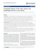
A renal biopsy was performed which showed diffuse pro-
liferative glomerulonephritis with marked IgA staining
compatible with Henoch-Schönlein nephritis (Figures 1
and 2). Over the following few days his serum creatinine
increased to 300 μmol/l, but subsequently returned to
151 μmol/l. His rash and the other systemic features also
resolved. In the absence of any crescents in the renal
biopsy, the cyclophosphamide was discontinued and he
was discharged home on oral prednisolone 50 mg daily.
However, because of severe indigestion despite taking
omeprazole 20 mg once daily, the prednisolone was
reduced to 30 mg once daily soon after.
Upon review 2 weeks later the patient's rash had returned
and he remained hypertensive with a BP of 145/95
mmHg. He was therefore prescribed azathioprine 100 mg
and ramipril 1.25 mg daily and the prednisolone was
increased again to 40 mg once daily. Unfortunately he was
again unable to tolerate the increased dose of steroids due
to gastrointestinal side effects and the prednisolone was
therefore discontinued.
One week later he presented to the accident and emer-
gency department with sudden onset of a severe head-
ache, right-sided weakness and expressive dysphasia,
followed by a generalized seizure. He was hypertensive
with a BP of 194/115 but with no papilloedema. Initial
investigations showed a serum creatinine concentration
of 438 μmol/l, haemoglobin of 11.2 g/dl, a white cell
count of 10.9 × 10
9
/litre, a platelet count of 2269 × 10
9
/
litre, prothrombin time of 14 seconds and activated par-
tial thromboplastin time of 28 seconds. He was trans-
ferred to the intensive therapy unit where he was
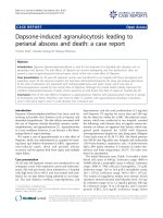
intubated and ventilated for 24 hours. A computed tom-
ography scan of his head confirmed a large left internal
capsule haemorrhage, but showed no mass effect (Figure
3). He was treated with four doses of intravenous methyl-
prednisolone 500 mg and one dose of intravenous cyclo-
phosphamide 750 mg, which was followed by oral
prednisolone 40 mg once daily and cyclophosphamide
100 mg once daily. Although his dysphasia and weakness
were improving, his renal function declined rapidly and
he required haemodialysis.
The cyclophosphamide was discontinued 2 weeks later
and the patient was discharged home on oral pred-
nisolone 25 mg once daily. Four weeks after the acute
admission he had made an almost complete recovery
from the intracerebral haemorrhage apart from some mild
Glomerulus demonstrating increased mesangial cellularity and endocapillary proliferationFigure 1
Glomerulus demonstrating increased mesangial cel-
lularity and endocapillary proliferation.
Immunostaining for IgAFigure 2
Immunostaining for IgA.
Journal of Medical Case Reports 2008, 2:200 />Page 3 of 4
(page number not for citation purposes)
dysphasia and his renal function improved enough not to
need renal replacement therapy. One year later he remains
well and dialysis-independent with a serum creatinine of
295 μmol/l, but has continued taking oral prednisolone
10 mg once daily, a dose which is gradually being
reduced. His PCR has improved to 87 and urine dipstick
has remained positive for blood 1+ and protein 1+.
Discussion
Intracerebral haemorrhage is a rare complication of HSP
that so far has mainly been described in children and
young adolescents [3-9]. Apart from this case, there is only
one other published report in an older patient, to the best
of our knowledge [10]. Such patients may develop acute
elevations in BP, and this is considered to be the primary
mechanism of intracerebral haemorrhage [11]. However,
other possible causes include the presence of cerebral vas-
culitis and the increased risk of haemorrhagic complica-
tions seen in patients with HSP. The increased risk of
bleeding in HSP has been attributed to reduced levels of
factor XIII [4] and prothrombin [5]. Other reported sites
of bleeding in patients with HSP include the gastrointesti-
nal tract, lungs, testicles and bladder [6]. Intracerebral
haemorrhage in patients with HSP has been successfully
treated in the past with surgical evacuation of the hae-
matoma [7], steroids [8] or plasmapheresis if cerebral vas-
culitis is confirmed by magnetic resonance imaging [9].
The underlying coagulopathy should also be corrected
[4,5].
HSP nephritis may affect as many as 80% of adult patients
with HSP and approximately 30% of them will develop
chronic kidney disease [2]. Adverse prognostic indicators
for progression of HSP nephritis are the presence of cres-
cents on biopsy, more than 1 g of proteinuria per 24 hours
and renal impairment on presentation [2]. The optimal
treatment of HSP nephritis remains unclear, because of
the lack of prospective randomized trials. Intravenous
pulse methylprednisolone followed by oral steroids has
been shown to be effective in the management of severe
HSP nephritis [12]. Other possible treatment regimens for
severe HSP nephritis include a combination of corticoster-
oids with cyclophosphamide, azathioprine or cyclosporin
[1,2].
Conclusion
Intracerebral haemorrhage is a rare complication of HSP
that may be caused by acute hypertension, cerebral vascu-
litis, and the increased risk of bleeding observed in this
disorder. Although our patient was severely hypertensive
at the time of presentation, both the HSP nephritis and his
neurological symptoms were successfully treated with
intravenous cyclophosphamide and methylprednisolone,
followed by a short course of oral cyclophosphamide and
long-term oral prednisolone. His renal function recovered
enough not to require renal replacement therapy.
Abbreviations
BP: blood pressure; HSP: Henoch-Schönlein purpura;
PCR: protein to creatinine ratio.
Competing interests
The authors declare that they have no competing interests.
Authors' contributions
LK wrote the initial draft of the manuscript. LL provided
the renal biopsy pictures and wrote the legends. AJW
revised and help to write the manuscript. All authors read
and approved the final manuscript.
Consent
Written informed consent was obtained from the patient
for publication of this case report and any accompanying
images. A copy of the written consent is available for
review by the Editor-in-Chief of this journal.
References
1. Saulsbury FT: Clinical update: Henoch-Schönlein purpura. Lan-
cet 2007, 369:976-978.
2. Kellerman PS: Henoch-Schönlein purpura in adults. Am J Kidney
Dis 2006, 48:1009-1016.
3. Sevcan AB, Mesiha E, Necmiye T, Gûlhis D, Erden İlhan, Tansel E:
Cerebral vasculitis in Henoch-Schönlein purpura. Nephrol Dial
Transplant 2000, 15:246-248.
4. Imai T, Okada H, Nanba M, Kawada K, Kusaka T, Itoh S: Henoch-
Schönlein purpura with intracerebral hemorrhage. Brain Dev
2002, 24:115-117.
Computed tomography scan of the head demonstrating left internal capsule haemorrhageFigure 3
Computed tomography scan of the head demon-
strating left internal capsule haemorrhage.
Publish with BioMed Central and every
scientist can read your work free of charge
"BioMed Central will be the most significant development for
disseminating the results of biomedical research in our lifetime."
Sir Paul Nurse, Cancer Research UK
Your research papers will be:
available free of charge to the entire biomedical community
peer reviewed and published immediately upon acceptance
cited in PubMed and archived on PubMed Central
yours — you keep the copyright
Submit your manuscript here:
/>BioMedcentral
Journal of Medical Case Reports 2008, 2:200 />Page 4 of 4
(page number not for citation purposes)
5. Clark JH, Fitzgerald JF: Hemorrhagic complications of Henoch-
Schönlein syndrome. J Pediatr Gastroenterol Nutr 1985, 4:311-315.
6. Chiaretti A, Caresta E, Piastra M, Pulitano S, Di Rocco C: Cerebral
hemorrhage in Henoch-Schönlein syndrome. Childs Nerv Syst
2002, 18:365-367.
7. Altinors N, Cepoglu C: Surgically treated intracerebral
hematoma in a child with Henoch-Schönlein purpura. J Neu-
rosurg Sci 1991, 35:47-49.
8. Ng CC, Huang SC, Huang LT: Henoch-Schönlein purpura with
intracerebral hemorrhage: case report. Pediatr Radiol 1996,
26:276-277.
9. Wen YK, Yang Y, Chang CC: Cerebral vasculitis and intracere-
bral hemorrhage in Henoch-Schönlein purpura treated with
plasmapheresis. Pediatr Nephrol 2005, 20:223-225.
10. Lévaif F, Szücs LH, Jászai Z: Cerebral hemorrhage and acute
glomerulonephritis in Schonlein-Henoch syndrome in old
age. Z Gesamte Inn Med 1971, 26:309-311.
11. Sang-Wuk J, Keun-Hwa J, Kon C, Hee-Joon B, Seung-Hoon L, Jae-Kyu
R: Clinical and radiologic differences between primary
intracerebral hemorrhage with and without microbleeds on
gradient-echo magnetic resonance images. Arch Neurol 2004,
61:905-909.
12. Niaudet P, Habib R: Methylprednisolone pulse therapy in the
treatment of severe forms of Schonlein-Henoch purpura
nephritis. Pediatr Nephrol 1998, 12:238-243.