Báo cáo y học: " Subcutaneous hydatid cysts occurring in the palm and the thigh: two case reports" ppt
Bạn đang xem bản rút gọn của tài liệu. Xem và tải ngay bản đầy đủ của tài liệu tại đây (338.67 KB, 4 trang )
BioMed Central
Page 1 of 4
(page number not for citation purposes)
Journal of Medical Case Reports
Open Access
Case report
Subcutaneous hydatid cysts occurring in the palm and the thigh:
two case reports
Abuzer Dirican, Bulent Unal*, Cuneyt Kayaalp and Vedat Kirimlioglu
Address: Department of General Surgery, Medical Faculty of Inonu University, Malatya, Turkey
Email: Abuzer Dirican - ; Bulent Unal* - ; Cuneyt Kayaalp - ;
Vedat Kirimlioglu -
* Corresponding author
Abstract
Introduction: Hydatid cyst disease is common in some regions of the world and is usually located
in the liver and lungs. This report presents two cases of primary hydatid cysts located
subcutaneously: one in the medial thigh and one in the left palm between the index and middle
fingers.
Case presentations: A 64-year-old male farmer visited our hospital because a swelling on the
right medial thigh had grown during the last year. Superficial ultrasound and computed tomography
revealed a lesion resembling a hydatid cyst. A germinative membrane was encountered during
surgical excision. Pathological examination was compatible with a hydatid cyst. The second case
involved a 67-year-old male farmer who complained of a swelling that had grown in his left palm in
the last year. The preliminary diagnosis was a lipoma. However, a hydatid cyst was diagnosed during
surgical excision and after the pathological examination. The patient did not have a history of
hydatid cyst disease and hydatid cysts were not detected in other organs. There has been no
disease recurrence after following both patients for 3 years.
Conclusion: A hydatid cyst should be considered in the differential diagnosis of subcutaneous
cystic lesions in regions where hydatid cysts are endemic, and should be excised totally, with an
intact wall, to avoid recurrence.
Introduction
A hydatid cyst is a parasitosis caused by the larval form of
Echinococcus granulosus or rarely Echinococcus alveolaris. The
main hosts for E. granulosus are predators such as dogs,
wolves, and foxes, while intermediate hosts include
sheep, goats, and cattle. Humans are a coincidental inter-
mediate host. The disease is more frequent in the Middle
East, Central Europe, Australia, and South America, where
the intermediate hosts are common. The organs affected
most often are the liver (70%) and lungs (10–15%).
Other locations are extremely rare [1]. Primary subcutane-
ous hydatid cyst is very rare and the incidence is
unknown. In this report, we present two cases of primary
hydatid cysts located subcutaneously: one in the medial
thigh and one in the hand.
Case presentations
A 64-year-old male farmer visited our clinic because of a
swelling on the medial thigh that had grown during the
last year. On physical examination, a mobile, painless,
fluctuant, 8 × 9 cm mass was palpated. The overlying skin
was normal. The only abnormality in the pre-operative
Published: 13 August 2008
Journal of Medical Case Reports 2008, 2:273 doi:10.1186/1752-1947-2-273
Received: 9 January 2008
Accepted: 13 August 2008
This article is available from: />© 2008 Dirican et al; licensee BioMed Central Ltd.
This is an Open Access article distributed under the terms of the Creative Commons Attribution License ( />),
which permits unrestricted use, distribution, and reproduction in any medium, provided the original work is properly cited.
Journal of Medical Case Reports 2008, 2:273 />Page 2 of 4
(page number not for citation purposes)
laboratory examination was an increased erythrocyte sed-
imentation rate (ESR 60 mm/hour). The patient had no
history of surgery for a hydatid cyst in another organ.
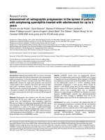
Ultrasound (US) and computed tomography (CT)
showed a lesion resembling a hydatid cyst (Fig. 1). During
surgical exploration under spinal anesthesia, the skin and
subcutaneous layers were incised and the cyst was
reached. Hypertonic saline (3% NaCl) was injected into
the cyst and after waiting for 10 min, the cyst was com-
pletely excised. A germinative membrane was seen during
excision (Fig. 2). We thought that the cyst was fertile as it
contained daughter cysts. The surgical site was irrigated
with 40% povidone iodine (Betadine
®
) and hypertonic
saline. The subcutaneous layers and skin were closed in
the standard manner.
Histopathological examination revealed a hydatid cyst,
but no additional hydatid cysts were observed on US or
CT of the abdomen and thorax; the indirect hemaggluti-
nation test for hydatid cysts was negative. The patient was
started on albendazole for 3 months (15 mg/kg/day). No
findings associated with local or systemic hydatid cysts
were detected during a 3-year follow-up period.
The other case involved a 67-year-old male farmer who
complained of a subcutaneous swelling inside the left
palm between the index and middle fingers. Physical
examination revealed a subcutaneous immobile 2 × 3 cm
mass on the palmar side of the left hand between the
thumb and index fingers. Surgical excision was planned
with a pre-operative diagnosis of lipoma. A hydatid cyst
was considered when a germinative membrane was seen
during excision under local anesthesia (Fig. 3). We also
thought that the cyst was fertile as it contained daughter
cysts as in the previous patient. The cyst space was irri-
gated with 40% povidone iodine (Betadine
®
) and hyper-
tonic saline. Total cyst excision and primary closure were
performed, and histopathological examination revealed a
hydatid cyst. The only abnormality in the pre-operative
laboratory examination was an increased ESR (60 mm/
hour). The patient had no history of surgery for a hydatid
cyst in another organ, and no additional cysts were
observed on US and CT of the abdomen and thorax. The
indirect hemagglutination test for hydatid cysts was nega-
tive, and the patient was placed on albendazole for 3
months (15 mg/kg/day). No findings associated with
Germinative membrane of cyst localized in the palmar site of the handFigure 3
Germinative membrane of cyst localized in the palmar site of
the hand.
Subcutaneous hydatid cyst in the right medial thigh, displacing the muscles laterallyFigure 1
Subcutaneous hydatid cyst in the right medial thigh, displacing
the muscles laterally.
Subcutaneous hydatid cyst in the right medial thighFigure 2
Subcutaneous hydatid cyst in the right medial thigh.
Journal of Medical Case Reports 2008, 2:273 />Page 3 of 4
(page number not for citation purposes)
local or systemic hydatid cysts were detected during a 3-
year follow-up period.
Discussion
Here we report two cases of primary subcutaneous
hydatid cysts both treated surgically. In a large series, the
distribution of hydatid cysts outside the liver and lungs
was reported as 9% of cases [2]. Chevalier et al. reported
that the incidence of subcutaneous hydatid cysts was 2%,
but some of the patients had hydatid cysts in other organs
too [3]. Subcutaneous hydatid cyst may be secondary or
primary. In secondary cysts, there is a primary location of
hydatid disease like liver, lung, or spleen that is operated
or not operated. Reports of primary subcutaneous hydatid
cysts are very rare [4-6], and we were unable to find a case
of a palmar hydatid cyst in a literature review. In our cases,
the hydatid cysts were located subcutaneously, the
patients had not undergone previous surgery for hydatid
cysts, and no hydatid cysts were found in other organs.
Therefore, our patients were diagnosed as having primary
subcutaneous hydatid cysts.
The mechanism of primary subcutaneous localization is
unclear. After being ingested orally, under the action of
gastric and intestinal enzymes, the oncosphere is released;
it penetrates the intestinal wall, joins the portal system
and reaches the liver. If the eggs attach to the liver, an
hepatic hydatid cyst takes shape. Parasite eggs can pass to
the systemic circulation and cause disease in other end
organs. Larvae must pass through two filters (liver and
lung) to form a solitary hydatid cyst, but that is very diffi-
cult. It is very possible that systemic dissemination via the
lymphatic route accounts for cases with solitary cysts in
uncommon sites [4]. Direct spread from adjacent sites
may be another mechanism of infection provided a
microrupture has occurred [7].
Diagnosing hydatid cysts is very difficult in patients living
outside the endemic regions. Because exposure to the con-
tents of the cyst can cause problems such as anaphylactic
reaction and local recurrence, making the diagnosis pre-
operatively is important. The diagnosis of a palmar
hydatid cyst was not considered in our second patient pre-
operatively since the mass was very small and this locali-
zation is very rare. When the cyst contents were seen dur-
ing excision, the possibility of a hydatid cyst was then
considered. No anaphylactic reaction developed in either
patient.
The radiological findings of a thick cyst wall, calcification,
daughter cysts, and a germinative membrane separate
from the cyst wall are findings specific to hydatid cysts [8].
Our first case was diagnosed according to the appearance
of the mass on superficial US and CT.
Serology is a useful tool for the diagnosis. The indirect
hemagglutination (IHA) test is positive in more than 80%
of liver hydatid cysts. However, false negative IHA results
can be higher in other located hydatid cyst. In those cases,
more specific serologic tests are mandatory. A positive
indirect hemagglutination test for hydatid cysts is signifi-
cant, although negative test results do not indicate the
absence of the disease, as in our patients. Therefore, the
most important diagnostic tool is the awareness of the
physician, particularly for the unusual presentation of the
disease.
The best treatment option is total surgical excision with-
out opening the cyst. If the cyst cannot be excised without
opening, the fluid contents should be removed, the lami-
nated membrane should be totally excised, and the cyst
pouch should be irrigated with protoscolicidal solutions
[9]. Subcutaneous located cysts are more prone to rupture
since they have not been diagnosed pre-operatively. We
performed total cyst excision in both cases and irrigated
the surgical areas with protoscolicidal agents. Identifying
postoperative recurrence of the cyst in endemic regions is
very difficult because the probability of formation of a
new cyst is high. However, since our patients were still free
of disease in the third postoperative year, any subsequent
hydatid cyst formation may be considered to be a new
infestation.
Conclusion
Hydatid cyst should be considered in the differential diag-
nosis of subcutaneous cysts in regions where hydatid cysts
are endemic. Total excision of the cyst with an intact wall
is the best treatment.
Competing interests
The authors declare that they have no competing interests.
Authors' contributions
AD is the consultant surgeon who drafted the article and
performed the operations. BU assisted in performing the
surgery, took the pictures and helped revise the article. CK
helped in acquisition of data and technical support. VK
performed the literature search and helped in revision. All
authors read, appraised and approved the final manu-
script.
Consent
Written informed consent was obtained from the patients
before publication of this case series and any accompany-
ing images. A copy of the written consent is available for
review by the Editor-in-Chief of this journal.
References
1. Kayaalp C: Hydatid cyst of the liver. In Surgery of the Liver, Biliary
Tract, and Pancreas 4th edition. Edited by: Blumgart LH, Belghiti RJ,
Publish with Bio Med Central and every
scientist can read your work free of charge
"BioMed Central will be the most significant development for
disseminating the results of biomedical research in our lifetime."
Sir Paul Nurse, Cancer Research UK
Your research papers will be:
available free of charge to the entire biomedical community
peer reviewed and published immediately upon acceptance
cited in PubMed and archived on PubMed Central
yours — you keep the copyright
Submit your manuscript here:
/>BioMedcentral
Journal of Medical Case Reports 2008, 2:273 />Page 4 of 4
(page number not for citation purposes)
DeMatteo RP, Chapman WC, Büchler MW, Hann LE, D'Angleca M.
Philadelphia, PA: Saunders Elsevier; 2007:952-970.
2. Prousalidis J, Tzardioglou K, Sgouradis L, Katsohis C, Aletras H:
Uncommon sites of hydatid disease. World J Surg 1998,
22:17-22.
3. Chevalier X, Rhomouni A, Bretagne S, Martigny J, Larget Piet B:
Hydatid cyst of the subcutaneous tissue without other
involvement: MR imaging features. AJR 1994, 163:645-646.
4. Engin O, Erdoğan M: Solitary subcutaneous hydatid cyst. Am J
Trop Med Hyg 2000, 62:583-584.
5. Öztürk S, Deveci M, Yıldırım S: Hydatid cyst in the soft tissue of
the face without any primary. Ann Plast Surg 2001, 46:170-173.
6. Ambo M, Adachi K, Okhawara A: Postoperative alveolar hydatid
disease with cutaneous involvement. J Dermatol 1999,
26:343-347.
7. Safioleas M, Nikiteas N, Stamatakos M, Safioleas C, Manti CH, Reve-
nas C, Safioleas P: Echinococcal cyst of the subcutaneous tis-
sue: A rare case report. Parasitol Int 2008, 57:236-238.
8. Fikry T, Harfaoui A, Sibai H, Zryoil BL: Echinococcose musculaire
primitive. J Chir 1997, 134:325-328.
9. Duncan GJ, Tooke SMT: Echinococcus infestation of the biceps
brachii. Clin Orthop 1990, 261:247-250.