Báo cáo y học: "Radiofrequency-induced thermotherapy of nasopharyngeal angiofibroma and immunohistochemical analysis of vessel proliferation: a case report" docx
Bạn đang xem bản rút gọn của tài liệu. Xem và tải ngay bản đầy đủ của tài liệu tại đây (1.12 MB, 5 trang )
BioMed Central
Page 1 of 5
(page number not for citation purposes)
Journal of Medical Case Reports
Open Access
Case report
Radiofrequency-induced thermotherapy of nasopharyngeal
angiofibroma and immunohistochemical analysis of vessel
proliferation: a case report
Mira Krstulja*
1
, Milodar Kujundžić
2
, Adelaida Halaj
3
, Tamara Braut
2
and
Niko Cvjetković
2
Address:
1
Department of Pathology, School of Medicine, University of Rijeka, Brace Branchetta, 51000, Rijeka, Croatia,
2
Clinic for Otolaryngology
and Head and Neck Surgery, School of Medicine, University of Rijeka, Brace Branchetta, 51000, Rijeka, Croatia and
3
Clinical Department of
Radiology, School of Medicine, University of Rijeka, Brace Branchetta, 51000, Rijeka, Croatia
Email: Mira Krstulja* - ; Milodar Kujundžić - ; Adelaida Halaj - ;
Tamara Braut - ; Niko Cvjetković -
* Corresponding author
Abstract
Introduction: Nasopharyngeal angiofibroma presents with symptoms of nasal obstruction and
epistaxis. The treatment of choice is embolization followed by surgery.
Case presentation: A 52-year-old man underwent surgery for nasopharyngeal angiofibroma after
adjuvant radiofrequency-induced thermotherapy. To the best of the authors' knowledge, this is the
first case of angiofibroma with clinical follow-up after thermocoagulation therapy supported by
quantitative, double immunohistochemistry. We found this case of angiofibroma to be of interest
owing to the presentation of symptoms leading to biopsy, the pathohistological observations
obtained with synchronous Ki67/cluster of differentiation 34 and Ki67/smooth muscle actin
immunohistochemistry and high pericyte proliferation.
Conclusion: Coagulation of angiofibroma vessels followed by acquisition of a thick mantle of
pericytes in a patient with a nasopharyngeal growth suggests that radiofrequency-induced
thermotherapy could be a useful, palliative therapy for bleeding nasopharyngeal angiofibroma,
supporting vessel maturation prior to surgical tumor removal.
Introduction
Nasopharyngeal angiofibroma is considered to be a reac-
tive, malformed, benign but aggressive neoplasm. Clinical
staging and tumor embolization reduce surgical morbid-
ity. The therapy protocol is influenced by hospital-related
factors. Radiofrequency-induced thermotherapy (RFITT)
is a minimally invasive surgical procedure that causes
thermal ablation through coagulation and is used in the
treatment of both head and neck diseases. We were unable
to find reported cases of angiofibroma that were treated
with RFITT, subjected to follow-up evaluation and had
documented histological changes with time.
We present an unusual case of a 52-year-old man with
nasopharyngeal angiofibroma that first appeared as a
nasal polyp. Coagulation, thrombosis, sclerosis and peri-
cyte proliferation occurred after RFITT. We looked for a
change in angiofibroma cell proliferation through biop-
Published: 16 August 2008
Journal of Medical Case Reports 2008, 2:278 doi:10.1186/1752-1947-2-278
Received: 30 December 2007
Accepted: 16 August 2008
This article is available from: />© 2008 Krstulja et al; licensee BioMed Central Ltd.
This is an Open Access article distributed under the terms of the Creative Commons Attribution License ( />),
which permits unrestricted use, distribution, and reproduction in any medium, provided the original work is properly cited.
Journal of Medical Case Reports 2008, 2:278 />Page 2 of 5
(page number not for citation purposes)
sies obtained before and after RFITT when the patient was
free of bleeding episodes. The cell origin of vessel forma-
tion after thermocoagulation therapy was investigated.
Our results are of interest for surgeons applying pre-oper-
ative thermal ablation therapy.
Case presentation
A 52-year-old white man, who experienced breathing dif-
ficulties and nasal speech for 15 months, was hospitalized
for nasal polyps. A radiograph of his paranasal sinuses (21
January 2005) showed a soft tissue lesion in the medios-
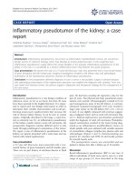
agittal line, suggesting a nasal polyp. A biopsy (18 Febru-
ary 2005) of the polyp revealed that it was immovable and
provoked bleeding. The provided tissue (0.5 cm
3
) was
diagnostic for nasopharyngeal angiofibroma after routine
hematoxylin and eosin (H&E) staining (Figure 1), the
stromal cells were negative for both cluster of differentia-
tion (CD) 34 antigen and smooth muscle actin (SMA)
antibodies and C-kit antibody was rarely detected in sin-
gle cells.
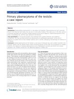
Digital subtraction angiography showed the pathological
vascularization of the tumor (8 March 2005; Figure 2A). A
computed tomography (CT) scan of the viscerocranium
with intravenous contrast revealed a 56 mm × 48 mm
large, soft tissue growth that filled the nasopharynx and
extended to the left nasal cavity (24 February 2005; Figure
2B). A multiple slice CT carotidography (10 May 2005)
revealed that there was blood supply to the tumor from
the external carotid vessels (Figure 2C).
With a diagnosis of nasopharyngeal angiofibroma (Rad-
kowski's stage Ib), the patient was subjected to RFITT
using a Celon AG medical instrument (radiofrequency
power, 15 to 20 W and a 5-minute application time). The
therapy was performed three times over a 2-month period
(1 June 2005, 9 June 2005 and 31 August 2005). The
lesion did not bleed but hardened. The second surgical
specimen (21 September 2005) was 5 cm
3
of angiofi-
broma tissue with multiple 2 to 3 mm centers of coagula-
tion (Figure 3). After RFITT, the clinical symptoms were
alleviated despite the incomplete reduction in tumor size.
Staining for Ki67 showed low overall proliferation in the
first biopsy but increased proliferation in the second (1%
and 10%, respectively). A control CT scan (29 September
2005) of the epipharynx revealed a residual tumor, an
enlarged left maxillary sinus with a missing medial wall,
thickened mucosa without post-contrast opacification
and no enlarged lymph nodes.
A third biopsy 10 months after RFITT provided 0.075 cm
3
of residual tumor with an overall Ki67 proliferation index
of 10%. Plump SMA-positive and predominantly Ki67-
negative cells were detached from the vessel wall and
formed sheets resembling angiomyofibroblastoma after
H&E staining. The second and third biopsies respected the
recovery time from RFITT and were not complicated by
hemorrhage.
One year after RFITT, angiography found no arteries feed-
ing the residual tumor. The patient underwent surgery at
another institution without prior embolization (no
hypertrophic feeding arteries were found at repeated ang-
iography before the operation).
The primary intention was to reduce the tumor and allevi-
ate the symptoms using RFITT before the operation. Dou-
ble immunostaining was planned later because of
increased Ki67 staining observed in the control biopsy
after RFITT. Ki67 is a proliferation marker providing
nuclear staining when the cell is in the S phase preparing
to enter mitosis. To determine which cell type is prolifer-
ating in a tissue, a second differentiation marker is added,
that is, CD34 for endothelial cells or SMA for pericytes.
The immunohistochemical analysis of all three angiofi-
broma biopsies was repeated with a double-staining tech-
nique for both Ki67/CD34 and Ki67/SMA to distinguish
between endothelial cell and pericyte proliferation over
time (Figure 4A, B and 4C). Three parameters were used to
quantify proliferation. The endothelial cell proliferation
index (EPI) and pericyte proliferation index (PEPI) were
defined as the percentage of Ki67-positive nuclei per 1000
cells for each cellular compartment. This was different
from routine, less expensive single Ki67 immunostaining
where the proliferation index takes into consideration all
the cells in the tissue without distinguishing between ves-
Angiofibroma prior to radiofrequency-induced thermother-apyFigure 1
Angiofibroma prior to radiofrequency-induced ther-
motherapy. Hematoxylin and eosin stain, magnification
×10.
Journal of Medical Case Reports 2008, 2:278 />Page 3 of 5
(page number not for citation purposes)
sel cells and stromal cells. The number of vessel sections
per field was obtained and the results were expressed as
microvessel density (MVD), which is the number of
lumina per square millimeter. The proliferating capillary
index (PCI) was defined as the percentage of vessel sec-
tions of any cell type whose nuclei stained positive for
Ki67. The proliferation analysis results are shown in Table
1.
Double immunohistochemical staining revealed higher
proliferation indices for cells of the vessel compartment
compared with single Ki67 staining of each routine
biopsy. The EPI slightly decreased while the PEPI
increased 10 months after RFITT. The third biopsy con-
tained a large number of detached SMA-positive cells.
There were scattered Ki67-positive nuclei of cells outside
the vessel wall that were defined by neither CD34 nor
SMA in all three biopsies. The MVD increased 20 days
after RFITT and further increased with time. The PCI also
increased with time. Measurements and images were
obtained using a BX-40 Olympus microscope, Sony CCD-
Iris color video camera and ISSA 3.1 software (Vamstec,
Zagreb).
Discussion
Nasopharyngeal angiofibroma is considered a malforma-
tion in juveniles [1-3], but does not exclude the unusual
presentation of the disease in mature patients, as con-
firmed by this report and occasional reports from other
authors [4]. While nasal polyps are not subjected rou-
tinely to CT or magnetic resonance imaging, these are
established pre-operative diagnostic tools for nasopharyn-
geal angiofibroma.
The case presented here is of interest from both the clini-
cal and the pathological points of view. The nasopharyn-
geal and sinonasal tracts are sites of different pathologies
prone to epistaxis, such as the angiofibroma, angiectatic
nasal polyp [5], and necrotizing angiocentric lesion. The
stroma is different in these lesions and quite typical in
Scans of a nasopharyngeal angiofibromaFigure 2
Scans of a nasopharyngeal angiofibroma. (A) Digital subtraction angiography (maximum intensity projection technique):
the terminal branch of the left maxillary artery is at the hilus of the pathological angiofibroma neovascularization. (B) Com-
puted tomography of the viscerocranium: nasopharyngeal angiofibroma seen with intravenous contrast. (C) The same tumor
seen with computed tomography carotidography (volume rendering technique).
Coagulation in angiofibroma (on the right), 3 weeks after radiofrequency-induced thermotherapyFigure 3
Coagulation in angiofibroma (on the right), 3 weeks
after radiofrequency-induced thermotherapy. Hema-
toxylin and eosin stain, magnification ×10.
Journal of Medical Case Reports 2008, 2:278 />Page 4 of 5
(page number not for citation purposes)
angiofibroma. SMA decorates the stromal cells in certain
nasal polyps. It is strongly positive in the vessel wall (per-
icytes) and occasionally in the stroma of angiofibromas
[1,6], which may help in differential diagnosis.
In our case, two pathologies were present synchronously,
a mucosal nasal polyp and an angiofibroma, making the
diagnosis more complex as noticed by other authors [6].
The association between inflammatory nasal polyps and
angiofibroma is not routinely expected, but once a biopsy
is obtained, there are criteria to distinguish between nasal
polyps arising through different pathogenic processes [7].
Nasopharyngeal angiofibroma is a rare event and biopsy
is not advised. The first biopsy of our patient resulted
from atypical extension of the tumor into the nasal cavity.
The dates for the second and third biopsy were chosen
with regards to the recovery period after RFITT. Although
not a new disease, nasopharyngeal angiofibroma remains
a clinical and scientific challenge. Thermocoagulation
should be considered as a possible pre-operative protocol
when embolization is not available.
The origin of angiofibroma is still under investigation.
Zhang et al. [2] presented arguments for primary stromal
change at the molecular level of angiofibroma organiza-
tion. However, the origin of vessel formation is uncertain
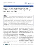
Proliferation of pericytes in angiofibromaFigure 4
Proliferation of pericytes in angiofibroma. (A) Prior to radiofrequency-induced thermotherapy. (B) Three weeks after
radiofrequency-induced thermotherapy. (C) Ten months after radiofrequency-induced thermotherapy, detachment of peri-
cytes from the vessel wall. Magnification ×20. Ki67/SMA double immunohistochemistry. Ki67-positive nuclei of cycling cells
were visualized using ChemMate DAB+ Chromogen. Cytoplasm of the endothelial cells and pericytes was visualized by fast red
staining.
Table 1: Variables of cell proliferation and vessel proliferation in angiofibroma with time
Variable Endothelial cell
proliferation index (%)
Pericyte proliferation
index (%)
Proliferating
capillary
index (%)
Microvessel
density per mm
2
Order of biopsy 1 2 3 1 2 3 1 2 3 1 2 3
Mean* 8.34 11.18 9.10 16.04 19.36 20.59 38.5 42 54 138 180 226
1, 2 and 3: The first, second and third biopsies. *25 microscopic fields per variable (microscopic field 0.0415265 mm
2
).
Journal of Medical Case Reports 2008, 2:278 />Page 5 of 5
(page number not for citation purposes)
[8,9] and pericyte behavior in angiofibroma may be of
interest. We were unable to find reports on pericyte prolif-
eration in nasopharyngeal angiofibroma treated with
RFITT. We find our observations of importance for the
investigation of angiogenesis, angiofibroma and post-
RFITT control biopsies. Our observations are in accord-
ance with the purpose of the therapy, that is, to impede
circulation and produce coagulation, thus reducing
growth. The lesion was successfully treated surgically
without pre-operative embolization, suggesting that
RFITT might function as a pre-operative adjuvant therapy.
Two years after RFITT, our patient is without symptoms or
nasopharyngeal growth.
Histologically, both endothelial cell and pericyte prolifer-
ation were more accurately expressed with double immu-
nohistochemistry compared with routine Ki67 staining.
Pericyte proliferation was stronger than endothelial cell
proliferation prior to therapy (PEPI 16.04%, EPI 8.34%).
While the PEPI increased upon coagulation and pro-
gressed with time, the EPI did not. These results support
the theory of angiofibroma as a maturing vasoformative
lesion. Vessel formation is observed in inflammation,
malformation, neovascularization of neoplasia and as a
neoplastic event. Proliferation in vascular malformations
has been studied previously [10,11]. Vessel formation in
inflammation is diffuse except in granulomas. Malforma-
tions and neoplasias, including angiofibromas, behave as
a 'body' in that they are fed and can be embolized, and
angiofibromas are not considered neoplastic events. Mal-
formations occurring with age are unusual but not unex-
pected. Zhang et al. [2] showed that angiofibroma stromal
cells might be neoplastic. Our investigation of angiofi-
broma using double immunohistochemistry showed neg-
ligible proliferation outside the vascular compartment.
Conclusion
We have presented a rare case of angiofibroma in a 52-
year-old man with pericyte proliferation, supporting the
maturation of the vessel compartment and revealing
active angiogenic machinery (cooperation between
endothelial cells and pericytes). We observed the diver-
gent behavior of endothelial cells and pericytes after
RFITT adjuvant therapy prior to surgery. Further studies of
RFITT related to vessel behavior are needed. We found
thrombosis and coagulation resulting from RFITT to func-
tion as equivalent to embolization prior to surgical ther-
apy for angiofibroma. An analysis of vessel cell
proliferation in tissues treated with thermal ablation
might have broader clinical impact across medicine.
Abbreviations
CD; Cluster of differentiation; CT: Computed tomogra-
phy; EPI: Endothelial cell proliferation index; H&E:
Hematoxylin and eosin; MVD: Microvessel density; PCI:
Proliferating capillary index; PEPI: Pericyte proliferation
index; RFITT: Radiofrequency-induced thermotherapy;
SMA: Smooth muscle actin.
Competing interests
The authors declare that they have no competing interests.
Authors' contributions
MKr is the author of this study and performed the quanti-
tative analysis of the double-stained immunohistological
slides. MKu, TB and NC are surgeons who treated and
observed the patient and provided the angiofibroma
biopsy specimens. AH is our radiologist responsible for
the acquisition of data and analysis and interpretation of
data.
Consent
Written informed consent was obtained from the patient
for publication of this case report and any accompanying
images. A copy of the written consent is available for
review by the Editor-in-Chief of this journal.
References
1. Liang J, Yi Z, Lianq P: The nature of juvenile nasopharyngeal
angiofibroma. Otolaryngol Head Neck Surg 2000, 123:475-481.
2. Zhang PJ, Weber R, Liang HH, Pasha TL, LiVolsi VA: Growth factors
and receptors in juvenile nasopharyngeal angiofibroma and
nasal polyps: an immunohistochemical study. Arch Pathol Lab
Med 2003, 127:1480-1484.
3. Beham A, Beham-Schmid C, Regauer S, Auböck L, Stammberger H:
Nasopharyngeal angiofibroma: true neoplasm or vascular
malformation? Adv Anat Pathol 2000, 7:36-46.
4. Celik B, Erisen L, Saraydaroglu O, Coskun H: Atypical angiofibro-
mas: a report of four cases. Int J Pediatr Otorhinolaryngol 2005,
69:415-421.
5. Yfantis HG, Drachenberg CB, Gray W, Papadimitriou JC: Angiec-
tatic nasal polyps that clinically simulate a malignant proc-
ess: report of 2 cases and review of the literature. Arch Pathol
Lab Med 2000, 124:406-410.
6. Wang QP, Escudier E, Roudot-Thoraval F, Abd-Al Samad I, Peynegre
R, Coste A: Myofibroblast accumulation induced by trans-
forming growth factor-beta is involved in the pathogenesis of
nasal polyps. Laryngoscope 1997, 107:926-931.
7. Baumgarten C, Kunkel G, Rudolph R, Staud RD, Sperner I, Gelderb-
lom H: Histopathological examinations of nasal polyps of dif-
ferent etiology. Arch Otorhinolaryngol 1980, 226:187-197.
8. Minasi MG, Riminucci M, De Angelis L, Borello U, Berarducci B, Inno-
cenzi A, Caprioli A, Sirabella D, Baiocchi M, De Maria R, Boratto R,
Jaffredo T, Broccoli V, Bianco P, Cossu G: The meso-angioblast: a
multipotent, self-renewing cell that originates from the dor-
sal aorta and differentiates into most mesodermal tissues.
Development 2002, 129:2773-2783.
9. DeRuiter MC, Poelmann RE, VanMunsteren JC, Mironov V, Markwald
RR, Gittenberger-de Groot AC: Embryonic endothelial cells
transdifferentiate into mesenchymal cells expressing
smooth muscle actins in vivo and in vitro. Circ Res 1997,
80:444-451.
10. Meijer-Jorna LB, Loos CM van der, de Boer OJ, Horst CM van der,
Wal AC van der:
Microvascular proliferation in congenital vas-
cular malformations of skin and soft tissue. J Clin Pathol 2007,
60:798-803.
11. Vargel I, Cil BE, Er N, Ruacan S, Akarsu AN, Erk Y: Hereditary
intraosseous vascular malformation of the craniofacial
region: an apparently novel disorder. Am J Med Genet 2002,
109:22-35.