Báo cáo y học: " Osteoid osteoma of a metacarpal bone: a case report and review of the literature" docx
Bạn đang xem bản rút gọn của tài liệu. Xem và tải ngay bản đầy đủ của tài liệu tại đây (276.29 KB, 4 trang )
BioMed Central
Page 1 of 4
(page number not for citation purposes)
Journal of Medical Case Reports
Open Access
Case report
Osteoid osteoma of a metacarpal bone: a case report and review of
the literature
Efstathios Chronopoulos, Fragiskos N Xypnitos, Vassilios S Nikolaou*,
Nicolas Efstathopoulos and Dimitrios Korres
Address: 2nd Orthopedic Department, Konstantopoulion Hospital, Athens University, Nea Ionia, Greece
Email: Efstathios Chronopoulos - ; Fragiskos N Xypnitos - ;
Vassilios S Nikolaou* - ; Nicolas Efstathopoulos - ; Dimitrios Korres -
* Corresponding author
Abstract
Introduction: Osteoid osteoma is a benign tumor of the growing skeleton. It presents with pain,
which is usually worse at night. The radiographic features consist of a central oval or round nidus
surrounded first by a radiolucent area followed by another area of sclerotic bone. In the hand,
osteoid osteoma is more commonly located in the phalanges and carpal bones. The metacarpals
are the least common sites for osteoid osteoma.
Case presentation: We present a case of an osteoid osteoma of the left third metacarpal bone
in a 36-year-old woman. The clinical and radiographic findings along with the surgical management
of the lesion are presented. The pain disappeared immediately after the operation. At the 2-year
follow-up, the patient was pain-free and there was no evidence of recurrence.
Conclusion: Physicians should be aware of the unusual presence and the atypical clinical
presentation of this benign lesion in the metacarpal bones of the hand.
Introduction
Osteoid osteoma is a benign bone tumor of the growing
skeleton representing approximately 10% of all benign
bone neoplasias [1]. It usually affects children and young
adults [1]. Heine in 1927 [2], Bergstrand in 1930 [3], and
Jaffe in 1935 [4] identified osteoid osteoma as a clinical
entity. Pain is often the only symptom of the disease and
is typically described as mild and intermittent at first,
becoming more constant and severe at night [5]. When
the lesions appear in the hand, diagnosis is challenging
for three reasons: first, the typical pain pattern may be
absent; second, lesions in the hand may have unusual
clinical signs and radiographic presentations; and third,
histologic features may differ from classic osteoid osteo-
mas, which occur in the long bones [6]. The metacarpals
in particular are not a common site for osteoid osteoma
and the diagnosis is often missed in the initial examina-
tion. We report a case of an osteoid osteoma in the third
metacarpal, and describe the clinical presentation, radio-
logical findings and successful outcome after surgical exci-
sion of the lesion.
Case presentation
A 36-year-old woman was referred to our clinic in May
2005 with a 1-year history of pain in her left hand. The
pain was constant but increased at night and after manual
labor, and was reduced by non-steroidal anti-inflamma-
tory agents. There was no history of injury.
Published: 27 August 2008
Journal of Medical Case Reports 2008, 2:285 doi:10.1186/1752-1947-2-285
Received: 1 January 2008
Accepted: 27 August 2008
This article is available from: />© 2008 Chronopoulos et al; licensee BioMed Central Ltd.
This is an Open Access article distributed under the terms of the Creative Commons Attribution License ( />),
which permits unrestricted use, distribution, and reproduction in any medium, provided the original work is properly cited.
Journal of Medical Case Reports 2008, 2:285 />Page 2 of 4
(page number not for citation purposes)
There was a tender swelling of the head of the third meta-
carpal bone in the dorsum of the left hand at physical
examination. The range of motion was not limited and
there were no sensory disturbances. The grip strength of
the left hand was slightly reduced, mainly due to pain.
Blood count and biochemical profile were within the ref-
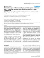
erence ranges. The radiograph showed an oval nidus sur-
rounded by a radiolucent ring (Fig. 1).
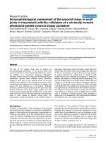
Computed tomography (CT) of the left hand clearly
showed an oval radiolucent zone at the head of the third
metacarpal bone and marked sclerosis around the lesion
(Fig. 2). The history and clinical and radiographic findings
pointed to the diagnosis of an osteoid osteoma of the
head of the third metacarpal bone in the left hand. The
patient was operated on 30 days later, by a dorsal
approach (Fig. 3a), under a brachial plexus block. An en
bloc excision of the nidus was performed using a small
curette. A high-speed burr was also used to remove the
sclerotic bone inside the lesion (Fig. 3b). The defect was
filled with an autogenous cancellous bone graft (Fig. 3c).
The hand was immobilized postoperatively with a splint.
Histological examination confirmed the diagnosis of oste-
oid osteoma. The pain disappeared immediately after the
operation. At the 2-year follow-up, the patient was pain-
free and there was no evidence of recurrence (Fig. 1).
Discussion
Osteoid osteoma is a benign bone tumor of the growing
skeleton representing approximately 10% of all benign
bone neoplasias. It usually affects children and young
adults. Normally the tumor does not exceed 1 cm in diam-
eter [7]. The radiographic characteristic of osteoid
osteoma is the central nidus, a 2 to 10 mm focus of oste-
oid nested in a more radiolucent fibrous stroma, sur-
rounded by marginal sclerosis.
Osteoid osteoma usually occurs in the second and third
decade of life. Male patients are more often affected than
female patients by a ratio of 2:1, and the tumor is rare in
the African-American population. It has a predilection for
the lower extremity, with half or more of the lesions
occurring in the femur and tibia, near the end of the shaft.
Of the remaining lesions, approximately 30% are equally
distributed among the spine, hand and foot [8].
Localization in the hand occurs with an incidence of only
about 8% of all reported cases. Nevertheless, osteoid
osteoma of the hand is well described in the literature.
Allieu and Lussiez [9] and Ambrosia et al. [10] reported
the largest series of hand osteoid osteomas. The phalanges
are the most frequent sites for osteoid osteoma in the
hand [11-13], followed by the carpal bones. The metacar-
pals are the least common sites for osteoid osteoma [14-
16].
Trauma has been considered to be a contributing factor,
although for others the correlation between injury and the
onset of osteoid osteoma remains unclear [11]. Carroll
[11] asserted that there is no direct correlation between
them, but many cases have been reported in which an
injury precedes the onset of the lesion. Kendrick and
Evarts [17] reported that 15 out of their 36 cases had had
an episode of initial trauma, and the incidence reported
by Bednar et al. [18] was 11 out of 46 cases. Baron et al.
[19] described 15 patients with post-traumatic osteoid
osteoma. Uda et al. [14] reported a case of an osteoid
osteoma of the metacarpal bone presenting after an
injury.
Clinically, patients usually present with pain and swell-
ing. The pain, which occurs in about 80% of patients, is
more severe at night and is often relieved with salicylates
Plain radiography of the left handFigure 1
Plain radiography of the left hand. A small, oval, radiolu-
cent lesion partially surrounded by sclerotic bone (left). No
signs of recurrence at the 2-year follow-up (right).
Computed tomography showing the radiolucent zone and the marked sclerosis around the lesion (arrow)Figure 2
Computed tomography showing the radiolucent
zone and the marked sclerosis around the lesion
(arrow).
Journal of Medical Case Reports 2008, 2:285 />Page 3 of 4
(page number not for citation purposes)
or other non-steroidal anti-inflammatory agents that
inhibit the production of prostaglandins by the lesion
[20]. Several hypotheses have been proposed to explain
the intensity of pain. Nerve endings might be stimulated
by the high pressure owing to the increased blood flow
within the tumor [21]. Nerve fibers, which are presumed
to be components of the autonomic nervous system, are
identified in the fibrous zone around the nidus [22]. Pros-
taglandins may directly stimulate free nerve endings
inside or close to the tumor by lowering the nociceptive
threshold [23]. A painless osteoid osteoma in a metacar-
pal has been reported by Basu et al. [15], nevertheless, all
other metacarpal osteoid osteomas reported to date have
presented with pain [7,9,10,12,13,23], as in our patient.
The diagnosis of an osteoid osteoma in the metacarpals
may be difficult and is usually based on clinical and radi-
ographic findings. Conventional radiographs can show
the nidus as a small lytic spot surrounded by a radiolucent
ring. However, about a quarter of osteoid osteomas are
not detected on plain radiographs alone. In such cases,
CT, bone scintigraphy, magnetic resonance imaging and
angiography are useful in making the correct diagnosis
[20]. Surgical treatment including excision of the nidus is
usually curative [7], and is the treatment of choice.
Recently, minimally invasive techniques, such as percuta-
neous trephine or drill resection [24,25], with or without
the subsequent injection of ethanol [26,27] and thermal
destruction with laser photocoagulation [28] or radiofre-
quency ablation [29], have been used for the removal or
destruction of the nidus.
Recurrence of an osteoid osteoma is likely due to incom-
plete excision [30,31]. Usually, such recurrences have
been recorded after curettage or drilling and rarely after an
en bloc excision. Carroll [11] has stressed the need for
careful radiological and microscopic control at the time of
operation. Patients may experience a symptom-free inter-
val after unsuccessful surgery. Recurrence of symptoms
may indicate the presence of a second osteoid osteoma.
Although such cases are rare, lesions with as many as three
distinct nidi have been reported [32]. Most recurrences
occur in the first 7 months after primary treatment [33]
and have been associated with a nidus diameter of 1.0 to
1.5 cm [34].
Conclusion
Osteoid osteomas of the hand are challenging to diagnose
for several reasons. First, the typical pain pattern may be
absent. Second, lesions in the hand may have unusual
clinical signs and radiographic presentations. Third, histo-
logic features may differ from classic osteoid osteomas,
which occur in the long bones.
Osteoid osteomas of the metacarpal bones, although unu-
sual, should be considered in the differential diagnosis of
chronic pain in the hand of a young patient, presenting
with or without a history of previous injury.
Abbreviations
CT: Computed tomography.
Competing interests
The authors declare that they have no competing interests.
Authors' contributions
EC carried out the operation and conceived of the idea of
presenting the case report. FNX assisted at the operation
and in the preparation and drafting of the manuscript.
VSN and NE assisted in the drafting of the manuscript. DK
made the final check and approval of the submitted man-
uscript. All authors read and approved the final manu-
script.
Consent
Written informed consent was obtained from the patient
for publication of this case report and any accompanying
Surgical procedureFigure 3
Surgical procedure. (a) Dorsal approach at the third metacarpal head. (b) Resection of the dorsal sclerotic bone. (c) The
defect filled with an autogenous cancellous bone graft.
Publish with BioMed Central and every
scientist can read your work free of charge
"BioMed Central will be the most significant development for
disseminating the results of biomedical research in our lifetime."
Sir Paul Nurse, Cancer Research UK
Your research papers will be:
available free of charge to the entire biomedical community
peer reviewed and published immediately upon acceptance
cited in PubMed and archived on PubMed Central
yours — you keep the copyright
Submit your manuscript here:
/>BioMedcentral
Journal of Medical Case Reports 2008, 2:285 />Page 4 of 4
(page number not for citation purposes)
images. A copy of the written consent is available for
review by the Editor-in-Chief of this journal.
References
1. Resnick D, Niwayama G: Tumors and tumor like diseases. In
Diagnosis of Bone Joint Disorders Philadelphia, PA: Saunders;
1988:3621-3635.
2. Heine J: Einheilender Knochensequester und der Grundpha-
lanx des Ringfingers. Arch Klin Chir 1927, 146:737.
3. Bergstrand H: Über eine eigenartige, wahrscheinlich bisher
nicht beschriebene osteoblastische Krankheit in den langen
Knochen der Hand und des Fusses. Acta Radiol 1930, 11:596.
4. Jaffe H: A benign osteoblastic tumor composed of osteoid and
atypical bone. Arch Surg 1935:709.
5. Greenspan A: Benign bone-forming lesions: osteoma, osteoid
osteoma, and osteoblastoma. Clinical, imaging, pathologic,
and differential considerations. Skeletal Radiol 1993, 22:485-500.
6. Burger IM, McCarthy EF: Phalangeal osteoid osteomas in the
hand: a diagnostic problem. Clin Orthop Relat Res 2004,
427:198-203.
7. De Smet L, Fabry G: Osteoid osteoma of the hand and carpus:
peculiar presentations and imaging. Acta Orthop Belg 1995,
61:113-116.
8. Kransdorf MJ, Stull MA, Gilkey FW, Moser RP Jr: Osteoid osteoma.
Radiographics 1991, 11:671-696.
9. Allieu Y, Lussiez B: Osteoid osteoma of the hand. Apropos of
46 cases. Ann Chir Main 1988, 7:298-304.
10. Ambrosia JM, Wold LE, Amadio PC: Osteoid osteoma of the hand
and wrist. J Hand Surg [Am] 1987, 12:794-800.
11. Carroll RE: Osteoid osteoma in the hand. J Bone Joint Surg Am
1953, 35A:888-893.
12. Muren C, Hoglund M, Engkvist O, Juhlin L: Osteoid osteomas of
the hand. Report of three cases and review of the literature.
Acta Radiol 1991, 32:62-66.
13. Wachtl SW, Exner GU, von Hochstetter A, Sennwald G: Osteoid
osteoma of the hand. Case representation with special refer-
ence to magnetic resonance tomography and literature
review.
Z Orthop Ihre Grenzgeb 1995, 133:76-78.
14. Uda H, Mizuzeki T, Tsuge K: Osteoid osteoma of the metacarpal
bone presenting after an injury. Scand J Plast Reconstr Surg Hand
Surg 2002, 36:238-242.
15. Basu S, Basu P, Dowell JK: Painless osteoid osteoma in a meta-
carpal. J Hand Surg [Br] 1999, 24:133-134.
16. Kallio E: Osteoid osteoma of the metacarpal and metatarsal
bones. Acta Orthop Scand 1963, 33:246-252.
17. Kendrick JI, Evarts CM: Osteoid-osteoma: a critical analysis of
40 tumors. Clin Orthop Relat Res 1967, 54:51-59.
18. Bednar MS, McCormack RR Jr, Glasser D, Weiland AJ: Osteoid
osteoma of the upper extremity. J Hand Surg [Am] 1993,
18:1019-1025.
19. Baron D, Soulier C, Kermabon C, Leroy JP, Le Goff P: Post-trau-
matic osteoid osteoma. Apropos of 2 cases and review of the
literature. Rev Rhum Mal Osteoartic 1992, 59:271-275.
20. Healey JH, Ghelman B: Osteoid osteoma and osteoblastoma.
Current concepts and recent advances. Clin Orthop Relat Res
1986, 204:76-85.
21. Golding JS: The natural history of osteoid osteoma; with a
report of twenty cases. J Bone Joint Surg Br 1954, 36B(2):218-229.
22. Sherman MS, McFarland G Jr: Mechanism of pain in osteoid
osteomas. South Med J 1965, 58:163-166.
23. Greco F, Tamburrelli F, Ciabattoni G: Prostaglandins in osteoid
osteoma. Int Orthop 1991, 15:35-37.
24. Towbin R, Kaye R, Meza MP, Pollock AN, Yaw K, Moreland M: Oste-
oid osteoma: percutaneous excision using a CT-guided coax-
ial technique. AJR Am J Roentgenol 1995, 164:945-949.
25. Ward WG, Eckardt JJ, Shayestehfar S, Mirra J, Grogan T, Oppenheim
W: Osteoid osteoma diagnosis and management with low
morbidity.
Clin Orthop Relat Res 1993, 291:229-235.
26. Adam G, Neuerburg J, Vorwerk D, Forst J, Gunther RW: Percuta-
neous treatment of osteoid osteomas: combination of drill
biopsy and subsequent ethanol injection. Semin Musculoskelet
Radiol 1997, 1:281-284.
27. Duda SH, Schnatterbeck P, Harer T, Giehl J, Bohm P, Claussen CD:
Treatment of osteoid osteoma with CT-guided drilling and
ethanol instillation. Dtsch Med Wochenschr 1997, 122:507-510.
28. Gangi A, Dietemann JL, Guth S, Vinclair L, Sibilia J, Mortazavi R, Steib
JP, Roy C: Percutaneous laser photocoagulation of spinal oste-
oid osteomas under CT guidance. AJNR Am J Neuroradiol 1998,
19:1955-1958.
29. de Berg JC, Pattynama PM, Obermann WR, Bode PJ, Vielvoye GJ,
Taminiau AH: Percutaneous computed-tomography-guided
thermocoagulation for osteoid osteomas. Lancet 1995,
346:350-351.
30. Norman A: Persistence or recurrence of pain: a sign of surgi-
cal failure is osteoid-osteoma. Clin Orthop Relat Res 1978,
130:263-266.
31. Voto SJ, Cook AJ, Weiner DS, Ewing JW, Arrington LE: Treatment
of osteoid osteoma by computed tomography guided exci-
sion in the pediatric patient. J Pediatr Orthop 1990, 10:510-513.
32. Glynn JJ, Lichtenstein L: Osteoid-osteoma with multicentric
nidus. A report of two cases. J Bone Joint Surg Am 1973,
55:855-858.
33. Vanderschueren GM, Taminiau AH, Obermann WR, Bloem JL: Oste-
oid osteoma: clinical results with thermocoagulation. Radiol-
ogy 2002, 224:82-86.
34. Lindner NJ, Ozaki T, Roedl R, Gosheger G, Winkelmann W, Wortler
K: Percutaneous radiofrequency ablation in osteoid
osteoma. J Bone Joint Surg Br 2001, 83:391-396.