Báo cáo y học: " Biliary peritonitis caused by a leaking T-tube fistula disconnected at the point of contact with the anterior abdominal wall: a case report" docx
Bạn đang xem bản rút gọn của tài liệu. Xem và tải ngay bản đầy đủ của tài liệu tại đây (561.86 KB, 4 trang )
BioMed Central
Page 1 of 4
(page number not for citation purposes)
Journal of Medical Case Reports
Open Access
Case report
Biliary peritonitis caused by a leaking T-tube fistula disconnected at
the point of contact with the anterior abdominal wall: a case report
Marko Nikolić
1
, Alan Karthikesalingam
1
, Senthil Nachimuthu
2
, Tjun Y Tang
2
and Adrian M Harris*
2
Address:
1
Cambridge University Hospitals NHS Foundation Trust, Hills Road, Cambridge CB2 2QQ, UK and
2
Department of General Surgery,
Hinchingbrooke Hospital NHS Trust, Hinchingbrooke Park, Huntingdon PE29 6NT, UK
Email: Marko Nikolić - ; Alan Karthikesalingam - ;
Senthil Nachimuthu - ; Tjun Y Tang - ;
Adrian M Harris* -
* Corresponding author
Abstract
Introduction: Operations on the common bile duct may lead to potentially serious complications
such as biliary peritonitis. T-tube insertion is performed to reduce the risk of this occurring
postoperatively. Biliary leakage at the point of insertion into the common bile duct, or along the
fistula, can sometimes occur after T-tube removal and this has been reported extensively in the
literature. We report a case where the site at which the T-tube fistula leaked proved to be the
point of contact between the fistula and the anterior abdominal wall, a previously unreported
complication.
Case presentation: A 36-year-old sub-Saharan African woman presented with gallstone-induced
pancreatitis and, once her symptoms settled, laparoscopic cholecystectomy was performed,
common bile duct stones were removed and a T-tube was inserted. Three weeks later, T-tube
removal led to biliary peritonitis due to the disconnection of the T-tube fistula which was
recannulated laparoscopically using a Latex drain.
Conclusion: This case highlights a previously unreported mechanism for bile leak following T-tube
removal caused by detachment of a fistula tract at its contact point with the anterior abdominal
wall. Hepatobiliary surgeons should be aware of this mechanism of biliary leakage and the use of
laparoscopy to recannulate the fistula.
Introduction
The placement of a T-tube to drain the biliary system is a
widely used alternative to primary closure of choledochot-
omy following Common Bile Duct (CBD) exploration,
especially in a non-dilated system. T-tubes are used to
ensure decompression of the biliary tree by creating a
fibrous fistula to the anterior abdominal wall. This per-
mits healing of the choledochotomy incision and reduces
the risk of bile leak and stricture formation [1,2]. A small
bile discharge from the dermal ostium of the fistula may
still be observed but usually stops within 24 hours after
removal of the tube without causing biliary peritonitis [3].
As long as there is no distal CBD obstruction, normal
intra-abdominal pressure will cause compression and
Published: 16 September 2008
Journal of Medical Case Reports 2008, 2:302 doi:10.1186/1752-1947-2-302
Received: 27 March 2008
Accepted: 16 September 2008
This article is available from: />© 2008 Nikolić et al; licensee BioMed Central Ltd.
This is an Open Access article distributed under the terms of the Creative Commons Attribution License ( />),
which permits unrestricted use, distribution, and reproduction in any medium, provided the original work is properly cited.
Journal of Medical Case Reports 2008, 2:302 />Page 2 of 4
(page number not for citation purposes)
obliteration of the fistula lumen. We describe a case where
the fistula tract failed to adhere to the anterior abdominal
wall, causing a leak after removal of the T-tube.
Case presentation
A 36-year-old sub-Saharan African woman presented to
the Accident and Emergency department with a 7-hour
history of vomiting and central abdominal pain radiating
to the back. There were no respiratory, cardiovascular or
urinary symptoms, and past medical history was unre-
markable. The blood results included an amylase of 3070
U/litre and an abdominal ultrasound showed multiple
tiny gallstones confined to a thin-walled gallbladder with
normal pancreas, liver, kidneys and spleen. A diagnosis of
gallstone-induced pancreatitis was made and laparo-
scopic cholecystectomy was performed 5 days later, once
her symptoms had settled. An on-table cholangiogram
demonstrated a filling defect at the distal end of the CBD
with no duodenal filling. Laparoscopic CBD exploration
was undertaken and two stones were removed from the
distal CBD using a Dormia basket through the choledo-
choscope. A Latex 12-Fr T-tube was inserted into the CBD
at the end of the procedure. The patient made an unevent-
ful recovery postoperatively and was discharged with the
T-tube spigotted and left in situ.
A T-tube cholangiogram 3 weeks later excluded any bile
duct obstruction or leakage and the T-tube was therefore
removed without difficulty. However, the patient soon
started vomiting and complained of increasingly severe
right upper quadrant abdominal pain. Following over-
night observation, ultrasonography for suspected bile leak
was inconclusive. Biliary peritonitis was clinically sus-
pected and an emergency diagnostic laparoscopy was per-
formed. This revealed that the fistula had become
disconnected at the point of contact with the anterior
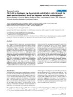
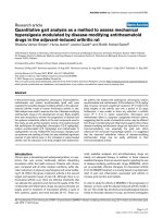
abdominal wall (Fig. 1a,b). Bile was clearly visible drain-
ing from the fistula opening (arrowed). The whole length
of the fistula was inspected and no other leak was found;
the proximal junction with the CBD was intact. The distal
fistula was recannulated with a 10 Fr Latex drain (Fig. 2)
and bile was observed to be draining freely from it. Fol-
lowing an uneventful recovery, cholangiography of the
cannulated tract with the Latex drain in-situ was repeated
after 5 weeks and no dye was able to pass down it. The
tract was therefore presumed to have closed. The Latex
drain was removed 24 hours later with good recovery to
date and no further complications.
Discussion
Biliary peritonitis is regarded as a rare but serious compli-
cation of elective T-tube removal after CBD exploration.
Incidence reported in the literature varies from 0.8 to 5%
in elective removal of T-tubes, rising to 24% in cases of
liver transplants [3].
Historically, a latex T-tube has always been used during
open exploration, specifically to encourage a vigorous
inflammatory reaction around it causing formation of a
biliary fistula. This makes T-tube removal much safer by
reducing the potential for intraperitoneal bile leak. The
fistula closes rapidly after removal of the T-tube as long as
there is no distal CBD obstruction. More recently, sili-
cone-coated or polyethylene T-tubes have become availa-
ble, but these are less irritant and the resulting fistula
tends to be less mature, increasing the risk of a leak after
T-tube removal. We do not recommend use of these newer
T-tubes after CBD exploration unless the patient has a
latex allergy.
This case is novel since the site of the bile leak was distal,
at the point of contact between fistula and anterior
abdominal wall. Usually biliary leakage occurs through
(a) T-tube fistula tract openingFigure 1
(a) T-tube fistula tract opening. Intraoperative laparo-
scopic photograph illustrating opening to T-tube fistula tract
(arrow) with diagrammatic representation of relation to bil-
iary anatomy. (b) Diagram of fistula pathway and leak mecha-
nism.
a
b
Journal of Medical Case Reports 2008, 2:302 />Page 3 of 4
(page number not for citation purposes)
lack of complete fibrous T-tube fistula formation or
through proximal fistula disruption during the removal
process [4]. Identification of the location of the leak as
described was important for three reasons: first, it pro-
vided an accurate diagnosis; second, it confirmed that the
usual leak point (i.e. the junction between fistula and
CBD) was intact and did not therefore require a further T-
tube placement, and third, it allowed a simple therapeutic
manoeuvre by re-intubating the fistula opening.
It may be suggested that the fistula was disrupted from the
abdominal wall during insufflation at the time of laparos-
copy. However, this does not explain the clinical presen-
tation before laparoscopy. We knew there was no leak
before T-tube removal because of a normal T-tube cholan-
giogram and lack of abdominal symptoms. The patient
suffered upper abdominal pain soon after removal of the
T-tube, developing biliary peritonitis along with raised
inflammatory markers (white cell count and C-reactive
protein). This indicates a leak at the time of T-tube
removal which was subsequently confirmed at laparos-
copy with a pool of bile on the surface of the abdominal
viscera. Bile was clearly observed emanating directly from
the distal fistula opening.
The literature has been reviewed in view of factors which
affect the risk of biliary leakage.
T-tubes versus choledochorrhaphy
The first option is to avoid T-tube insertion altogether and
perform a choledochorrhaphy (primary closure of the
choledochotomy). In the past, this was rarely advised as it
was thought to increase the risk of stricture formation and
prevent postoperative CBD decompression. However,
research has recently suggested that primary closure may
be as safe as T-tube usage [5], although it should be
avoided if the CBD is not significantly dilated. The other
benefit of T-tubes is the ease of postoperative visualisation
of retained CBD stones (T-tube cholangiogram).
Duration of T-tube insertion
Many factors may affect the risk of symptomatic bile leak-
age following T-tube removal. Ellis [2] originally sug-
gested that T-tubes should be removed 10 days after
operation. It has been suggested that leaving T-tubes in
situ for longer periods allows maturation of the temporary
biliary cutaneous fistula, thus potentially reducing the risk
of leakage [4]. However, there is no experimental evidence
to prove this hypothesis. Indeed, one study has shown
that leaving T-tubes in situ for longer periods, such as 1
month postoperatively does not provide protection
against increased rates of bile leakage [1]. In this case, the
T-tube was removed after 3 weeks, in line with common
practice in the UK.
T-tube material
Experimental evidence demonstrates that the material
used for manufacturing T-tubes affects the quality of
fibrous fistula formed [6,7]. This finding is supported by
clinical evidence that polyvinyl chloride (PVC) or hypoal-
lergenic latex T-tubes (such as those coated with silicon)
increase rates of biliary peritonitis compared to red rubber
or normal latex T-tubes, as the former take longer to form
a mature tract [8]. In our case, therefore, the standard latex
T-tube used is unlikely to be of aetiological significance.
Immune system
The hypothesis that an increased inflammatory response
leads to the formation of a stronger fistulous tract may
explain the increased rates of symptomatic bile leaks in
immunocompromised patients, such as those undergoing
liver transplantation [3]. In our case, there was no past
medical history of diabetes or steroid use and no medical
evidence of occult immunosuppressive pathologies,
although an HIV test was not performed.
T-tube morphology
It has been suggested that the morphology of the T-tube or
its placement could reduce leakage [2], although other
authors have pointed out that there is no experimental
evidence for this theory. Figure 3 illustrates the morphol-
ogy of the T-tube used in this patient, designed to mini-
mise trauma during T-tube removal and thus potentially
the risk of biliary leakage [9]. Some authorities recom-
mend cutting a notch in the short 'crossbar', opposite the
drainage tube, to further facilitate removal by allowing the
two 'wings' to fold more easily. If this is done, care must
be taken not to make the resulting bridge of material too
Cannulation of T-tube fistulaFigure 2
Cannulation of T-tube fistula. Intraoperative laparo-
scopic photograph illustrating cannulation of T-tube fistula
tract with 10-Fr Latex drain.
Publish with BioMed Central and every
scientist can read your work free of charge
"BioMed Central will be the most significant development for
disseminating the results of biomedical research in our lifetime."
Sir Paul Nurse, Cancer Research UK
Your research papers will be:
available free of charge to the entire biomedical community
peer reviewed and published immediately upon acceptance
cited in PubMed and archived on PubMed Central
yours — you keep the copyright
Submit your manuscript here:
/>BioMedcentral
Journal of Medical Case Reports 2008, 2:302 />Page 4 of 4
(page number not for citation purposes)
thin as the wings may then detach during the removal
process.
T-tube removal technique
Goodwin et al. [10] reported a significant reduction in bile
leakage and subsequent biliary peritonitis after T-tube
removal in liver transplant patients when the tube was
removed along a wire (Seldinger method). This technique
is generally only recommended in high-risk patients in
whom bile leakage is anticipated following T-tube
removal, especially in immunocompromised patients fol-
lowing liver transplantation.
Conclusion
This case and our review of the literature highlight a pre-
viously unreported mechanism for bile leak following T-
tube removal caused by dehiscence of a fistula tract at its
contact point with the anterior abdominal wall. Hepato-
biliary surgeons should be aware of this mechanism of
biliary leakage and the use of laparoscopy to recannulate
the fistula with a satisfactory outcome.
Consent
Written informed consent was obtained from the patient
for publication of this case report and any accompanying
images. A copy of the written consent is available for
review by the Editor-in-Chief of this journal.
Competing interests
The authors declare that they have no competing interests.
Authors' contributions
MN and AK were involved in design of the case report,
drafted the manuscript and performed critical literature
review. AH, SN and TT conceived the original idea of the
case report, conducted the operations detailed and have
been involved in critically revising the manuscript.
References
1. Wills VL, Gibson K, Karihaloot C, Jorgensen JO: Complications of
biliary T-tubes after choledochotomy. ANZ J Surg 2002,
72(3):177-180.
2. Ellis H: Choledocholithiasis. In Maingot's Abdominal Operations
Edited by: Schwartz S, Ellis H. Norwalk, CT: Appleton-Century-
Crofts; 1985:1883-1907.
3. Lazaridis C, Papaziogas B, Patsas A, Galanis I, Paraskevas G, Argiria-
dou H, Papaziogas T: Detection of tract formation for preven-
tion of bile peritonitis after T-tube removal. Case report.
Acta Chir Belg 2005, 105(2):210-212.
4. Maghsoudi H, Garadaghi A, Jafary GA: Biliary peritonitis requiring
reoperation after removal of T-tubes from the common bile
duct. Am J Surg 2005, 190(3):430-433.
5. Gurusamy KS, Samraj K: Primary closure versus T-tube drain-
age after open common bile duct exploration. Cochrane Data-
base Syst Rev 2007.
6. Koivusalo A, Eskelinen M, Wolff H, Talva M, Mäkisalo H: Develop-
ment of T-tube tracts in piglets: effect of insertion method
and material of T-tubes. Res Exp Med (Berl) 1997, 197(1):53-56.
7. Apalakis A: An experimental evaluation of the types of mate-
rial used for bile duct drainage tubes. Br J Surg 1976,
63(6):440-445.
8. Winstone NE, Golby MG, Lawson LJ, Windsor CW: Biliary perito-
nitis: a hazard of polyvinyl chloride T-tubes. Lancet 1965,
1:843-844.
9. Sakorafas GH, Stafyla V, Tsiotos GG: Biliary peritonitis due to fis-
tulous tract rupture following a T-tube removal. N Z Med J
2005, 118(1217):U1522.
10. Goodwin SC, Bittner CA, Patel MC, Noronha MA, Chao K, Sayre JW:
Technique for reduction of bile peritonitis after T-tube
removal in liver transplant patients. J Vasc Interv Radiol 1998,
9:986-990.
T-tube morphologyFigure 3
T-tube morphology. A gutter is cut out of the cross arm
to lower resistance during T-tube removal and thus reduce
the risk of traumatic fistula disruption.