Báo cáo y học: " Double primary bronchogenic carcinoma of the lung and papillary thyroid carcinoma: a case report" pps
Bạn đang xem bản rút gọn của tài liệu. Xem và tải ngay bản đầy đủ của tài liệu tại đây (512.75 KB, 5 trang )
BioMed Central
Page 1 of 5
(page number not for citation purposes)
Journal of Medical Case Reports
Open Access
Case report
Double primary bronchogenic carcinoma of the lung and papillary
thyroid carcinoma: a case report
Jen-Hsun Cheng
1
, Ying-Chieh Huang
1,2
, Chih Kuo
3
, Yih-Shyong Lai
3
, Tzu-
Ching Wu
4
, Thomas Chang-Yao Tsao
4
, Shi-Ping Luh*
1,2
and Chong-
Bin Tsai*
1
Address:
1
Department of Thoracic Medicine and Surgery, Chia-Yi Christian Hospital, Chia-Yi 600, Taiwan,
2
National Chung-Cheng University,
Min-Hsiung, Chia-Yi 621, Taiwan,
3
Department of Pathology, Chung-Shan Medical University Hospital, Taichung City, Taiwan and
4
Department
of Medicine, Chung-Shan Medical University Hospital, Taichung City, Taiwan
Email: Jen-Hsun Cheng - ; Ying-Chieh Huang - ; Chih Kuo - ; Yih-
Shyong Lai - ; Tzu-Ching Wu - ; Thomas Chang-Yao Tsao - ;
Shi-Ping Luh* - ; Chong-Bin Tsai* -
* Corresponding authors
Abstract
Introduction: Double primary bronchogenic carcinoma and papillary carcinoma of the thyroid are
extremely rare. We describe the case of a patient who underwent surgical resection for these two
cancers.
Case presentation: A 56-year-old man presented to our hospital complaining of a cough with
blood-tinged sputum. A slowly growing mass in the left lobe of the lung had been noted for about
1 year. He underwent video-assisted thoracic surgery of the left lower lobe and mediastinal lymph
node dissection through an 8 cm utility incision. Pathology revealed a well-differentiated
adenocarcinoma and the dissected lymph nodes were negative for malignancy. He also complained
of a mass in his neck, which had grown slowly for over 5 years. A computed tomography scan of
the neck revealed a left thyroid mass compressing the trachea towards the right side. There was
no cervical lymphadenopathy. A left thyroid lobectomy was performed and pathology revealed a
papillary carcinoma. Thus, he underwent a second operation to remove the right lobe of the
thyroid. He underwent subsequent adjuvant chemotherapy.
Conclusion: In a review of the literature, it appears that there has only been one previously
reported case of these two cancers, which was in Japan. The relationship between these two
cancers is still unclear, and more case reports are required to determine this relationship.
Introduction
The incidence of multiple primary malignancies has
increased in recent years [1]. Commonly occurring malig-
nancies accompanying primary lung cancer are found in
the lung, upper respiratory tract, breast, esophagus, colon,
rectum, stomach and cervix [2]. Double primary thyroid
and lung cancers have rarely been reported [3-5]. Here we
describe a case of a patient with double primary lung and
thyroid cancers who underwent curative surgical resec-
tion.
Published: 23 September 2008
Journal of Medical Case Reports 2008, 2:309 doi:10.1186/1752-1947-2-309
Received: 10 December 2007
Accepted: 23 September 2008
This article is available from: />© 2008 Cheng et al; licensee BioMed Central Ltd.
This is an Open Access article distributed under the terms of the Creative Commons Attribution License ( />),
which permits unrestricted use, distribution, and reproduction in any medium, provided the original work is properly cited.
Journal of Medical Case Reports 2008, 2:309 />Page 2 of 5
(page number not for citation purposes)
Case presentation
A 56-year-old man, who was well except for hypertension
of over 10 years duration for which he received regular
treatment, presented to our hospital complaining of inter-
mittent chest tightness for a month. The chest tightness,
which had been aggravated in the previous week, was
located in the left precordal area, and was persistent in
character and induced by exercise. On examination, the
patient was slightly anxious but generally well. A mass was
noted over the left side of his neck and he stated that this
had been present for more than 4 years. He did not pay
attention to it initially because it had been growing very
slowly. However, he had noted mild labor on respiration
in recent months. No abnormal breath sounds or heart
murmurs were noted. The hemogram and blood chemis-
try were normal. Chest X-ray revealed a mass in the left
lower lung field (Figure 1). Computed tomography (CT)
revealed a nodule, 3.5 cm in diameter, in the left lower
lobe of the lung with pleural retraction (Figure 2A), and
also a mass, 5 cm in diameter, within the left lobe of the
thyroid (Figure 2B). Fiberoptic bronchoscopy was nega-
tive for any intraluminal lesions. An adenocarcinoma of
the lung was confirmed by CT-guided biopsy. A whole
body bone scan was negative for skeletal metastasis. A
fluorodeoxyglucose-positron emission tomography
revealed a hypermetabolic focus in the left lower lobe of
the lung and in the left lobe of the thyroid. He was admit-
ted for further evaluation and treatment.
The patient underwent a left lower lobectomy to remove
the pulmonary mass and mediastinal lymph node dissec-
tion through video-assisted thoracic surgery with a
minithoracotomy. The resected specimen revealed a 3.5 ×
3 × 2.5 cm elastic-firm, high-cellular mass with pleural
retraction. All of the nine dissected mediastinal lymph
nodes were negative. Grossly, the localized lung tumor
was a well-differentiated adenocarcinoma which was
shown pushed against the pleura with lymphocytic infil-
tration, but not penetrating it. Microscopically, the tumor
was arranged in a glandular structure, composed of neo-
plastic cells with irregularly enlarged and hyperchromatic
nuclei. Some papillary configuration and fused glands
were present. The lung adenocarcinoma revealed on
immunohistochemistry surfactant apoprotein A positivity
for tumor cells as well as normal alveolar cells. The
patient's postoperative course was uneventful and he was
discharged 9 days after the operation.
He was later readmitted and underwent a left thyroid
lobectomy for what appeared to be a nodular goiter.
Microscopy revealed a papillary structure with a ground-
glass appearance of tumor cell nuclei. Some colloid
within neoplastic follicles was evident. Immunohisto-
chemical staining was positive for tumor cells. The patient
underwent a residual radical thyroidectomy. No residual
tumor was found in the resected thyroid, parathyroids or
cervical lymph nodes. During follow-up, his thyroglobu-
lin level remained low. Hypothyroidism and hypoparath-
yroidism were noted after the radical thyroidectomy and
these symptoms were controlled by thyroid hormone and
calcium supplements. The pathology findings confirmed
the diagnosis of a double primary pulmonary adenocarci-
noma and thyroid papillary carcinoma (see figure 3).
Discussion
Patients with lung cancer have a high risk of multiple pri-
mary malignancies. Other potential sites for multiple pri-
mary cancers include the nasopharynx, lungs, large bowel
and mammary glands [6]. The incidence of multiple pri-
mary malignancies for patients with overall and resected
non-small cell lung carcinoma (NSCLC) was 11% and 7–
7.4%, respectively [7]. Liu et al. [1] reported that the most
common tumors accompanying lung cancer were in the
upper aerodigestive tract, followed by colorectal and cer-
vical malignancies. Hsieh et al. [8] reported from the same
database that the order of frequency of malignancies for
the upper aerodigestive tract was larynx, nasopharynx,
esophagus, oral cavity and hypopharynx.
Chest X-ray showing a mass shadow over the left lower lung field (arrow)Figure 1
Chest X-ray showing a mass shadow over the left
lower lung field (arrow).
Journal of Medical Case Reports 2008, 2:309 />Page 3 of 5
(page number not for citation purposes)
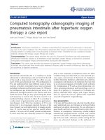
Computed tomography (CT) showing a nodule, 3.5 cm in diameter, within the left lower lobe of the lung with pleural retrac-tion (A), and a mass 5 cm in diameter within the left lobe of the thyroid (B)Figure 2
Computed tomography (CT) showing a nodule, 3.5 cm in diameter, within the left lower lobe of the lung with
pleural retraction (A), and a mass 5 cm in diameter within the left lobe of the thyroid (B).
A
B
Journal of Medical Case Reports 2008, 2:309 />Page 4 of 5
(page number not for citation purposes)
Double primary thyroid and lung carcinomas have been
reported only rarely in the literature [3-5]. Shinozaki et al.
[9] reported that thyroid carcinoma occurred in 9.7% of
patients with multiple primary malignancies, and the
most frequent sites for the associated cancers were the
breast, uterine cervix and uterine body in women, and the
stomach and larynx in men. However, thyroid carcinoma
was found with a higher rate of second malignancy
(22.7%) than average (4.2%) in autopsy findings, and fol-
licular carcinoma was more frequent among the cancers
associated with another tumor (12 out of 20 cases), while
in general papillary carcinoma was the most frequent (48
out of 88 cases) [10].
Differential diagnosis for the patient in our case included
pulmonary metastasis from the thyroid cancer or vice
versa, and both these situations have been reported previ-
ously [11]. Pathological iodine-131 uptake will occur in
both the primary lung tumor as well as in metastases from
the thyroid gland, thus it is not reliable for making a diag-
nosis [12].
Double primary cancer is the most reasonable diagnosis
in our case because there was no evidence of either medi-
astinal or cervical lymph node metastasis, and the tumors
from the two sites had different pathological characteris-
tics.
The associations between these two cancers are still
unclear. Mutating proto-oncogenes associated with thy-
roid carcinoma, such as the ret oncogene, have not been
found in patients with lung carcinoma [13]. Furthermore,
the environmental factors associated with lung carci-
noma, such as smoking or air pollution, have not been
not correlated with thyroid carcinoma [14]. Therefore,
coincidence is possible in this patient, but further related
studies are required to determine where there is an associ-
ation between these two cancers.
Surgical resection is indicated for either thyroid papillary
carcinoma or early to mid stage (before Stage IIIa) non-
small cell lung carcinomas (NSCLCs). Therapeutic strate-
gies for the management of double primary thyroid and
lung carcinomas, in general, follow their separate guide-
lines. However, since the progression of a thyroid papil-
lary carcinoma is much slower than that of NSCLCs, in
some patients with limited survival removal of the thyroid
neoplasm may not be considered appropriate [4]. In the
patient described in this case report, since there were no
lymph nodes involved or distant metastasis, surgical
resection of both lesions was the therapy of choice.
Conclusion
A patient with a double primary thyroid papillary carci-
noma and pulmonary adenocarcinoma was successfully
treated by surgical resection of both tumours. Reports of
related cases in the previous literature are rare.
Abbreviations
CT: computed tomography; NSCLC: non-small cell lung
carcinoma
Competing interests
The authors declare that they have no competing interests.
Well differentiated pulmonary adenocarcinomaFigure 3
Well differentiated pulmonary adenocarcinoma. (A). The tumor is arranged in glandular structure, composed of neo-
plastic cells with irregularly enlarged and hyperchromatic nuclei. Some papillary configuration and fused glands are present. (H
& E stain, 200×). Histopathology of the thyroid tumor reveals papillary structure with ground glass appearance of tumor cell
nuclei (B). Some colloid within neoplastic follicles is evident (H & E stain, 200×).
A B
Publish with BioMed Central and every
scientist can read your work free of charge
"BioMed Central will be the most significant development for
disseminating the results of biomedical research in our lifetime."
Sir Paul Nurse, Cancer Research UK
Your research papers will be:
available free of charge to the entire biomedical community
peer reviewed and published immediately upon acceptance
cited in PubMed and archived on PubMed Central
yours — you keep the copyright
Submit your manuscript here:
/>BioMedcentral
Journal of Medical Case Reports 2008, 2:309 />Page 5 of 5
(page number not for citation purposes)
Authors' contributions
S-PL was the attending doctor, carried out the surgical pro-
cedure and literature review and wrote the manuscript. CK
and Y-SL performed the pathological examination and
assisted in writing the report. T-CW and TC-YT were the
chest physicians providing care to this patient. Y-CH and
C-BT revised and provided comments on the manuscript.
J-HC collected the data and literature review, and wrote
the manuscript.
Consent
Written informed consent was obtained from the patient
for publication of this case report and any accompanying
images. A copy of the written consent is available for
review by the Editor-in-Chief of this journal.
References
1. Liu YY, Chen YM, Yen SH, Tsai CM, Perng RP: Multiple primary
malignancies involving lung cancer-clinical characteristics
and prognosis. Lung Cancer 2002, 35:189-194.
2. Yi SZ, Zhang DC, Wang YG, Sun KL: Clinical features and prog-
nosis of multiple primary tumors of lung combined with
other organs – report of 281 cases. Ai Zheng 2006, 25:731-735.
3. Higashiyama M, Kodama K, Yokouchi H, Takami K, Motomura K, Inaji
H, Koyama H: Mediastinal lymph node involvement as the ini-
tial manifestation of occult thyroid cancer in the surgical
treatment of lung cancer: report of a case. Surg Today 1999,
29:670-674.
4. Hamada Y, Takise A, Uno D, Itoh H, Ichikawa H, Morishta Y: Syn-
chronous primary triple cancers including the lung, stomach,
and thyroid: a case report. Kyobu Geka 2000, 53:101-105.
5. Arimura T, Niwa K, Mitani N, Hagiwara I, Kawaida T, Shimazu H: A
resected case of triple cancer in the uterus, lung and thyroid.
Nippon Kyobu Geka Gakkai Zasshi 1989, 37:1233-1237.
6. Li W, Zhan Y, Li G: Double cancers: a clinical analysis of 156
cases. Zhonghua Zhong Liu Za Zhi 1996, 18:296-298.
7. Brock MV, Alberg AJ, Hooker CM, Kammer AL, Xu L, Roig CM, Yang
SC: Risk of subsequent primary neoplasms developing in lung
cancer patients with prior malignancies. J Thorac Cardiovasc
Surg 2004, 127:1119-1125.
8. Hsieh WC, Chen YM, Perng RP: Temporal relationship between
cancers of the lung and upper aerodigestive tract. Jpn J Clin
Oncol 1997, 27:63-66.
9. Shinozaki N, Sakamoto A, Kasai N, Uchida M, Sakurai K: Multiple
primary malignancies associated with thyroid cancer. Gan No
Rinsho 1983, 29:1385-1391.
10. Tiszlavicz L, Varga Z: Primary malignant tumours associated
with thyroid carcinoma in autopsy material. Acta Morphol Hung
1992, 40:95-102.
11. Samuel AM, Rajashekharrao B, Shah DH: Pulmonary metastases
in children and adolescents with well-differentiated thyroid
cancer. J Nucl Med 1998, 39:1531-1536.
12. Zohar Y, Strauss M: Occult distant metastases of well-differen-
tiated thyroid carcinoma. Head Neck 1994, 16:438-442.
13. Wang YL, Wang JC, Wu Y, Zhang L, Huang CP, Shen Q, Zhu YX, Li
DS, Ji QH: Incidentally simultaneous occurrence of RET/PTC,
H4-PTEN and BRAF mutation in papillary thyroid carci-
noma. Cancer Lett 2008, 263:44-52.
14. Guignard R, Truong T, Rougier Y, Baron-Dubourdieu D, Guenel P:
Alcohol drinking, tobacco smoking, and anthropometric
characteristics as risk factors for thyroid cancer: a country-
wide case-control study in New Caledonia. Am J Epidemiol
2007, 166:1140-1149.