Báo cáo y học: "Dermatological conditions during TNF-α-blocking therapy in patients with rheumatoid arthritis: a prospective study" pps
Bạn đang xem bản rút gọn của tài liệu. Xem và tải ngay bản đầy đủ của tài liệu tại đây (353.98 KB, 11 trang )
Open Access
Available online />R666
Vol 7 No 3
Research article
Dermatological conditions during TNF-α-blocking therapy in
patients with rheumatoid arthritis: a prospective study
Marcel Flendrie
1
, Wynand HPM Vissers
2
, Marjonne CW Creemers
1
, Elke MGJ de Jong
2
,
Peter CM van de Kerkhof
2
and Piet LCM van Riel
1
1
Department of Rheumatology, Radboud University Nijmegen Medical Centre, Nijmegen, the Netherlands
2
Department of Dermatology, Radboud University Nijmegen Medical Centre, Nijmegen, the Netherlands
Corresponding author: Marcel Flendrie,
Received: 3 Jan 2005 Revisions requested: 20 Jan 2005 Revisions received: 25 Feb 2005 Accepted: 1 Mar 2005 Published: 4 Apr 2005
Arthritis Research & Therapy 2005, 7:R666-R676 (DOI 10.1186/ar1724)
This article is online at: />© 2005 Flendrie et al.; licensee BioMed Central Ltd.
This is an Open Access article distributed under the terms of the Creative Commons Attribution License ( />2.0), which permits unrestricted use, distribution, and reproduction in any medium, provided the original work is properly cited.
Abstract
Various dermatological conditions have been reported during
tumor necrosis factor (TNF)-α-blocking therapy, but until now no
prospective studies have been focused on this aspect. The
present study was set up to investigate the number and nature
of clinically important dermatological conditions during TNF-α-
blocking therapy in patients with rheumatoid arthritis (RA). RA
patients starting on TNF-α-blocking therapy were prospectively
followed up. The numbers and natures of dermatological events
giving rise to a dermatological consultation were recorded. The
patients with a dermatological event were compared with a
group of prospectively followed up RA control patients, naive to
TNF-α-blocking therapy and matched for follow-up period. 289
RA patients started TNF-α-blocking therapy. 128
dermatological events were recorded in 72 patients (25%)
during 911 patient-years of follow-up. TNF-α-blocking therapy
was stopped in 19 (26%) of these 72 patients because of the
dermatological event. More of the RA patients given TNF-α-
blocking therapy (25%) than of the anti-TNF-α-naive patients
(13%) visited a dermatologist during follow-up (P < 0.0005).
Events were recorded more often during active treatment (0.16
events per patient-year) than during the period of withdrawal of
TNF-α-blocking therapy (0.09 events per patient-year, P <
0.0005). The events recorded most frequently were skin
infections (n = 33), eczema (n = 20), and drug-related eruptions
(n = 15). Other events with a possible relation to TNF-α-
blocking therapy included vasculitis, psoriasis, drug-induced
systemic lupus erythematosus, dermatomyositis, and a
lymphomatoid-papulosis-like eruption. This study is the first
large prospective study focusing on dermatological conditions
during TNF-α-blocking therapy. It shows that dermatological
conditions are a significant and clinically important problem in
RA patients receiving TNF-α-blocking therapy.
Introduction
The introduction of biological agents such as TNF-α-blocking
agents has dramatically changed the therapeutic approach to
rheumatic diseases in recent years. TNF-α-blocking therapy
has had a remarkable effect on disease activity in an increas-
ing number of rheumatic diseases, including rheumatoid arthri-
tis (RA) [1-3], juvenile idiopathic arthritis [4], ankylosing
spondylitis [5,6], and psoriatic arthritis [7]. At present, two
monoclonal anti-TNF-α antibodies (infliximab and adalimumab)
and one soluble p75 TNF-α receptor (etanercept) are being
used in rheumatological practice.
Various skin conditions have been reported in clinical trials,
including urticaria, rash, and stomatitis (during infliximab ther-
apy) [8]; rash and injection-site reactions (during adalimumab
therapy) [3,9]; and injection-site reactions (during etanercept
therapy) [2].
However, clinical trials are not designed to provide information
about the occurrence of rare adverse events associated with
TNF-α-blocking therapy. More severe cutaneous reactions,
such as erythema multiforme, discoid and subacute cutaneous
lupus erythematosus, atopic dermatitis, necrotizing vasculitis,
and bullous skin lesions, have been reported, mostly as single-
case observations [10-15]. Larger observational studies such
CI = confidence interval; DAS28 = disease activity score including 28-joint counts; DMARD = disease-modifying antirheumatic drug; ELISA =
enzyme-linked immunosorbent assay; pt-yr = patient-year; RA = rheumatoid arthritis; Th1/Th2 = T helper cell type 1/2; TNF = tumor necrosis factor.
Arthritis Research & Therapy Vol 7 No 3 Flendrie et al.
R667
as biological registries are needed to provide information on
the nature and number of such dermatological adverse events
during TNF-α-blocking therapy.
The aim of this study was to investigate whether dermatologi-
cal conditions after TNF-α-blocking therapy are a significant
and clinically important problem in RA patients receiving TNF-
α-blocking therapy.
Materials and methods
Study design
In a prospective cohort study, all consecutive patients with a
diagnosis of RA according to the criteria of the American
Rheumatism Association [16] who were starting on TNF-α-
blocking therapy at the Department Of Rheumatology of the
Radboud University Nijmegen Medical Centre were followed
as part of a Biological Registry [17]. Approval was obtained by
the hospital's ethics committee.
Patients were required to meet the criteria set out in the Dutch
guidelines for biological therapies: a moderate to high disease
activity score (DAS) based on 28 joints (DAS28 ≥ 3.2), and
failure or intolerability of at least two disease-modifying
antirheumatic drugs (DMARDs), including methotrexate, in
adequate dosage regimens. Besides therapy with registrated
TNF-α-blocking agents – infliximab, etanercept, and adalimu-
mab – some patients were treated in clinical trials with lener-
cept, a soluble p55 TNF-α-receptor [18].
The number and nature of dermatological conditions that led
patients in this cohort to consult a dermatologist during follow-
up were investigated. The RA patients treated with TNA-α-
blocking agents who experienced dermatological events was
compared with a control group of patients who had RA but
had never had TNF-α-blocking therapy. The control patients
were selected from the Nijmegen inception cohort, in which
500 RA patients have been followed since 1985 [19]. Each
control was paired with a TNF-α-treated patient for duration
and season of the follow-up period, within a 2-month window.
Variables
Data collected at the start of TNF-α-blocking therapy were
age, sex, duration of disease, presence or absence of rheuma-
toid factor (measured by ELISA; considered positive if results
showed >10 IU/ml), antinuclear antibody (tested for by
immunofluorescence on Hep-2 cells), number of DMARDs
previously used, and start date of TNF-α-blocking therapy.
Baseline information obtained included erythrocyte sedimen-
tation rate (ESR), 28-joint counts for swelling and tenderness,
and general wellbeing as indicated on a visual analogue scale,
and the disease activity score (DAS28) was calculated [20].
Variables about which information was collected during TNF-
α-blocking therapy were the use of concomitant DMARDs and
prednisolone, dose and interval changes of TNF-α-blocking
agents and, if appropriate, date and reason for
discontinuation.
All patients who visited a dermatologist during follow-up were
identified. Clinically important dermatological events were
defined as any new manifestation or any exacerbation of pre-
existing skin disease during follow-up. A standardized chart
review form was used to record the following: start date of
event, dermatological history, medication, morphological
description, localization, histopathological and immunohisto-
logical information if available, working diagnosis, additional
investigations, topical and systemic therapeutic actions, out-
come of event, and any available information on rechallenge.
Drug-related eruptions were defined as skin reactions with a
probable or definite relation to the use of TNF-α-blocking
agents, based on a time relation with the administration of the
agent, morphological pattern, and/or histological information.
Drug-related eruptions were classified morphologically
according to the criteria of Fitzpatrick and colleagues [21].
Events were also classified as major or minor, major events
being any requiring hospitalization.
Patient-years of follow-up were calculated for total follow-up,
time on active therapy, and time after discontinuation of ther-
apy (time off therapy). The number of events per year of follow-
up was calculated for each RA patient for total time of follow-
up, time on active treatment, and time off treatment, if
appropriate.
In the control group, the following baseline characteristics
were collected: age, sex, disease duration, rheumatoid factor,
antinuclear antibody, DAS28, the number of DMARDs previ-
ously used, and prednisolone use. All visits to a dermatologist
during follow-up were identified. Events were not recorded in
the control group.
Statistical analyses
The baseline characteristics of RA patients on TNF-α-blocking
therapy were compared according to whether or not the
patients experienced dermatological events. The chi-square
test was applied for dichotomous variables and Student's t-
test was used for continuous variables. Nonparametric tests
were applied when appropriate. The Wilcoxon signed rank test
was used to compare the number of events per patient-year of
follow-up in patients receiving and patients not receiving
active TNF-α-blocking therapy. Univariate and multivariate
logistic regression analyses were performed to identify possi-
ble predictive factors for the occurrence of a dermatological
visit (independent variable, dichotomous) in RA patients on
TNF-α-blocking therapy. Dependent variables tested were
sex, age at diagnosis, rheumatoid factor, antinuclear antibody,
disease duration, DAS28 at baseline, prior number of
DMARDs, use of prednisolone, and duration of follow-up.
Available online />R668
Odds ratios (ORs) and 95% confidence intervals (95% CIs)
were calculated.
The number of patients who visited a dermatologist was com-
pared between RA patients on TNF-α-blocking therapy and
controls, using the chi-square test. P values and ORs were
calculated.
All tests were two-sided, with P < 0.05 considered statistically
significant. Statistical analyses were performed using SPSS
statistical software (v 12.0.1, SPSS Inc, USA).
Results
Patients
A total of 289 RA patients started TNF-α-blocking therapy
between June 1994 and December 2003. Their baseline char-
acteristics are shown in Table 1.
The median follow-up time was 2.3 years (range 0.02 to 9.6).
The total follow-up time was 911 patient-years, with 627
patient-years representing active therapy. Seventy of the 289
RA patients (24%) received more than one TNF-α-blocking
agent and 8 (3%) received more than two agents. Infliximab
was administered to 167 patients, adalimumab to 108, etaner-
cept to 78, and lenercept to 31.
Dermatological events were recorded in 72 of the 289 RA
patients (25%) receiving TNF-α-blocking therapy and in 37
(13%) of the control group (n = 289). The odds ratio (OR) of
TNF-α-blocking therapy for a dermatological referral was 2.26
(95%CI 1.46 to 3.50, P < 0.0005). In patients on TNF-α-
blocking therapy fifty-six instances of dermatological condi-
tions were recorded in 34 patients (47%) and included,
among others, 10 drug reactions – while the patient was
receiving gold (7), nonsteroidal anti-inflammatory drugs (2), or
methotrexate (1) – 10 cases of eczema, 9 of mycosis, 3 of
other infections, and 5 of chronic venous insufficiency.
Predictive factors
In univariate analyses, duration of follow-up (OR 1.27, 95%CI
1.14 to 1.41, P < 0.0005) and of disease (OR 1.03, 95%CI
1.003 to 1.07, P < 0.05) were statistically significant predic-
tive factors for a dermatological event. In a multivariate model,
only duration of follow-up was a statistically significant predic-
tive factor (OR 1.30, 95%CI 1.12 to 1.52, P < 0.001).
Dermatological events
One hundred and twenty-eight dermatological events were
recorded during follow-up in RA patients on TNF-α-blocking
therapy (0.14 event per patient-year), as listed in Table 2. The
event per patient-year ratio was 0.16 during active treatment
and 0.10 off treatment (P < 0.001). The number of events
recorded during or after treatment was 56 for adalimumab
(0.12 event per patient-year), 49 for infliximab (0.14 per
patient-year), 16 for etanercept (0.13 per patient-year), and 13
for lenercept (0.07 per patient-year). TNF-α-blocking therapy
was permanently withdrawn because of dermatological events
21 times in 19 patients.
Infections
Thirty-three infections were recorded in 27 patients, consist-
ing of 20 fungal, 11 bacterial, and 2 viral infections (see Table
3). Two patients had had a previous episode of
dermatomycosis. None of the patients required hospitalization.
One patient, who temporarily discontinued adalimumab mon-
otherapy twice because of elective surgery, developed a bac-
terial superinfection of pre-existing eczema after every restart.
Table 1
Baseline characteristics of patients with rheumatoid arthritis (RA) studied
Given TNF-α-blocking therapy Controls
a
Characteristic All patients N = 289 Patients with dermatological
events N = 72
N = 289
Male sex, no. (%) 89 (31) 20 (28) 110 (38)
Age (yr) at diagnosis, mean (SD) 44.5 (14.7) 43.4 (12.7) 54.6 (14.1)**
RF-positive, no. (%) 249 (87) 68 (94) 205 (71)*
Disease duration (yr) at baseline, median (range) 9.2 (0.1–44.9) 10.3 (0.3–44.9)
†
6.2 (0.0–12.6)**
DAS28 at baseline, mean (SD) 5.9 (1.1) 6.1 (1.1) 3.6 (1.4)**
ANA-positive at baseline, no. (%)
b
112 (50) 33 (49) 118 (41)
Prior DMARDs, median (range) 4 (1–10) 5 (2–8) 1 (0–6)**
Prednisolone at baseline, no. (%) 112 (39) 34 (47) 21 (7)**
a
Not given TNF-α-blocking therapy.
b
ANA at start was present in respectively 261 and 67 patients on TNF-α-blocking therapy. *P < 0.001, **P <
0.0001, compared with RA patients on TNF-α-blocking therapy;
†
P < 0.001 compared with RA patients on TNF-α-blocking therapy who
experienced no dermatological events. ANA, antinuclear antibody; DAS28, disease activity score based on 28 joints; DMARD, disease-modifying
antirheumatic drug; RF, rheumatoid factor; SD, standard deviation; TNF, tumor necrosis factor.
Arthritis Research & Therapy Vol 7 No 3 Flendrie et al.
R669
Eczema
Eczema was diagnosed 20 times in 19 patients and appeared
in various morphological patterns. Most events were
described as erythematosquamous (n = 8) or erythematous (n
= 3) lesions or plaques, localized on hands and feet (n = 3),
arms and legs (n = 5), face (n = 1), neck (n = 1), and buttocks
(n = 1). A vesicular rash on hands and feet was described five
times. A papular rash was described in three cases, with local-
ization around the eyes, on the back, and once on the back and
lower legs. Diagnoses comprised dyshidrotic (n = 5), contact
(n = 4), nummular (n = 1), atopic (n = 1), papular (n = 1), and
nonspecific eczema (n = 8). Two patients had a prior history
of dyshidrotic eczema.
Biopsies were performed in five events. Histology showed der-
matitis and spongiosis in all cases, with high dermal perivascu-
lar infiltration in three. One biopsy also showed mild
psoriasiform acanthosis and another showed additional kerat-
inocyte necrosis.
Three patients stopped TNF-α-blocking therapy because of
the dermatological event, after which the lesions resolved.
Hospitalization was necessary for treatment of eczema in one
patient. In another patient the eczematous lesions recurred
after adalimumab therapy was restarted. Adalimumab was
continued and topical steroids were applied with good effect.
TNF-α-blocking therapy had already been stopped in 4
patients before the onset of eczema and was continued in 13
patients, of whom 7 had persisting or recurring lesions. Ther-
apy consisted mostly of topical corticosteroids.
Drug-related eruptions
Drug-related eruptions occurred frequently during the first 5
months of TNF-α-blocking therapy and were caused by all four
TNF-α-blocking agents (see Table 4). In two cases, a general-
ized drug-related eruption followed subcutaneous injection of
etanercept. In two cases, the eruption developed during infu-
sion (patients numbers 8 and 11, Table 4). In the other cases
the time of onset ranged between 2 and 57 days after the
most recent infusion.
Most drug-related eruptions consisted of a combination of
morphological patterns, including exanthema, urticarial erup-
tions, lichenoid skin lesions, and purpura. In four patients, an
eczematous drug-related eruption was seen. Classification as
drug-related eruption was based on a time relation with admin-
istration of the TNF-α-blocking agent, the morphological pat-
tern, and/or histological information. Two patients had
Table 2
Dermatological events in patients with rheumatoid arthritis (RA) given TNF-α-blocking therapy
Nature of event Events Time to event (months) Events during
treatment
Major events Histology DMARDs
b
Prednisolone
b
Permanent
withdrawal of anti-
TNF-α
c
No. (%) Median
a
Range No. (%) No. (%) No. (%) No. (%) No. (%) No. (%)
Infection 33 (25.8) 9.1 1.1–61.1 24 (73) 0 5 (15) 20 (61) 21 (64) 4 (12)
Eczema 20 (15.6) 7.1 0.2–49.9 16 (80) 1 (5) 4 (20) 8 (40) 7 (35) 3 (15)
Drug-related eruption 15 (11.7) 1.9 0.1–18.8 15 (100) 1 (7) 12 (80) 6 (40) 6 (40) 7 47)
Ulcers 9 (7.0) 13.6 0.3–52.5 3 (33) 1 (11) 2 (22) 7 (78) 4 (44) 1 (11)
Skin tumor, benign 7 (5.5) 12.9 2.0–18.1 7 (100) 0 2 (29) 5 (71) 4 (57) 0
Skin tumor, malignant 5 (3.9) 4.5 1.1–38.0 4 (80) 0 5 (100) 2 (40) 2 (40) 1 (20)
Xerosis cutis 6 (4.7) 8.9 4.2–26.3 6 (100) 0 1 (16) 4 (67) 1 (17) 1 (17)
Vasculitis 5 (3.9) 12.0 1.5–49.9 4 (80) 0 4 (80) 3 (60) 5 (100) 1 (20)
Actinic keratosis 5 (3.9) 26.3 4.5–112.9 2 (40) 0 3 (60) 5 (100) 2 (40) 0
CVI/varices 4 (3.0) 24.0 1.7–33.6 3 (75) 0 0 3 (75) 2 (50) 0
Psoriasis/
psoriasiform
3 (2.3) 15.5 8.4–50.1 3 (100) 0 3 (100) 0 2 (67) 1 (33)
Edema 3 (2.2) 8.2 4.0–39.6 2 (67) 0 1 (33) 1 (33) 1 (33) 0
Stasis dermatitis 3 (2.2) 17.5 14.6–42.1 3 (100) 0 1 (33) 1 (33) 1 (33) 0
Seborrheic dermatitis 2 (1.5) 0.4, 19.8 – 2 (100) 0 0 0 0 0
Other event 8 (6.0) 5.0 1.9–25.9 6 (75) 0 4 (50) 4 (50) 2 (25) 2 (25)
Total 128 (100) 9.1 0.1–112.9 100 (78) 3 (2) 47 (37) 69 (54) 60 (47) 21 (16)
a
Median and range given for three cases or more; individual data given for two cases or fewer.
b
Number of patients with concomitant DMARDs and
prednisolone at the time of event.
c
Permanent discontinuation of TNF-α-blocking therapy because of the event. DMARD, disease-modifying anti-
rheumatic drug; TNF, tumor necrosis factor.
Available online />R670
experienced a previous drug-induced eruption (1 dermatitis in
response to gold, 1 dermatitis after indomethacin).
The histological findings were compatible with the diagnosis in
all cases. Perivascular infiltrations – predominantly lym-
phocytic – epidermal exocytosis, and hyperorthokeratosis
were described. Interface dermatitis was described in three
instances. One biopsy revealed focal infiltrations with marked
vascular and endothelial proliferation.
Seven patients stopped and 8 patients continued therapy; 6
of them had a positive rechallenge and recurring lesions. One
major event was recorded: an RA patient was hospitalized for
an extensive eczematous eruption with urticaria on arms and
legs (Fig. 1, and Patient no. 6 in Table 4). Treatment consisted
mostly of topical application of corticosteroids and sometimes
of systemic antihistamines.
Tumors and actinic keratosis
Events of skin malignancies were recorded five times, in four
patients. One RA patient developed three basal cell carcino-
mas simultaneously on her left arm, right nostril, and right eye-
lid after 2.7 years of adalimumab therapy, which was
subsequently stopped. One 74-year-old RA patient developed
Bowen's disease on his right hand 2 years after adalimumab
therapy had been stopped. The same patient later developed
a squamous cell carcinoma on the left earlobe after the start of
etanercept therapy. Other skin malignancies recorded were a
squamous cell carcinoma (earlobe) after 1.5 months of adali-
mumab therapy and a low-grade basalioma (Pinkus epitheli-
oma) on the leg after 6 months of adalimumab therapy. In all
cases, histology confirmed the diagnosis and therapy con-
sisted of excision. No recurrences were seen.
Actinic keratosis was recorded in five patients (three receiving
adalimumab, one infliximab, and one lenercept). Excision or
cryotherapy was successful in four. One patient had recurring
actinic lesions on the scalp.
Benign tumors were recorded seven times during TNF-α-
blocking therapy. One patient experienced an increased
growth of a facial telangiectatic nevus, present since child-
hood, 2 months after starting etanercept therapy. Seborrheic
keratosis (n = 3), oral hyperkeratosis (n = 1), histiocytoma (n
= 1), and fibroma (n = 1) were also recorded.
Vasculitis
Vasculitis was recorded five times: four during and one after
cessation of TNF-α-blocking therapy. The diagnosis was con-
firmed by biopsy in four cases. One patient developed a super-
ficial necrotizing leukocytoclastic vasculitis with ulceration
after 7 months of infliximab therapy, with complete recovery
after discontinuation of infliximab. One patient developed a
papular erythema in the groins after 5 years of adalimumab
therapy. Histological examination was compatible with vascu-
litis with infiltration of mononuclear cells and presence of eosi-
Table 3
Skin infections in patients with rheumatoid arthritis (RA) given TNF-α-blocking therapy
Time to event
Infection No. of events Median Range Drug
a
(no.) Active treatment
b
(no.)
Rechallenge
(no.)
Permanent
withdrawal of anti-
TNF-α
c
(no.)
Biopsy
(no.)
Cultured species
Fungal 20 8.7 1.1–61.1
Dermatomycosis 9 A 3, I 4, E 2 7 0 1 Trichophyton
verrucosum (1) T.
rubrum (1)
Onychomycosis 3 A 3 3 0 0
Combination 5 A 3, I 1, L 1 4 0 1 Trichophyton
rubrum (3) T.
mentagrofytes (1)
Candidiasis 3 I 3 2 0 0 Candida spp. (2)
Bacterial 11 9.5 1.4–52.5
Folliculitis 5 A 3, E 2 4 yes, negative 1 2 Staphylococcus
aureus (1)
Erysipelas 3 E 2, I 1 3 yes, negative 2 1
Bacterial
superinfection of
eczema
2 A 1, I 1 1 yes, positive 1 0
Furuncle 1 I 1 0 0 0
Viral – herpes zoster 2 17.3, 40.9
d
A 1, I 1 0 0 0
a
A, adalimumab; I, infliximab; E, etanercept; L, lenercept.
b
During active treatment with TNF-α-blocking therapy.
c
Permanent discontinuation of TNF-
α-blocking therapy due to the event.
d
Individual values
Arthritis Research & Therapy Vol 7 No 3 Flendrie et al.
R671
nophilic granulocytes. One patient developed a purpuric
vasculitis on the legs after 1.5 months of lenercept therapy,
improving spontaneously despite continuation of lenercept.
One patient developed isolated digital vasculitis on his toes
Table 4
Drug-related skin eruptions in patients with rheumatoid arthritis (RA) given TNF-α-blocking therapy
Patient
no.
Age
(yr)
Sex Drug
a
Route Type of eruption Clinical
description
Localization Time to
event
(mo)
Biopsy Comedication
b
Therapy Rechallenge Permanent
withdrawal
of anti-
TNF-α
Course
1 62 f A i.v. Eczematous Erythematosq
uamous
plaques and
papules
Neck/
axillary/
legs
4.5 Yes naproxen Local positive No Recurring
2 71 m A i.v. Exanthematous
lichenoid
Maculopapula
r exanthema
Generalized 0.7 Yes prednisolone,
naproxen,
paracetamol
Local positive Yes Recovery
3 77 m E s.c. Exanthematous Macular
exanthema
Generalized 6.8 Yes prednisolone,
naproxen,
omeprazole
Local positive No Recurring
4 67 m E s.c. Lichenoid Macular
exanthema,
purpura
Generalized 1.5 Yes diclofenac,
omeprazole,
triamterene,
furosemide,
candesartan
Topical,
systemic
No Yes Recovery
5 69 f I i.v. Eczematous Erythematous
plaque
Right cheek 0.1 Yes MTX, pantoprazole,
atenolol, calcium,
hydrochlorothiazi
de
Topical positive No Recurring
6 88 f I i.v. Eczematous
urticarial
Erythematosq
uamous
macula,
purpura
Lower
arms/legs
3.9 Yes leflunomide,
carbasalate
calcium,
omeprazole,
furosemide,
simvastatin,
paracetamol
Topical No Yes Recovery
7 68 f I i.v. Eczematous
urticarial
Erythematosq
uamous
plaques,
urticaria,
excoriations,
lichenificatio
n, purpura
Generalized 10.3 No AZA, furosemide,
oxazepam,
enalapril,
spironolactone,
metoprolol,
flixotide,
formoterol
Topical,
systemic
negative No Recovery
8 60 f I i.v. Exanthematous Stippled
exanthema
Generalized 0.5 Yes naproxen,
omeprazole
Topical No Yes Recovery
9 53 f I i.v. Exanthematous Exanthema Upper
arms/legs
0.2 No indomethacin Topical positive No Recurring
10 73 f I i.v. Exanthematous,
with purpura
Exanthema,
purpura
Lower legs 18.8 No MTX, folic acid,
prednisolone,
morphine,
loperamide,
latanoprost
Topical No Yes Recovery
11 70 f I i.v. Exanthematous
urticarial
Exanthema,
urticaria
Arms/ trunk 16.6 Yes leflunomide None positive No Recurring
12 35 f I i.v. Exanthematous
urticarial, with
purpura
Macular
exanthema,
uricaria,
purpura
Trunk/
axillary/
groins
1.9 Yes none Topical - Yes Recovery
13 58 f I i.v. Lichenoid Erythema,
hyperpigme
ntation,
atrophy
Upper legs 15.5 Yes leflunomide,
meloxicam,
metoclopramide,
acenocoumarol,
digoxin
None No Yes Recovery
14 58 f L i.v. Exanthematous Papular
exanthema
Generalized 0.4 Yes none Topical positive No Recurring
15 68 m L i.v. Exanthematous
lichenoid
Maculopapula
r exanthema
Generalized 1.7 Yes prednisolone,
paracetamol
Topical negative No Recovery
Events numbers 5 and 11 occurred in the same patient, as did events numbers 2, 3, and 15.
a
A, adalimumab; Age = age ar event; I, infliximab; E,
etanercept; L, lenercept.
b
MTX, methotrexate; AZA, azathioprine. f, female; i.v., intravenous; m, male; s.c., subcutaneous.
Available online />R672
after one year of adalimumab therapy, which was continued.
The lesions persisted. No biopsy was performed. One patient
developed a generalized urticarial exanthema after therapy
with etanercept 2 years earlier. Current therapy consisted of
hydroxychloroquine and prednisolone. Histology showed a
mild leukocytoclastic vasculitis.
Ulcers
The nine events with ulcers included four pressure ulcers, two
ulcers due to dependency edema, one traumatic ulcer, one
ulcer secondary to an unguis incarnatus, and one ulcer without
further specification. Biopsies were taken in two patients, but
no signs of vasculitis were found. A patient had a pressure
ulcer with secondary infection and a fistula on his ankle, which
contained osteosynthetic material. The patient was admitted
to the hospital for intravenous antibiotic therapy and infliximab
was stopped for several months. After recovery, the patient
restarted infliximab without recurrence of his skin problems.
TNF-α-blocking therapy was continued in the other eight
patients, and in four of these the ulcers recovered; follow-up
was missing in the other four.
Stasis dermatitis, edema, varices and chronic venous
insufficiency
In 10 patients, a dermatological consultation was recorded for
stasis dermatitis (n = 3), edema (n = 3), varices (n = 2), or
chronic venous insufficiency (n = 2). In one patient with exten-
sive varices, infliximab therapy was stopped temporarily
because of a complicating thrombophlebitis. One patient had
edema of both legs of unknown cause, with livid discoloration
and induration. One patient had lymphedema secondary to
RA. All other events were considered to be related to comor-
bidity, other than RA.
Psoriasis and psoriasiform eruptions
Psoriatic or psoriasiform eruptions were recorded in three RA
patients. One developed a vesiculopustular erythematosqua-
mous rash on hands and feet after 9 months of adalimumab
therapy. Histology showed a mixed psoriasiform and spongi-
otic dermatitis. A second RA patient developed psoriasis gut-
tata-like eruptions on her lower legs after 4 years of therapy
with adalimumab. The lesions diminished after adalimumab
was withdrawn. A third patient developed a psoriasiform
eruption on arms and legs after 16 months of adalimumab
therapy. Histology obtained in the latter two patients was con-
sistent with psoriasis.
Other dermatological conditions
Other dermatological conditions that occurred during or after
TNF-α-blocking therapy included, among others, dermatomy-
ositis (1), drug-induced systemic lupus erythematosus (1), and
lymphomatoid papulosis-like eruption (1). Details are shown in
Table 5.
One RA patient developed a macular rash on the inner sides
of the upper arms and legs after 2.5 months of lenercept mon-
otherapy. A skin biopsy showed a nonspecific chronic derma-
titis. A soft-tissue biopsy, including skin, fascia, and muscle,
showed fascial and muscular infiltration, consistent with
dermatomyositis.
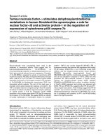
Figure 1
Eczematous drug-related eruption a patient with rheumatoid arthritis after infliximab therapy: Eczematous eruptions on the left arm (top left) and right arm (top right) and erythematous eruptions with purpura on the left leg (bottom left) and right leg (bottom right)Eczematous drug-related eruption a patient with rheumatoid arthritis after infliximab therapy: Eczematous eruptions on the left arm (top left) and right
arm (top right) and erythematous eruptions with purpura on the left leg (bottom left) and right leg (bottom right).
Arthritis Research & Therapy Vol 7 No 3 Flendrie et al.
R673
One RA patient developed a drug-induced systemic lupus ery-
thematosus after 20 months of infliximab therapy in combina-
tion with methotrexate, consisting of discoid lupus
erythematosus lesions on her hands and scalp, aphthous
lesions, conversion to antinuclear antibody positivity, and a
positive anti-double stranded-DNA (titer 60 U/L). The skin
lesions flared within one week after infusion and disappeared
after discontinuation of infliximab.
A third RA patient developed macular erythematosquamous
lesions on her lower arms, upper legs and trunk after 2.6
months of adalimumab monotherapy. Histology showed a der-
mal infiltration with CD30-positive atypical T cells. Although
the lesions appeared to be lymphomatoid papulosis, they com-
pletely disappeared within 6 weeks. Adalimumab was not
stopped. This patient developed a large-cell anaplastic non-
Hodgkin lymphoma 2 years later.
Discussion
The present study is the first large prospective study focusing
on dermatological conditions in RA patients on TNF-α-block-
ing therapy. Of the patients studied, 25% needed a dermato-
logical consultation, compared with 13% in a RA control
group, naive to TNF-α-blocking therapy. The number of
dermatological events per patient-year was significantly higher
during treatment than after treatment with TNF-α-blocking
therapy. Dermatological events led to withdrawal of TNF-α-
blocking therapy in 19 patients of 72 patients (26%). The
events recorded most frequently were skin infections, eczema,
and drug-related eruptions. Some other interesting events
were recorded, such as psoriasis, drug-induced systemic
lupus erythematosus, dermatomyositis, and a lymphomatoid-
papulosis like eruption.
RA is known to be associated with dermatological conditions
such as vasculitis, nodulosis, palmar erythema, and bullous
pemphigoid, among others [22,23]. At present, information on
the incidence and prevalence of dermatological conditions in
RA mainly originates from cross-sectional or retrospective
studies [24-26]. Few prospective studies have been con-
ducted focusing on specific conditions affecting the skin
[27,28].
Table 5
Other dermatological events in patients with rheumatoid arthritis (RA) given TNF-α-blocking therapy
Patient
no.
Age (yr) Sex Diagnosis Drug
a
Active
treatment
Event Clinical
description
Localization Time to
event
Biopsy Comedication
b
Permanent
withdrawal
anti-TNF-α
Therapy Course
1 56 f RA A Yes Lymphomatoid
papulosis-
like eruption
Macular
erythematos
quamous
lesions
Lower
arms,
upper
legs and
trunk
2.6 Yes naproxen No None Recovery
2 53 f RA A Yes Rosacea Diffuse
erythema,
scaling,
telangiectasi
as
Head and
face
1.9 Yes prednisolone,
captopril,
indomethaci
n, midazolam
No Topical Persisting
3 74 f RA E Yes Pruritus Itch Trunk 2.5 No None No Topical Unknown
4 61 f RA I No Ecchymoses Ecchymoses Hands and
feet
25.9 No AZA,
prednisolone
No Topical Partial
recovery
5 58 f RA I Yes Drug-induced
systemic
lupus
emythemato
sus
Discoid
erythematou
s lesions,
aphthous
lesions, ANA
positive, anti-
ds-DNA
positive
Hands,
face,
scalp
20.0 No MTX Yes Topical
and
systemic
Recovery, no
rechallenge
6 68 m RA I Yes Transient
swelling of
unknown
cause
Transient
swelling 2 ×
3 cm
Scalp 20.0 No MTX, folic
acid,
naproxen
No None Recovery
7 52 f RA L Yes Dermatomyosit
is
Livid erythema,
raised CPK,
decreased
proximal
muscular
strength
Inner upper
arms and
legs
2.5 Yes None Yes None Recovery
8 53 m RA L No Erythema
nodosum
Painful
erythematou
s nodules
Lower legs 7.4 Yes AZA,
naproxen,
paracetamol
No Topical Partial
recovery
a
A, adalimumab; I, infliximab; E, etanercept; L, lenercept.
b
MTX, methotrexate; AZA, azathioprine. CPK, creatinine phosphokinase; f, female, m, male.
Available online />R674
In establishing a relation between the use of a drug and the
occurrence of dermatological conditions, various factors must
be considered. Information on clinical and histological pat-
terns, time and dose relation, dechallenge and rechallenge,
and analogy with previously reported cases can provide sup-
port in assessing the plausibility of such a relation [29]. The
underlying disease and concomitant medication also need
careful consideration, as they can provide alternative
explanations.
In this study the largest group of dermatological events con-
sisted of skin infections, mostly fungal infections and folliculi-
tis. The use of TNF-α-blocking therapy has raised concerns
regarding an increased susceptibility to infections, as TNF-α
plays an important role in host-defence mechanisms [30]. An
increased incidence of tuberculosis has been described [31],
as well as a growing number of serious infections with fungal,
mycobacterial, and intracellular bacterial pathogens [32-34].
Infections of the skin have not been the subject of report in
clinical trials and observational studies with TNF-α-blocking
therapy. Cases of severe necrotizing fasciitis have been
described [35,36].
Skin infections have been reported frequently in the normal
population and especially in RA patients [24-26]. Host-
defence impairments resulting from the underlying disease
might play a role in an increased susceptibility to skin infec-
tions in RA patients, as well as the use of corticosteroids and
DMARDs such as methotrexate [28,37], which were recorded
frequently in the present study (see Table 2). They could pro-
vide an alternative explanation for the occurrence of skin infec-
tions. However, most infections occurred during active
treatment with TNF-α-blocking therapy, a finding that could
suggest at least a relative contribution to an increased vulner-
ability to skin infections in the study population. In one patient,
a bacterial superinfection of eczema occurred twice immedi-
ately after restart of adalimumab, showing a clear time relation.
For the description of the recorded drug-related eruptions, a
clinico-morphological classification was chosen [21]. Four
eruptions with a time relation and clinically or histological dis-
tinct drug-induced patterns also showed an eczematous
appearance, both clinically and histologically. This is an
unusual presentation for a drug-induced eruption and warrants
further investigation.
Two drug-related eruptions occurred during infusion with inf-
liximab or adalimumab, whereas all the others occurred after
infusion. This will most likely not reflect the true ratio between
acute and delayed reactions involving the skin, since acute
reactions with skin involvement occur in 4% of the infusions
and are usually treated by the rheumatologist without derma-
tological consultation [38].
Eczema was reported frequently in this study, even with vari-
ous dermatitis conditions, such as xerosis cutis, stasis
eczema, and seborrheic eczema, classified as separate enti-
ties. Previous studies have reported RA, in which Th1 (T helper
cell type 1) immune responses dominate, to be negatively
associated with Th2-cell-mediated atopic disorders, such as
eczema [39-41], although a similar incidence of eczema in RA
and non-RA patients has also been reported [42]. TNF-α-
blocking therapy down-regulates Th1 immune responses [43],
which might induce a shift of the Th1/Th2 balance towards
Th2-dominated immune responses and which might promote
an increased susceptibility to atopic disorders, such as
eczema.
Although the time between the initiation of TNF-α-blocking
therapy and the onset of dermatological conditions varied, a
probable relation was seen in various events. These included,
besides drug-related eruptions, events of cutaneous vasculitis,
drug-induced systemic lupus erythematosus, dermatomyosi-
tis, and a lymphomatoid papulosis-like eruption.
An association between the use of TNF-α-blocking therapy
and the induction of systemic lupus erythematosus and dis-
coid lupus erythematosus is strongly suggested by the
number of cases that have been published [10,11,13,44-46].
One case of discoid lupus erythematosus has been described
on both etanercept and infliximab in the same RA patient [47].
Analogy with previous reports is also present for cutaneous
vasculitis [13,47-49], although it is a known extra-articular
manifestation of RA [22,23]. In the first case described, a
probable relation with infliximab was present, based on the
time relation and positive dechallenge. The other cases
described were considered possibly related (Results section,
Vaculitis, cases 2 and 3) and unlikely (cases 4 and 5). Almost
all reported ulcers were considered secondary to other
causes, as described.
Dermatomyositis has been reported previously, although the
patient affected in that case had a different presentation, with
raised creatinine phosphokinase, muscle atrophy, mechanic's
hands, and vasculitis [17].
Another interesting finding was the occurrence of psoriasiform
eruptions in three patients on TNF-α-blocking therapy. This
observation is particularly interesting, since etanercept has
received and infliximab is close to receiving FDA approval for
treatment of psoriasis, after remarkable efficacy results in clin-
ical trials [7,50,51]. The occurrence of guttate psoriasis has
been reported after initiation of etanercept therapy for psoria-
sis in a placebo-controlled trial [51]. Another case report
described the occurrence of psoriasiform eruptions with histo-
logically a lichenoid dermatitis pattern in a patient with Crohn's
disease [52].
Arthritis Research & Therapy Vol 7 No 3 Flendrie et al.
R675
An exacerbation of psoriasis was also seen in a patient with
psoriatic arthritis receiving infliximab therapy. An additional
analysis showed that 28 patients with various non-RA rheu-
matic diseases, including 12 juvenile idiopathic arthritis, 6 pso-
riatic arthritis, and 3 ankylosing spondylitis, had been treated
with TNF-α-blocking therapy in the study centre. Five patients
(18%) had visited a dermatologist for a dermatological condi-
tion during or after TNF-α-blocking therapy. The events
included a drug-related eruption, eczema, and a facial mycosis
in three patients with juvenile idiopathic arthritis and a superfi-
cial spreading melanoma in a patient with ankylosing spondyli-
tis. This indicates that the occurrence of dermatological events
during TNF-α-blocking therapy is not restricted to RA patients.
In the present study the control patients were matched for
sartdate and duration of follow-up period in order to control for
time-related effects. A statistically significant relation between
the use of TNF-α-blocking therapy and the occurrence of der-
matological visits was shown. The two groups studied differed
for most baseline characteristics. These differences result
from the indication for TNF-α-blocking agents, which were
reserved for patients who fulfilled criteria for active disease
and DMARD failure (see methods section; study design), had
a longer disease duration, and whose disease was perhaps
more refractory.
However, it is considered unlikely that these factors influenced
the relation between the use of TNF-α-blocking therapy and
dermatological visits. In a multivariate regression model, no
baseline characteristic showed a predictive value for the
occurrence of a dermatological event in RA patients on TNF-
α-blocking therapy. Also, a statistically significantly higher
number of dermatological events was recorded during active
treatment with TNF-α-blocking therapy than after the therapy
had been stopped.
Conclusion
This is the first prospective study showing a relation between
TNF-α-blocking therapy and the occurrence of dermatological
conditions. Future prospective studies are needed to investi-
gate the incidence and the pathogenesis of the encountered
events, because they are a clinically significant problem in RA
patients receiving TNF-α-blocking therapy.
Competing interests
The author(s) declare that they have no competing interests.
Authors' contributions
MF participated in the study design, carried out the data col-
lection and statistical analysis, and drafted the manuscript.
WV participated in the study design, carried out the data col-
lection, and helped to write the manuscript. MC participated in
the study design and coordination and helped in the writing
and revision of manuscript. EdJ participated in the study
design and the data collection and helped to write the manu-
script. PvdK and PvR helped to write and critically revise the
manuscript and gave final approval of the manuscript. MF and
WV contributed equally to the article. All authors read and
approved the final manuscript.
References
1. Lipsky PE, van der Heijde DM, St Clair EW, Furst DE, Breedveld
FC, Kalden JR, Smolen JS, Weisman M, Emery P, Feldmann M, et
al.: Infliximab and methotrexate in the treatment of rheumatoid
arthritis. Anti-Tumor Necrosis Factor Trial in Rheumatoid
Arthritis with Concomitant Therapy Study Group. N Engl J Med
2000, 343:1594-1602.
2. Moreland LW, Schiff MH, Baumgartner SW, Tindall EA, Fleis-
chmann RM, Bulpitt KJ, Weaver AL, Keystone EC, Furst DE,
Mease PJ, et al.: Etanercept therapy in rheumatoid arthritis. A
randomized, controlled trial. Ann Intern Med 1999,
130:478-486.
3. Weinblatt ME, Keystone EC, Furst DE, Moreland LW, Weisman
MH, Birbara CA, Teoh LA, Fischkoff SA, Chartash EK: Adalimu-
mab, a fully human anti-tumor necrosis factor alpha mono-
clonal antibody, for the treatment of rheumatoid arthritis in
patients taking concomitant methotrexate: the ARMADA trial.
Arthritis Rheum 2003, 48:35-45.
4. Lovell DJ, Giannini EH, Reiff A, Jones OY, Schneider R, Olson JC,
Stein LD, Gedalia A, Ilowite NT, Wallace CA, et al.: Long-term
efficacy and safety of etanercept in children with polyarticular-
course juvenile rheumatoid arthritis: interim results from an
ongoing multicenter, open-label, extended-treatment trial.
Arthritis Rheum 2003, 48:218-226.
5. Brandt J, Khariouzov A, Listing J, Haibel H, Sorensen H, Grass-
nickel L, Rudwaleit M, Sieper J, Braun J: Six-month results of a
double-blind, placebo-controlled trial of etanercept treatment
in patients with active ankylosing spondylitis. Arthritis Rheum
2003, 48:1667-1675.
6. Braun J, Brandt J, Listing J, Zink A, Alten R, Golder W, Gromnica-
Ihle E, Kellner H, Krause A, Schneider M, et al.: Treatment of
active ankylosing spondylitis with infliximab: a randomised
controlled multicentre trial. Lancet 2002, 359:1187-1193.
7. Mease PJ, Goffe BS, Metz J, VanderStoep A, Finck B, Burge DJ:
Etanercept in the treatment of psoriatic arthritis and psoriasis:
a randomised trial. Lancet 2000, 356:385-390.
8. Maini R, St Clair EW, Breedveld F, Furst D, Kalden J, Weisman M,
Smolen J, Emery P, Harriman G, Feldmann M, et al.: Infliximab
(chimeric anti-tumour necrosis factor alpha monoclonal anti-
body) versus placebo in rheumatoid arthritis patients receiv-
ing concomitant methotrexate: a randomised phase III trial.
ATTRACT Study Group. Lancet 1999, 354:1932-1939.
9. Keystone EC, Kavanaugh AF, Sharp JT, Tannenbaum H, Hua Y,
Teoh LS, Fischkoff SA, Chartash EK: Radiographic, clinical, and
functional outcomes of treatment with adalimumab (a human
anti-tumor necrosis factor monoclonal antibody) in patients
with active rheumatoid arthritis receiving concomitant meth-
otrexate therapy: a randomized, placebo-controlled, 52-week
trial. Arthritis Rheum 2004, 50:1400-1411.
10. Bleumink GS, ter Borg EJ, Ramselaar CG, Stricker BHC: Etaner-
cept-induced subacute cutaneous lupus erythematosus.
Rheumatology 2001, 40:1317-1319.
11. Brion PH, Mittal HA, Kalunian KC: Autoimmune skin rashes
associated with etanercept for rheumatoid arthritis [letter].
Ann Intern Med 1999, 131:634.
12. Kent PD, Davis JM, Davis MDP, Matteson EL: Bullous skin
lesions following infliximab infusion in a patient with rheuma-
toid arthritis. Arthritis Rheum 2002, 46:2257-2258.
13. Misery L, Perrot JL, Gentil PA, Pallot PB, Cambazard F, Alexandre
C: Dermatological complications of etanercept therapy for
rheumatoid arthritis. Br J Dermatol 2002, 146:334-335.
14. Vergara G, Silvestre JF, Betlloch I, Vela P, Albares MP, Pascual JC:
Cutaneous drug eruption to infliximab: Report of 4 cases with
an interface dermatitis pattern. Arch Dermatol 2002,
138:1258-1259.
15. Wright RC: Atopic dermatitis-like eruption precipitated by
infliximab. JAm Acad Dermatol 2003, 49:160-161.
16. Arnett FC, Edworthy SM, Bloch DA, McShane DJ, Fries JF, Cooper
NS, Healey LA, Kaplan SR, Liang MH, Luthra HS, et al.: The Amer-
Available online />R676
ican Rheumatism Association 1987 revised criteria for the
classification of rheumatoid arthritis. Arthritis Rheum 1988,
31:315-324.
17. Flendrie M, Creemers MC, Welsing PM, den Broeder AA, van Riel
PL: Survival during treatment with tumour necrosis factor
blocking agents in rheumatoid arthritis. Ann Rheum Dis 2003,
62(Suppl 2):ii30-ii33.
18. Rau R, Sander O, van Riel PL, van de Putte LB, Hasler F, Zaug M,
Kneer J, van der AP, Stevens RM: Intravenous human recom-
binant tumor necrosis factor receptor p55-Fc IgG1 fusion pro-
tein Ro 45-2081 (lenercept): a double blind, placebo controlled
dose-finding study in rheumatoid arthritis. J Rheumatol 2003,
30:680-690.
19. Welsing PMJ, van Riel PLCM: The Nijmegen inception cohort of
early rheumatoid arthritis. J Rheumatol 2004, 31:14-21.
20. Prevoo ML, van 't Hof MA, Kuper HH, van Leeuwen MA, van de
Putte LB, van Riel PL: Modified disease activity scores that
include twenty-eight-joint counts. Development and validation
in a prospective longitudinal study of patients with rheumatoid
arthritis. Arthritis Rheum 1995, 38:44-48.
21. Stern R, Wintroub BU: Cutaneous reactions to drugs. In Fitz-
patrick's Dermatology in General Medicine 5th edition. Edited by:
Freedberg IM, Eisen A, Wolff K, Austen KF, Goldsmith LA, Katz SI.
New York: McGraw-Hill; 1999:1634-1642.
22. Jorizzo JL, Daniels JC: Dermatologic conditions reported in
patients with rheumatoid arthritis. J Am Acad Dermatol 1983,
8:439-457.
23. Sibbitt WL, Williams RC: Cutaneous manifestations of rheuma-
toid arthritis. Int J Dermatol 1982, 21:563-572.
24. Yamamoto T, Ohkubo H, Nishioka K: Skin manifestations asso-
ciated with rheumatoid arthritis. J Dermatol 1995, 22:324-329.
25. Bicer A, Tursen U, Cimen OB, Kaya TI, Ozisik S, Ikizoglu G,
Erdogan C: Prevalence of dermatophytosis in patients with
rheumatoid arthritis. Rheumatol Int 2003, 23:37-40.
26. Doran MF, Crowson CS, Pond GR, O'Fallon WM, Gabriel SE: Fre-
quency of infection in patients with rheumatoid arthritis com-
pared with controls: a population-based study. Arthritis Rheum
2002, 46:2287-2293.
27. Wilkinson SM, Smith AG, Davis MJ, Mattey DL, Dawes PT: Sus-
pected cutaneous drug toxicity in rheumatoid arthritis – an
evaluation. Br J Rheumatol 1993, 32:798-803.
28. van der Veen MJ, van der HA, Kruize AA, Bijlsma JW: Infection
rate and use of antibiotics in patients with rheumatoid arthritis
treated with methotrexate. Ann Rheum Dis 1994, 53:224-228.
29. Miller FW, Hess EV, Clauw DJ, Hertzman PA, Pincus T, Silver RM,
Mayes MD, Varga J, Medsger TA, Love LA: Approaches for iden-
tifying and defining environmentally associated rheumatic
disorders. Arthritis Rheum 2000, 43:243-249.
30. Bresnihan B, Cunnane G: Infection complications associated
with the use of biologic agents. Rheum Dis Clin North Am 2003,
29:185-202.
31. Keane J, Gershon S, Wise RP, Mirabile-Levens E, Kasznica J,
Schwieterman WD, Siegel JN, Braun MM: Tuberculosis associ-
ated with infliximab, a tumor necrosis factor alpha-neutralizing
agent. N Engl J Med 2001, 345:1098-1104.
32. Hyrich KL, Silman AJ, Watson KD, Symmons DP: Anti-tumour
necrosis factor {alpha} therapy in rheumatoid arthritis: an
update on safety. Ann Rheum Dis 2004, 63:1538-1543.
33. Ellerin T, Rubin RH, Weinblatt ME: Infections and anti-tumor
necrosis factor alpha therapy. Arthritis Rheum 2003,
48:3013-3022.
34. Netea MG, Radstake T, Joosten LA, van der Meer JW, Barrera P,
Kullberg BJ: Salmonella septicemia in rheumatoid arthritis
patients receiving anti-tumor necrosis factor therapy: associa-
tion with decreased interferon-gamma production and Toll-
like receptor 4 expression. Arthritis Rheum 2003,
48:1853-1857.
35. Chan AT, Cleeve V, Daymond TJ: Necrotising fasciitis in a
patient receiving infliximab for rheumatoid arthritis. Postgrad
Med J 2002, 78:47-48.
36. Baghai M, Osmon DR, Wolk DM, Wold LE, Haidukewych GJ, Mat-
teson EL: Fatal sepsis in a patient with rheumatoid arthritis
treated with etanercept. Mayo Clin Proc 2001, 76:653-656.
37. Hernandez-Cruz B, Cardiel MH, Villa AR, Alcocer-Varela J: Devel-
opment, recurrence, and severity of infections in Mexican
patients with rheumatoid arthritis. A nested case-control
study. J Rheumatol 1998, 25:1900-1907.
38. Wasserman MJ, Weber DA, Guthrie JA, Bykerk VP, Lee P, Key-
stone EC: Infusion-related reactions to infliximab in patients
with rheumatoid arthritis in a clinical practice setting: relation-
ship to dose, antihistamine pretreatment, and infusion
number. J Rheumatol 2004, 31:1912-1917.
39. Hartung AD, Bohnert A, Hackstein H, Ohly A, Schmidt KL, Bein G:
Th2-mediated atopic disease protection in Th1-mediated
rheumatoid arthritis. Clin Exp Rheumatol 2003, 21:481-484.
40. Hilliquin P, Allanore Y, Coste J, Renoux M, Kahan A, Menkes CJ:
Reduced incidence and prevalence of atopy in rheumatoid
arthritis. Results of a case-control study. Rheumatology 2000,
39:1020-1026.
41. Rudwaleit M, Andermann B, Alten R, Sorensen H, Listing J, Zink A,
Sieper J, Braun J: Atopic disorders in ankylosing spondylitis
and rheumatoid arthritis. Ann Rheum Dis 2002, 61:968-974.
42. Olsson AR, Wingren G, Skogh T, Svernell O, Ernerudh J: Allergic
manifestations in patients with rheumatoid arthritis. APMIS
2003, 111:940-944.
43. Krueger JG: The immunologic basis for the treatment of pso-
riasis with new biologic agents. J Am Acad Dermatol 2002,
46:1-23.
44. Carlson E, Rothfield N: Etanercept-induced lupus-like syn-
drome in a patient with rheumatoid arthritis. Arthritis Rheum
2003, 48:1165-1166.
45. Favalli E, Sinigaglia L, Varenna M, Arnoldi C: Drug-induced lupus
following treatment with infliximab in rheumatoid arthritis.
Lupus 2002, 11:753-755.
46. Shakoor N, Michalska M, Harris CA, Block JA: Drug-induced sys-
temic lupus erythematosus associated with etanercept
therapy. Lancet 2002, 359:579-580.
47. Geborek P, Crnkic M, Petersson IF, Saxne T: Etanercept, inflixi-
mab, and leflunomide in established rheumatoid arthritis: clin-
ical experience using a structured follow up programme in
southern Sweden. Ann Rheum Dis 2002, 61:793-798.
48. Jarrett SJ, Cunnane G, Conaghan PG, Bingham SJ, Buch MH,
Quinn MA, Emery P: Anti-tumor necrosis factor-alpha therapy-
induced vasculitis: case series. J Rheumatol 2003,
30:2287-2291.
49. McCain ME, Quinet RJ, Davis WE: Etanercept and infliximab
associated with cutaneous vasculitis. Rheumatology 2002,
41:116-117.
50. Chaudhari U, Romano P, Mulcahy LD, Dooley LT, Baker DG, Gott-
lieb AB: Efficacy and safety of infliximab monotherapy for
plaque-type psoriasis: a randomised trial. Lancet 2001,
357:1842-1847.
51. Gottlieb AB, Matheson RT, Lowe N, Krueger GG, Kang S, Goffe
BS, Gaspari AA, Ling M, Weinstein GD, Nayak A, et al.: A rand-
omized trial of etanercept as monotherapy for psoriasis. Arch
Dermatol 2003, 139:1627-1632.
52. Verea MM, Del Pozo J, Yebra-Pimentel MT, Porta A, Fonseca E:
Psoriasiform eruption induced by infliximab. Ann Pharmacother
2004, 38:54-57.