Báo cáo y học: " Pseudo-Foster Kennedy Syndrome due to unilateral optic nerve hypoplasia: a case report" pdf
Bạn đang xem bản rút gọn của tài liệu. Xem và tải ngay bản đầy đủ của tài liệu tại đây (216.88 KB, 2 trang )
BioMed Central
Page 1 of 2
(page number not for citation purposes)
Journal of Medical Case Reports
Open Access
Case report
Pseudo-Foster Kennedy Syndrome due to unilateral optic nerve
hypoplasia: a case report
Shveta Bansal*, Timothy Dabbs and Vernon Long
Address: Department of Ophthalmology, St James' University Hospital, Leeds, UK
Email: Shveta Bansal* - ; Timothy Dabbs - ;
Vernon Long -
* Corresponding author
Abstract
Introduction: Pseudo-Foster Kennedy Syndrome is described as unilateral optic disc swelling with
contralateral optic atrophy in the absence of an intracranial mass causing compression of the optic
nerve. This occurs typically due to bilateral sequential optic neuritis or ischaemic optic neuropathy.
Case Presentation: We describe a case of pseudo-Foster Kennedy Syndrome in a two year old
boy with unilateral papilloedema due to a congenital optic disc anomaly in one eye preventing
transmission of raised intracranial pressure to the optic nerve.
Conclusion: From our findings we conclude that congenital optic nerve hypoplasia is a cause of
pseudo-Foster Kennedy Syndrome.
Introduction
Foster Kennedy Syndrome is unilateral optic disc swelling
with contralateral optic atrophy, usually due to a frontal
lobe tumour compressing the optic nerve on one side and
resulting in papilloedema contralaterally. In the absence
of an intracranial mass these findings may be labelled as
pseudo-Foster Kennedy Syndrome.
Case presentation
A two year old boy with panhypopituitarism, hydroceph-
alus, developmental delay and obesity was referred for an
ophthalmic opinion regarding concerns of recent severe
visual deterioration. There were no other symptoms elic-
ited of possible raised intra-cranial pressure. Magnetic res-
onance imaging showed Chiari malformation, ventricular
dilatation and a small pituitary gland. Bedside fundos-
copy was very difficult as the child kept moving. Prior to
this presentation there was no documentation of baseline
visual function. An examination under anaesthetic was
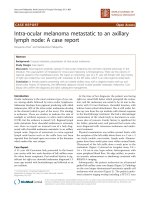
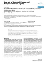
performed and right-sided severe papilloedema and a
hypoplastic left optic disc were found (Figures 1 and 2).
The findings were indicative of raised intracranial pressure
and the patient was urgently managed by the neurosur-
geons with a ventriculoperitoneal shunt operation.
Discussion
Hypoplastic optic disc is a congenital abnormality which
may be unilateral or bilateral and is a characterised by a
reduced diameter of the optic nerve head. Although clini-
cally distinct from optic atrophy, it has been suggested
that it is merely a type of non progressive optic atrophy
acquired before the full development of the eye [1].
The appearance of unilateral optic disc swelling with con-
tralateral optic disc atrophy has been described as the Fos-
ter Kennedy Syndrome. In "true" Foster-Kennedy
Syndrome unilateral disc swelling is caused by a tumour
on the inferior surface of the frontal lobe, compressing the
Published: 18 March 2008
Journal of Medical Case Reports 2008, 2:86 doi:10.1186/1752-1947-2-86
Received: 4 November 2007
Accepted: 18 March 2008
This article is available from: />© 2008 Bansal et al; licensee BioMed Central Ltd.
This is an Open Access article distributed under the terms of the Creative Commons Attribution License ( />),
which permits unrestricted use, distribution, and reproduction in any medium, provided the original work is properly cited.
Publish with BioMed Central and every
scientist can read your work free of charge
"BioMed Central will be the most significant development for
disseminating the results of biomedical research in our lifetime."
Sir Paul Nurse, Cancer Research UK
Your research papers will be:
available free of charge to the entire biomedical community
peer reviewed and published immediately upon acceptance
cited in PubMed and archived on PubMed Central
yours — you keep the copyright
Submit your manuscript here:
/>BioMedcentral
Journal of Medical Case Reports 2008, 2:86 />Page 2 of 2
(page number not for citation purposes)
optic nerve on one side with papilloedema contralaterally
[2]. In the absence of an intracranial mass these findings
may be labelled as pseudo-Foster Kennedy Syndrome,
typically due to bilateral sequential optic neuritis or
ischaemic optic neuropathy [3,4].
Explanations for the unilateral disc swelling in Foster
Kennedy syndrome include failure of transmission of the
intracranial pressure to the optic disc secondary to pres-
sure on the vaginal sheath; or closure of the vascular bed
of the optic disc [5]. Our case demonstrates that this find-
ing may be observed in patients with unilateral optic disc
hypoplasia and is thus another differential cause of
pseudo-Foster Kennedy Syndrome.
Conclusion
In this case the finding of unilateral papilloedema was
due to a congenital abnormality of the left optic disc, pre-
venting transmission of the raised intracranial pressure to
the optic nerve head. This is important to bear in mind
when examining children with optic nerve hypoplasia.
Competing interests
The author(s) declare that they have no competing inter-
ests.
Authors' contributions
SB was the lead author involved in carrying out the litera-
ture search, study design and writing the case report. TD
assisted with writing the paper, supervising and managing
the case. VL supervised the management of the case and
participated in its design and approval. All authors have
been involved in approving the final manuscript.
Consent
The authors obtained written informed consent from the
parents of this patient for the publication of this case
report along with images. A copy of the written consent is
available for review by the Editor-in-Chief of this journal.
References
1. Frisen L, Holmegaard L: Spectrum of optic nerve hypoplasia.
British Journal of Ophthalmology 1978, 62:7-15.
2. Massey EW, Schoenberg B: Foster Kennedy Syndrome. Archives
of Neurology 1984, 41:658-659.
3. Watnick RL, Trobe JD: Bilateral optic nerve compression as a
mechanism for the Foster Kennedy Syndrome. Ophthalmology
1989, 96:1793-1798.
4. Shatz N, Smith J: Non tumour causes of the Foster Kennedy
syndrome. Journal of Neurosurgery 1967, 27:37.
5. Primrose J: Mechanism of production of papilloedema. British
Journal of Ophthalmology 1964, 48:19-29.
Fundal photograph showing a hypoplastic optic disc in the left eyeFigure 2
Fundal photograph showing a hypoplastic optic disc in the left
eye.
Fundal photograph showing severe papilloedema in the right eyeFigure 1
Fundal photograph showing severe papilloedema in
the right eye.