Báo cáo y học: " Coliform pyosalpinx as a rare complication of appendicectomy: a case report and review of the literature on best practice" pdf
Bạn đang xem bản rút gọn của tài liệu. Xem và tải ngay bản đầy đủ của tài liệu tại đây (280.72 KB, 3 trang )
BioMed Central
Page 1 of 3
(page number not for citation purposes)
Journal of Medical Case Reports
Open Access
Case report
Coliform pyosalpinx as a rare complication of appendicectomy: a
case report and review of the literature on best practice
Deepak Singh-Ranger*, Abayomi Sanusi and Ishrak Hamo
Address: Department of General Surgery, Queen's Hospital, Rom Valley Way, Romford, Essex, UK
Email: Deepak Singh-Ranger* - ; Abayomi Sanusi - ; Ishrak Hamo -
* Corresponding author
Abstract
Introduction: Coliform pyosalpinx is a rare entity. We report a case that occurred three months
after appendicectomy for gangrenous appendicitis. There follows a literature review on best
practice for the treatment of pyosalpinx.
Case presentation: A seventeen year old girl presented with an acute abdomen three months
after an appendicectomy for gangrenous appendicitis. Intraoperative findings were bilateral
pyosalpinx treated by aspiration, saline and Betadine irrigation and intravenous antibiotics.
Conclusion: Microbiological analysis of the pus revealed Escherichia coli and anaerobes. Chlamydia
and Candida were not isolated. This is the first known reported case of Coliform Pyosalpinx
following appendicectomy. The best treatment does not necessarily involve salpingectomy
especially in women of reproductive age where fertility may become compromised.
Introduction
Pyosalpinx, in the majority of cases, is a sequela of pelvic
inflammatory disease. The ramifications of this condition
are important and include tubal infertility and ectopic
pregnancy [1]. There have been cases where a non-sexu-
ally transmitted cause for pyosalpinx has been described.
Notable examples are pyosalpinx following in vitro fertili-
zation [2] and infection by streptococcus pneumoniae [3]
and coliforms [4]. Only one case of spontaneous coliform
pyosalpinx has been published; that case involved a nine
year old girl [5].
We report a case of coliform pyosalpinx in a seventeen
year old girl following a recent appendicectomy. The best
treatment for pyosalpinx in pre-menopausal females is
discussed.
Case presentation
A seventeen year old girl presented as an emergency with
a two-day history of lower abdominal and back pain. She
experienced rigors and appetite loss but no nausea, vom-
iting, dysuria, cystitis or vaginal discharge. Three months
previously, she had undergone immediate appendicec-
tomy for a gangrenous retrocaecal appendix. Other intra-
operative findings at the time were a macroscopically
normal right ovary and fallopian tube.
There was no history of recent sexual activity or pelvic
inflammatory disease. Menstrual cycles were regular and
every 28 days and the patient was mid-cycle at the time of
presentation.
On examination, she had a temperature of 38.5°C, pulse
of 100 beats per minute and blood pressure of 114/59.
Lower abdominal rebound tenderness, guarding and
Published: 2 April 2008
Journal of Medical Case Reports 2008, 2:97 doi:10.1186/1752-1947-2-97
Received: 17 August 2007
Accepted: 2 April 2008
This article is available from: />© 2008 Singh-Ranger et al; licensee BioMed Central Ltd.
This is an Open Access article distributed under the terms of the Creative Commons Attribution License ( />),
which permits unrestricted use, distribution, and reproduction in any medium, provided the original work is properly cited.
Journal of Medical Case Reports 2008, 2:97 />Page 2 of 3
(page number not for citation purposes)
absent bowel sounds were present. The patient had a leu-
cocytosis of 16.4 × 10
9
.l
-1
and C-reactive protein concen-
tration of 322 mg.l
-1
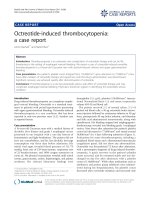
. A pregnancy test was negative and an
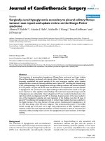
emergency computerized tomographic scan showed a
complex pelvic mass associated with or near to the right
ovary and overriding, but not connected to the uterus
(Figure 1). She subsequently underwent an emergency
laparotomy. The right fallopian tube was found in the
midline above the uterus. It was grossly enlarged, measur-
ing 10 × 5 cm, with multiple necrotic areas oozing pus.
The fimbrial end was oedematous with a radius of 2 cm.
The left fallopian tube was slightly enlarged and was
found postrolateral to the uterus, adherent to the sigmoid
colon by fibrinous adhesions. There was no visible enter-
osalpinx fistula and no appendicular stump leak. The left
salpinx was released by blunt dissection and pus drained
from both fallopian tubes by retrograde "milking". Both
tubes were irrigated generously with a 0.9% saline and
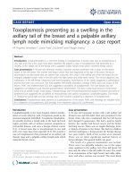
Betadine mixture. Microbiological analysis of the pus
revealed Escherichia coli and anaerobes but not Chlamydia
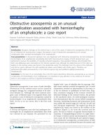
or Candida spp. A postoperative Gastrografin enema did
not reveal an occult fistula (Figure 2).
The patient was treated postoperatively with intravenous
Co-Amoxiclav and Metronidazole for a week and made an
uneventful recovery. However, she now faces the long-
term sequelae of potential infertility, ectopic pregnancy
and chronic pelvic pain.
Discussion
Coliform pyosalpinx is very rare, and coliform pyosalpinx
following gangrenous appendicitis treated by appendicec-
tomy has not been reported in the literature. This is the
first report ever of this disease entity.
Pyosalpinx following appendicectomy may be one expla-
nation for the small association between perforated
appendicitis and sterility [6,7]. When encountered, it is
vital for the trainee surgeon to be aware of the best treat-
ment, with the least morbidity. This encompasses a wide
range of interventions varying from intravenous antibiot-
ics, laparoscopic aspiration or laparoscopic salpingotomy
with saline irrigation, image-guided aspiration and/or
drainage [8,9] to salpingectomy. The latter should be con-
sidered as last resort in premenopausal females. Repeat
laparoscopy of patients who have undergone irrigation
have shown no recurrence [10]. A randomized trial has
Emergency computerized tomographic scan: a right ovarian mass is visualizedFigure 1
Emergency computerized tomographic scan: a right
ovarian mass is visualized.
Postoperative Gastrografin enema did not show an enterotu-bal fistulaFigure 2
Postoperative Gastrografin enema did not show an
enterotubal fistula.
Publish with BioMed Central and every
scientist can read your work free of charge
"BioMed Central will be the most significant development for
disseminating the results of biomedical research in our lifetime."
Sir Paul Nurse, Cancer Research UK
Your research papers will be:
available free of charge to the entire biomedical community
peer reviewed and published immediately upon acceptance
cited in PubMed and archived on PubMed Central
yours — you keep the copyright
Submit your manuscript here:
/>BioMedcentral
Journal of Medical Case Reports 2008, 2:97 />Page 3 of 3
(page number not for citation purposes)
shown that transvaginal sonographic drainage with intra-
venous antibiotics produces a faster resolution of symp-
toms than intravenous antibiotics alone; hospital stay and
need for surgery were also lower in the study cohort.
The role of transvaginal drains and the effect of intra-fal-
lopian antibiotic instillation on fertility still remains
unclear.
One possible way to assess fertility is by performing a
repeat diagnostic laparoscopy. This may demonstrate
tubal features (e.g. occlusion, adhesions) that are linked
to infertility [11,12]. The ideal time for the procedure is
varied and ranges from between two to 33 weeks [13,14].
Tubal function may also be assessed by salpingography
and/or salpingoscopy. A "cobblestone" appearance of the
tubal mucosa is suggestive of patchy loss and damage to
ciliated mucosal cells [13].
In premenopausal females, salpingectomy or laparotomy
is not encouraged as subsequent infertility is said to be
high [14].
In summary, coliform pysosalpinx may be a complication
of acute gangrenous appendicitis and/or may follow
appendicectomy. If diagnosed preoperatively sonographic
or laparoscopic drainage is advocated. The small risk of
infertility following open appendicectomy for perforated
or gangrenous appendicitis may also be one argument for
all premenopausal females to undergo a laparoscopic pro-
cedure for this condition.
Conclusion
This is the first documented case of coliform pyosalpinx
following appendicectomy for gangrenous appendicitis. It
may be one reason for the association between perforated
appendicitis and sterility [5,6]. In order to decrease the
risk of infertility, minimally invasive treatment options
should be used which endeavour to preserve the fallopian
tubes in young females. Tubal patency and mucosal archi-
tecture can be assessed subsequently, by salpingography
and salpingoscopy. Repeat diagnostic laparoscopy may
also be useful in assessment of premenopausal females
who have had appendicectomy but who are unable to
conceive.
Competing interests
The author(s) declare that they have no competing inter-
ests.
Authors' contributions
DSR was involved in postoperative care during both
admissions and drafted the manuscript. AZ obtained the
Gastrografin radiological images, participated in revising
the manuscript and was involved in the postoperative care
during the second admission. IH was the consultant in-
charge of the patient, performed the second operation,
and has given approval of the manuscript.
Consent
Written informed consent was obtained from the patient
for publication of this Case report and accompanying
images. A copy of the written consent is available for
review by the Editor-in-Chief of this journal.
Acknowledgements
No funding was received.
References
1. Paavonen J: Pelvic inflammatory disease. From diagnosis to
prevention. Dermatol Clin 1998, 16:747-56. xii.
2. Kihaile P, Misumi J, Utsunomiya T: Peritonitis after a ruptured
left pyosalpinx in a patient undergoing in vitro fertilization.
Fertil Steril 2003, 79:1034-6.
3. Meis JF, Festen C, Hoogkamp-Korstanje JA: Pyosalpinx caused by
Streptococcus pneumoniae in a young girl. Pediatr Infect Dis J
1993, 12:539-40.
4. Habek D, Vranko NN, Sklebar I, Grabovac S, Cerkez HJ: Rupture of
coliform pyosalpinx in a nine-year old girl. Zentralbl Gynakol
2002, 124:220-2.
5. Geryk B, Kubikova E, Stolzova E: [Female sterility after appendi-
citis in childhood]. Bratisl Lek Listy 1992, 93:541-4.
6. Mueller BA, Daling JR, Moore DE, Weiss NS, Spadoni LR, Stadel BV,
Soules MR: Appendectomy and the risk of tubal infertility. N
Engl J Med 1986, 315:1506-8.
7. Corsi PJ, Johnson SC, Gonik B, Hendrix SL, McNeeley SG Jr, Diamond
MP: Transvaginal ultrasound-guided aspiration of pelvic
abscesses. Infect Dis Obstet Gynecol 1999, 7:216-21.
8. Teisala K, Heinonen PK, Punnonen R: Transvaginal ultrasound in
the diagnosis and treatment of tubo-ovarian abscess. Br J
Obstet Gynaecol 1990, 97:178-80.
9. De Wilde R, Hesseling M: [Organ preserving endosurgery in
pyosalpingitis]. Zentralbl Gynakol 1992, 114:459-62.
10. Wolner-Hanssen P, Westrom L: Second-look laparoscopy after
acute salpingitis. Obstet Gynecol 1983, 61:702-4.
11. Gerber B, Krause A: A study of second-look laparoscopy after
acute salpingitis. Arch Gynecol Obstet 1996, 258:193-200.
12. Pouly JL, Mage G, Dupre B, Canis M, Bruhat MA: [Celioscopy in the
early follow-up of salpingitis]. J Gynecol Obstet Biol Reprod (Paris)
1985, 14:989-95.
13. Lang EK, Dunaway HE Jr: Salpingographic demonstration of
"cobblestone" mucosa of the distal tubes is indicative of irre-
versible mucosal damage. Fertil Steril 2001, 76:342-5.
14. Parsons AK: Regarding the best approach to the pyosalpinx.
Ultrasound Obstet Gynecol 1996, 7:398-400.