Báo cáo y học: " Hypertrophic osteoarthropathy as the cause of a super scan of the bone in a patient with prostate cancer: a case report" pps
Bạn đang xem bản rút gọn của tài liệu. Xem và tải ngay bản đầy đủ của tài liệu tại đây (422.67 KB, 6 trang )
BioMed Central
Page 1 of 6
(page number not for citation purposes)
Journal of Medical Case Reports
Open Access
Case report
Hypertrophic osteoarthropathy as the cause of a super scan of the
bone in a patient with prostate cancer: a case report
Boris L Kanen*
1,2
and Ruud JLF Loffeld
1
Address:
1
Department of Internal Medicine, Zaans Medical Center, Zaandam, The Netherlands and
2
Department of Endocrinology, VU University
Medical Center, Amsterdam, The Netherlands
Email: Boris LJ Kanen* - ; Ruud JLF Loffeld -
* Corresponding author
Abstract
Introduction: Prostate cancer is known to have a tendency to metastasize to bone. Skeletal
scintigraphy can be used to show multiple lesions. Diffuse metastasis, which is not infrequent in
prostate cancer, can also be suspected on the basis of a 'super scan'. However, this phenomenon
in nuclear medicine has several other causes that need to be considered.
Case presentation: A patient with a history of prostate cancer presented with pleural fluid,
peripheral edema and bone pain. A super scan of the bone was found which suggested diffuse
skeletal metastasis of the prostate cancer but the patient also had a prostate specific antigen level
which was not compatible with this diagnosis. Further investigations revealed the paraneoplastic
phenomenon of hypertrophic osteoarthropathy, related to an incurable carcinoma of the lung, to
be the cause of the super scan.
Conclusion: A super scan is characterized by a high bone to soft tissue ratio on skeletal
scintigraphy, with a uniform symmetrical increase in bone uptake and diminished to absent renal
visualization ('absent kidney sign'). It can be seen in a variety of diseases in which there is a diffusely
increased bone turnover. Diffuse skeletal metastasis of a well-differentiated prostate carcinoma is
unlikely to be the cause of a super scan when the prostate specific antigen level is not elevated. This
is the first report of a super scan due to pulmonary hypertrophic osteoarthropathy which can be
seen in lung carcinoma and other pulmonary diseases.
Introduction
Prostate cancer has a high tendency of metastasizing to
the skeleton. In fact, many cases of this cancer are diag-
nosed because of the detection of bone metastasis from a
primary tumor of unknown origin at the time of presenta-
tion. The majority of patients with metastatic prostate
cancer will have multiple skeletal lesions. However, dif-
fuse metastases are also described. These patients have a
so-called super scan of the bone. Presence of a super scan
is not pathognomic for diffuse bone metastasis. The dif-
ferential diagnosis is wider as is described in this case
report.
Case presentation
A 81-year-old man with an adenocarcinoma of the pros-
tate diagnosed one year earlier presented with a five
month history of gradually progressive complaints of dys-
pnea. At the time of diagnosis of the prostate cancer, there
had been no signs of metastases and since it was an
asymptomatic grade 2 prostate cancer in a man of
Published: 7 April 2008
Journal of Medical Case Reports 2008, 2:104 doi:10.1186/1752-1947-2-104
Received: 15 July 2007
Accepted: 7 April 2008
This article is available from: />© 2008 Kanen and Loffeld; licensee BioMed Central Ltd.
This is an Open Access article distributed under the terms of the Creative Commons Attribution License ( />),
which permits unrestricted use, distribution, and reproduction in any medium, provided the original work is properly cited.
Journal of Medical Case Reports 2008, 2:104 />Page 2 of 6
(page number not for citation purposes)
advanced age, a watchful waiting policy was followed. The
medical history revealed hypertension and a transurethral
resection of the prostate six years before presentation. The
patient complained of dyspnea, progressive peripheral
edema, orthopnea, and painful knees and thighs which
made walking extremely difficult. There had been weight
loss of ten kilograms over six months, with associated loss
of appetite. No thoracic pain, hemoptysis or other pulmo-
nary or cardiac complaints were present. The patient had
been a heavy smoker for fifty years.
On admission his blood pressure was 150/80 mmHg with
an irregular pulse of 96 per minute, temperature 36.2°C,
and he had a normal central venous pressure. The heart
sounds were normal. Percussion and auscultation of the
left lower lung revealed dullness with diminished breath
sounds. These signs were indicative of pleural effusion.
The liver was not enlarged. There was pitting edema espe-
cially at the lower extremities, but also of both hands,
which were also noted to be remarkably large. Percussion
of, and axial pressure on, the vertebrae was not painful.
The patient refused rectal examination because of painful
earlier experiences.
Laboratory examination revealed the following data: ESR
35 mm in the first hour (normal: <7), CRP 134 mg/l (nor-
mal <10), hemoglobin 6.3 mmol/l (normal: 8.9–10.7)
with a MCV of 82 fl (normal: 80–100), leukocytes 8.6 ×
10
9
/l (normal: 4.5–10.0) with 90% neutrophilic granulo-
cytes (normal 40–70), normal blood platelets, electrolytes
and liver enzymes. Creatinine was 77 μmol/l (normal:
64–108), alkaline phosphatase was elevated at 285 U/l
(normal: 40–120), calcium was 1.95 mmol/l (normal:
2.15–2.68) with an albumin of 23.9 g/l (normal: 35–50 g/
l) and a normal phosphate. Blood gas analysis showed a
chronic compensated respiratory acidosis with an oxygen
saturation of 80%.
Electrocardiography showed atrial fibrillation with a left
bundle branch block, similar to earlier ECGs. Chest X-ray
revealed a large amount of pleural fluid on the left side
and an enlarged heart without signs of vascular redistribu-
tion. There were no signs of tumor or pulmonary metasta-
sis on chest X-ray.
Analysis of the pleural fluid was performed. A total
amount of 4.5 liters was evacuated. Cytological and bio-
chemical analysis showed only lymphocytosis with no
signs of malignancy or bacterial infection. Auramin and
Löwenstein cultures were negative. An echocardiography
showed good left ventricular function. Ultrasound inves-
tigation of the abdomen showed a dilated inferior caval
vein without other abnormalities. The entire presentation
was compatible with right-sided heart failure in a patient
with probable pulmonary hypertension. On Computed
Tomography Angiography (CTA) there were no pulmo-
nary embolisms visible but a large amount of pleural fluid
was seen in the left pleural cavity.
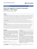
Because of the elevated alkaline phosphatase, the bone
pains and the previously diagnosed prostate cancer, skele-
tal scintigraphy was performed. It showed a 'super scan',
meaning there was diffuse uptake throughout the entire
skeleton. This was judged as fitting diffuse skeletal metas-
tasis of the prostate cancer [Figure 1]. However the pros-
tate specific antigen was within the normal range at 1.4
μg/l (normal < 4.4)
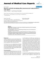
With the remarkably large hands in mind, additional
investigations were carried out [Figure 2]. A bone marrow
examination showed no marrow disease nor malignancy.
X-ray of the hands, humeri, femora and pelvis revealed
extensive subperiosteal bone appositions compatible with
generalized hypertrophic osteoarthropathy [Figure 3a/b].
Repeat of the earlier performed CTA indeed now showed
a fluid-containing cavity in the lower left lobe surrounded
by a large amount of pleural fluid at that side suggestive of
a lung cancer. Bronchoscopy confirmed this diagnosis.
The left main bronchus was stenotic with tumor totally
occluding the left lower lobe and almost occluding the left
upper lobe. Histological examination was not possible
due to technical difficulties during the procedure. The
diagnosis of incurable bronchial carcinoma with hyper-
trophic osteoarthropathy was made with the prostate can-
cer as an "innocent" bystander. Since the patient was
rapidly deteriorating palliative care was given. The patient
died several weeks after admission. Post mortum exami-
nation confirmed the clinical diagnosis. There was a large
undifferentiated non-small cell lung carcinoma with a
diameter of 10 cm and extension in the adventitia of the
esophagus and lymphatic metastasis in the hili and medi-
astinum. Three liters of tumor-positive pleural fluid and
extensive hypertrophic osteoarthropathy was seen with-
out distant metastasis.
Discussion
A super scan is characterized by a strikingly high bone to
soft tissue ratio on skeletal scintigraphy, with a uniform
symmetrical increase in bone uptake and diminished to
absent renal visualization ('absent kidney sign'). It can be
seen in a variety of diseases in which there is diffusely
increased bone turn over. Diffuse skeletal metastasis, as
can be observed from primary tumors of the breast, lung,
prostate, bladder and lymph nodes, is the most frequent
cause. Other causes are secondary hyperparathyroidism,
Paget disease, myelofibrosis and metabolic bone disease.
Technetium-99m-labeled methylene diphosphonate
(
99m
Tc-MPD) bone scintigraphy performed in patients
presenting with prostate cancer shows metastases in 10–
Journal of Medical Case Reports 2008, 2:104 />Page 3 of 6
(page number not for citation purposes)
50%. It has a false negative rate of 1–5%, mostly being
due to a super scan [1]. When caused by prostate cancer,
super scans are found exclusively in histologically high-
grade forms.
Hypertrophic Osteoarthropathy (HOA), also known as
the classical Pierre Marie-Bamberger syndrome, is a sys-
temic disorder of the bones, joints and soft tissues that
develops in association with other disease processes. It is
characterized by several or all of the following signs [2]:
clubbing of the digits, periosteal new bone formation,
particularly involving the long bones of the distal extrem-
ities, symmetric arthritis-like changes in the joints and
periarticular tissues (ankles, knees, wrists, and elbows),
increased thickness of the subcutaneous soft tissues in the
distal one-third of the arms and legs and sometimes of the
facial tissues, which may simulate acromegaly [3] and
finally, neurovascular changes of the hands and feet
including chronic erythema, paresthesia and increased
sweating.
Most commonly it is associated with an intrathoracic
malignancy, which can be carcinoma of the lung as well as
pulmonary metastasis of other tumors and Hodgkin's dis-
ease involving the mediastinum. HOA is also frequently
seen in severe cystic fibrosis, bronchiectasis, chronic
empyema and lung abscess and occasionally in certain
liver disorders' [4]. In some instances it may present with-
out any underlying illness when it is called primary, idio-
pathic or the hereditary form of HOA in which bone and
Skeletal scintigraphy showing diffusely increased uptake and the absent kidney sign (a super scan)Figure 1
Skeletal scintigraphy showing diffusely increased uptake and the absent kidney sign (a super scan).
Journal of Medical Case Reports 2008, 2:104 />Page 4 of 6
(page number not for citation purposes)
joint pain tends to be less, and the furrowing of the face
and scalp tends to be more severe. Diagnosis must be
based on global assessment of the clinical, laboratory and
radiographic findings rather than the presence of one
abnormality, since there are well-documented cases that
lacked radiographically detectable periostitis [5]. Blood
studies are usually unaffected by HOA except that often an
elevated ESR of more than 50 mm/h is seen and in
advanced cases an elevated alkaline phosphatase level can
be found [6]. The incidence of clinically apparent HOA in
patients diagnosed with lung cancer is approximately 4–
5% [7]. The etiology is still poorly understood. Several
pathogenetic theories have focused on the vascular
changes and proliferation that might be caused by circu-
lating growth factors that normally are inactivated in the
lungs. Pulmonary shunting caused by the several disease
processes that are associated with HOA causes a faulty
pulmonary clearance of macrothrombocytes, which
release growth factors in the systemic circulation. Elevated
levels of platelet-derived growth factor (PDGF), endothe-
lin-1 (ET-1), β-thromboglobulin (β-TG) and vascular
endothelial growth factor (VEGF) [8] have all been shown
to be elevated in patients with HOA.
HOA has no prognostic significance and early detection
may lead to detection of potentially resectable lung carci-
noma. Subclinical cases can be diagnosed by radiographs
or, with more sensitivity, by skeletal scintigraphy with an
incidence in bronchogenic carcinoma of up to 20% [6].
Usually scintigraphic abnormalities are found in the
peripheral skeleton and are not easily mistaken for diffuse
skeletal metastasis. Its appearance can range from
increased 'bracelet-like' appearance to more diffusely
increased uptake at the distal ends of the long bones.
Although usually located in the peripheral skeleton, it can
also affect the skull, claviculae, ribs and scapulae [9,10].
Sometimes along the cortical margins, a 'parallel track
sign' due to the periosteal bone formation can be seen. To
the best of our knowledge this is the first report of a super
scan as the presenting feature of HOA.
This case report clearly illustrates the pitfalls of diagnostic
tests. The most important is the interpretation of the bone
scan. Although the clinical presentation almost entirely
fitted the suggested diagnosis of diffusely metastasized
prostate carcinoma as an explanation for the symptoms,
signs and bone scintigraphy, it was the normal PSA, which
indicated that an alternative diagnosis should be sought.
Studies report that a PSA of less than 20 ng/ml has a neg-
ative predictive value of 92–95% for the absence of skele-
tal metastases in patients with well-differentiated (grade 1
and 2) or clinically localized (stages T1–2) prostate can-
cer. In patients with poorly differentiated (grade 3) or
clinically advanced (stages T3–4) tumors it has a negative
predictive value of only 70 and 50%, respectively [10,11].
And although advanced technologies are at our disposal,
in almost 4.5 liters of pleural fluid no malignant cells were
found. Imaging technologies are so refined that peripheral
embolism on a CTA can be detected, but gross abnormal-
ities like pleural fluid and atelectasis can cover up a malig-
nant tumor of 10 cm in diameter.
A. Large sized right hand with edematous swellingFigure 2
A. Large sized right hand with edematous swelling. B. Periungual erythema and clubbing.
Journal of Medical Case Reports 2008, 2:104 />Page 5 of 6
(page number not for citation purposes)
Conclusion
This case illustrates that a super scan of the bone is a dis-
tinct entity in nuclear medicine that can point towards
several different bone disorders. Malignancy is a frequent
cause, but the most obvious cause in that setting, namely
diffuse skeletal metastasis, is not the only one that should
be considered. In prostate carcinoma, especially when
well-differentiated with a low PSA level, skeletal metasta-
sis is unlikely. As in this case, non-metastatic paraneoplas-
tic, or even benign diagnoses should be considered. To
our knowledge this is the first report of a super scan due
to extensive HOA. Usually scintigraphic abnormalities are
confined to the peripheral skeleton.
In modern day medical practice, a clinician is often con-
fronted with the results of several diagnostic modalities. It
remains the task of the clinician to be critical and to inter-
pret the different aspects and findings in relation to each
other.
Competing interests
The author(s) declare that they have no competing inter-
ests.
Authors' contributions
BK and RL have both been involved in the management of
the patient as well as writing the case report. Both authors
have read and approved the manuscript.
Consent
Informed and verbal consent was obtained from the
patient for both publication and use of the clinical photo-
graphs. Written consent was not obtained before death.
The patient had no relatives or partner who could be
asked to provide written consent.
Acknowledgements
No funding was received.
References
1. Bruwer G, Heyns CF, Allen FJ: Influence of local tumour stage
and grade on reliability of serum prostate-specific antigen in
predicting skeletal metastases in patients with adenocarci-
noma of the prostate. Eur Urol 1999, 35:223-227.
2. COURY C: Hippocration fingers and hypertrophic osteoar-
thropathy. A study of 350 cases. Br J Dis Chest 1960, 54:202-209.
3. HAMMARSTEN JF, O'LEARY J: The features and significance of
hypertrophic osteoarthropathy. AMA Arch Intern Med 1957,
99:431-441.
4. Kuloglu Z, Kansu A, Ekici F, Demirceken F, Fitoz S, Tutar E, Girgin N:
Hypertrophic osteoarthropathy in a child with biliary
atresia. Scand J Gastroenterol 2004, 39:698-701.
5. Horn CR: Hypertrophic osteoarthropathy without radio-
graphic evidence of new bone formation. Thorax 1980, 35:479.
6. Koischwitz D, Dewes W, Bahre M, Schmidt RF: [Correlation of
scintigraphic and x-ray findings in Marie-Bamberger second-
ary hypertrophic osteoarthropathy]. Rofo 1986, 144:681-688.
7. Morgan B, Coakley F, Finlay DB, Belton I: Hypertrophic osteoar-
thropathy in staging skeletal scintigraphy for lung cancer.
Clin Radiol 1996, 51:694-697.
X-ray of the femur showing extensive characteristic perio-steal bone appositionFigure 3
X-ray of the femur showing extensive characteristic
periosteal bone apposition.
Publish with BioMed Central and every
scientist can read your work free of charge
"BioMed Central will be the most significant development for
disseminating the results of biomedical research in our lifetime."
Sir Paul Nurse, Cancer Research UK
Your research papers will be:
available free of charge to the entire biomedical community
peer reviewed and published immediately upon acceptance
cited in PubMed and archived on PubMed Central
yours — you keep the copyright
Submit your manuscript here:
/>BioMedcentral
Journal of Medical Case Reports 2008, 2:104 />Page 6 of 6
(page number not for citation purposes)
8. Vandemergel X, Decaux G: [Review on hypertrophic osteoar-
thropathy and digital clubbing]. Rev Med Brux 2003, 24:88-94.
9. Ali A, Tetalman MR, Fordham EW, Turner DA, Chiles JT, Patel SL,
Schmidt KD: Distribution of hypertrophic pulmonary osteoar-
thropathy. AJR Am J Roentgenol 1980, 134:771-780.
10. Buckley O, O'Keeffe S, Geoghegan T, Lyburn ID, Munk PL, Worsley
D, Torreggiani WC: 99mTc bone scintigraphy superscans: a
review. Nucl Med Commun 2007, 28:521-527.
11. Chybowski FM, Keller JJ, Bergstralh EJ, Oesterling JE: Predicting
radionuclide bone scan findings in patients with newly diag-
nosed, untreated prostate cancer: prostate specific antigen
is superior to all other clinical parameters. J Urol 1991,
145:313-318.