Báo cáo y học: " Atopic dermatitis, cutaneous steroids and cataracts in children: two case reports" pptx
Bạn đang xem bản rút gọn của tài liệu. Xem và tải ngay bản đầy đủ của tài liệu tại đây (247.98 KB, 4 trang )
BioMed Central
Page 1 of 4
(page number not for citation purposes)
Journal of Medical Case Reports
Open Access
Case report
Atopic dermatitis, cutaneous steroids and cataracts in children: two
case reports
Andrew Tatham
Address: Leicester Royal Infirmary, Infirmary Square, Leicester LE1 5WW, UK
Email: Andrew Tatham -
Abstract
Introduction: Atopic dermatitis is a chronic, pruritic, eczematous skin disease mediated through
an immediate (type I) hypersensitivity reaction. Posterior sub-capsular cataracts are a recognised
complication of atopic dermatitis in adults; however they are rare in children. The management of
atopic dermatitis is based on the exclusion of allergens, the use of emollients, and on topical
corticosteroids for disease exacerbations. Cataracts may be due to atopic dermatitis but may also
occur secondary to the use of corticosteroids.
Case presentation: We describe two children with atopic dermatitis, treated with cutaneous
corticosteroids, both of whom were diagnosed with bilateral posterior sub-capsular cataracts.
Conclusion: These cases demonstrate that atopic dermatitis and topical corticosteroids may be
associated with cataracts in children as well as adults. The cause of cataracts in atopic dermatitis is
not known, however, it has been suggested that habitual tapping and rubbing of the face may play
a role. Care needs to be taken when prescribing corticosteroids. Inadequate treatment of atopic
dermatitis may lead to other ocular complications such as keratitis and permanent visual loss.
Introduction
Atopic dermatitis (AD) is a chronic, pruritic, eczematous
skin disease mediated through an immediate (type I)
hypersensitivity reaction. It primarily affects the flexural
surfaces and lesions exhibit a red, elevated, scaly and often
excoriated appearance. AD is typically manifest in infants
aged 1 to 6 months and 90% of eventual sufferers have
had their first outbreak by age 5 years. Ocular complica-
tions of AD in adults include blepharitis, keratoconjuncti-
vitis, keratoconus, uveitis, sub-capsular cataract and
retinal detachment. Cataracts secondary to AD may occur
in 25 to 50% of adults but are rare in adolescents and
young adults [1]. The most common ocular finding in
children is a papillofollicular conjunctivitis [1]. Two main
types of cataract are seen in patients with AD, an anterior
sub-capsular plaque and anterior and posterior sub-cap-
sular opacities.
The management of AD is based on the exclusion of aller-
gens, the use of emollients and on topical corticosteroids
for disease exacerbations. Cataracts may be due to AD but
may also occur secondary to the use of corticosteroids.
The cataract associated with corticosteroids tends to be
posterior sub-capsular. Ocular complications of corticos-
teroids may occur following intravenous, oral, inhaled or
ocular administration. Although corticosteroids are com-
monly used in the treatment of dermatological diseases
there are few reports of cataracts occurring following the
use of cutaneous corticosteroids [1,2].
Published: 28 April 2008
Journal of Medical Case Reports 2008, 2:124 doi:10.1186/1752-1947-2-124
Received: 18 December 2007
Accepted: 28 April 2008
This article is available from: />© 2008 Tatham; licensee BioMed Central Ltd.
This is an Open Access article distributed under the terms of the Creative Commons Attribution License ( />),
which permits unrestricted use, distribution, and reproduction in any medium, provided the original work is properly cited.
Journal of Medical Case Reports 2008, 2:124 />Page 2 of 4
(page number not for citation purposes)
Case presentation
We report two cases of children with AD who developed
posterior sub-capsular cataracts.
Patient 1
A 13-year-old boy presented to the paediatric ophthal-
mology clinic with a 2-year history of progressive blurring
of vision in the right eye. He had a past history of AD,
which was diagnosed at age 2 years. He had required reg-
ular hydrocortisone 1% cream to control his symptoms.
Typically he was using a 30 g tube every 3 to 4 weeks on
his arms and face. On examination, his vision was 2/60 in
the right eye and 6/5 in the left eye. He was noted to have
a posterior sub-capsular opacity of the right lens (Figure
1A). There was no family history of note and his parents
had normal ocular examinations. A cataract extraction
and intra-ocular lens implantation was performed but
unfortunately his surgery was complicated by staphyloco-
ccal endophthalmitis which presented on day 5 postoper-
atively. Despite this major setback, he made a good
recovery and his vision 3 years later was 6/9 in the right
eye.
Over the following 2 years, he developed a posterior sub-
capsular cataract in his left eye (Figure 1B) and his vision
reduced to light perception only. Cataract extraction and
intra-ocular lens insertion was successfully performed on
his left eye and his vision in now 6/6 in this eye. He con-
tinues to require 0.5% hydrocortisone to his face and 1%
hydrocortisone on his arms.
Patient 2
An 8-year-old boy presented to the paediatric ophthal-
mology clinic complaining of gradual onset of blurred
vision in both eyes. On examination, his vision was 6/36
in the right eye and 6/9 in the left eye, with no improve-
ment with pinhole. On slit-lamp examination, he was
noted to have bilateral posterior sub-capsular cataract,
which was worse in the right eye (Figure 2). He was tested
for spectacles with which his vision improved to 6/9 in the
right eye and 6/6 in the left. The child had being using
topical steroids for the previous 2 years after being diag-
nosed with widespread AD of the face, neck, trunk and
limbs. Given his good corrected visual acuity and follow-
ing discussion with his parents, cataract extraction has not
yet been performed.
Discussion
The cataracts in these patients may have been due to the
underlying disease, the treatment, or a combination of
both. Cataracts secondary to AD are rare in children. In a
series of 59 children, there was just one case of cataract
[1]. Why cataracts develop in patients with AD is not
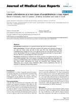
Posterior sub-capsular cataract in the right (A) and left eye (B) in patient 1Figure 1
Posterior sub-capsular cataract in the right (A) and left eye (B) in patient 1.
Posterior sub-capsular cataract in the right eye in patient 2Figure 2
Posterior sub-capsular cataract in the right eye in
patient 2.
Journal of Medical Case Reports 2008, 2:124 />Page 3 of 4
(page number not for citation purposes)
known, however, habitual tapping and rubbing of the
face, a common problem in pruritic conditions, may play
a role [3]. Indeed, the presence of facial skin lesions in AD
correlates with progression of the cataract [3]. An alterna-
tive hypothesis suggests that the cataract is secondary to
compromise of the blood-aqueous barrier. Patients with
AD have been found to have higher levels of protein flare
in the aqueous humour than controls [4]. Posterior sub-
capsular cataracts may also be caused by corticosteroids
used in the treatment of AD.
The association between systemic corticosteroids and pos-
terior sub-capsular cataracts was first noted by Black et al.
[5]. Subsequent studies have shown that corticosteroid-
induced cataracts may develop following even small doses
of steroids, particularly in children. Posterior sub-capsular
cataract may occur at a faster rate and lower dosage in chil-
dren [6]. In addition to systemic steroids, cataracts have
also been associated with ocular topical steroids, inhaled
steroids and topical steroid creams [7,8]. When steroids
are applied topically to the skin, the degree of systemic
absorption depends on factors such as drug potency, the
duration of application and whether the skin is thin or
damaged. Even low potency steroid creams applied to the
eyelids may result in increased intra-ocular pressure and
cataract [9].
The mechanism of corticosteroid-induced cataract is not
known but may be due to osmotic imbalance, oxidative
damage or disrupted lens growth factors [2]. The osmotic
theory suggests that corticosteroids interfere with the
ionic composition of the lens. The oxidative theory pro-
poses that corticosteroids inhibit the normal mechanisms
that protect the lens from oxidative stress. Another theory
of cataract formation proposes that steroids influence
lens-related growth factors. Normal lens growth is medi-
ated by growth factors such as fibroblast growth factor-2
present in the aqueous and vitreous humour [2]. Corticos-
teroids may influence lens epithelial cell behaviour by
interfering with the normal production of growth factors.
This effect may result in undifferentiated anterior epithe-
lial cells migrating and accumulating at the posterior pole
forming a posterior sub-capsular cataract [2].
Cataract extraction in children with atopic cataract can
produce excellent visual results, however, it is important
to consider the presence of a coexisting retinal detach-
ment. Retinal detachment has been reported in 8% of
patients with AD and in one series, 25% of eyes with
atopic cataract had retinal breaks or detachment noted
pre-operatively [10]. A rapidly progressing cataract may
mask the presence of a shallow retinal detachment and an
unrecognised retinal detachment can cause a mild cataract
to progress faster. B-scan ultrasonography is a useful
investigation to evaluate the anatomy of the retina in
these eyes. Retinal detachment may also be associated
with panuveitis or hypotony [11].
Conclusion
Corticosteroids are known to cause posterior sub-capsular
cataract by most routes of administration. After diabetes,
myopia and glaucoma, steroid use is the fourth leading
risk factor for secondary cataract and accounts for 4.7% of
all cataract extractions [2]. Care needs to be taken when
prescribing corticosteroids, particularly in children who
have a lower susceptibility to side effects. Corticosteroids
are frequently required to adequately treat AD, to limit
pruritus and prevent complications such as keratitis that
can lead to permanent visual loss. Exacerbations of AD
need to be treated aggressively. Many patients and parents
have negative perceptions regarding the use of steroids,
which may lead to inadequate treatment of the skin dis-
ease and increased eye rubbing. The increasing use of
alternative specific immunosuppressants that lack the side
effect profile of corticosteroids may reduce the incidence
of cataracts in these patients.
Abbreviations
AD: atopic dermatitis.
Competing interests
The author declares that they have no competing interests.
Authors' contributions
AT examined the patients, conducted the literature review
and wrote the manuscript.
Consent
Written informed consent was obtained from the patients'
parents for publication of this case report and accompany-
ing images. A copy of the written consent is available for
review by the Editor-in-Chief of this journal.
References
1. Carmi E, Defossez-Tribout C, Ganry O: Ocular complications of
atopic dermatitis in children. Acta Derm Venereol 2006,
86:515-517.
2. Jobling AI, Augusteyn RC: What causes steroid cataracts? A
review of steroid-induced posterior subcapsular cataracts.
Clin Exp Optom 2002, 85:61-75.
3. Nagaki Y, Hayasaka S, Kadoi C: Cataract progression in patients
with atopic dermatitis. J Cataract Refract Surg 1999, 25:96-99.
4. Matsuo T, Saito H, Matsuo N: Cataract and aqueous flare levels
in patients with atopic dermatitis. Am J Ophthalmol 1997,
124:36-39.
5. Black RL, Oglesby RB, von Sallman L, Bunim JJ: Posterior subcapsu-
lar cataracts induced by corticosteroids in patients with
rheumatoid arthritis. JAMA 1960, 174:166-171.
6. Kaye LD, Kalenak JW, Price RL, Cunningham R: Ocular implica-
tions of long-term prednisolone therapy in children. J Pediatr
Ophthalmol Strabismus 1993, 30:142-144.
7. McLean CJ, Lobo RF, Brazier DJ: Cataracts, glaucoma and femo-
ral avascular necrosis caused by topical corticosteroid oint-
ment. Lancet 1995, 345:330.
8. Cumming RG, Mitchell P, Leeder SR: Use of inhaled corticoster-
oids and the risk of cataracts. N Engl J Med 1997, 337:8-14.
Publish with BioMed Central and every
scientist can read your work free of charge
"BioMed Central will be the most significant development for
disseminating the results of biomedical research in our lifetime."
Sir Paul Nurse, Cancer Research UK
Your research papers will be:
available free of charge to the entire biomedical community
peer reviewed and published immediately upon acceptance
cited in PubMed and archived on PubMed Central
yours — you keep the copyright
Submit your manuscript here:
/>BioMedcentral
Journal of Medical Case Reports 2008, 2:124 />Page 4 of 4
(page number not for citation purposes)
9. Garrott HM, Walland MJ: Glaucoma from topical corticoster-
oids to the eyelids. Clin Experiment Ophthalmol 2004,
32(2):224-226.
10. Hayashi H, Igarashi C, Hayashi K: Frequency of ciliary body or
retinal breaks and retinal detachment in eyes with atopic
cataract. Br J Ophthalmol 2002, 86:898-901.
11. Lim WK, Chee SP: Retinal detachment in atopic dermatitis can
masquerade as acute panuveitis with rapidly progressive cat-
aract. Retina 2004, 24:953-956.