Báo cáo y học: " Morphologically and immunohistochemically undifferentiated gastric neoplasia in a patient with multiple metastatic malignant melanomas: a case report" pot
Bạn đang xem bản rút gọn của tài liệu. Xem và tải ngay bản đầy đủ của tài liệu tại đây (345.25 KB, 5 trang )
BioMed Central
Page 1 of 5
(page number not for citation purposes)
Journal of Medical Case Reports
Open Access
Case report
Morphologically and immunohistochemically undifferentiated
gastric neoplasia in a patient with multiple metastatic malignant
melanomas: a case report
Federico Alghisi
1
, Pietro Crispino
2
, Andrea Cocco
1
, Antonio G Richetta
2
,
Francesco Nardi
3
, Paolo Paoluzi
1
and Danilo Badiali*
1
Address:
1
Gastroenterology Unit, Department of Clinical Sciences, Policlinico Umberto I, University 'La Sapienza', Viale del Policlinico, 00161
Rome, Italy,
2
Department of Dermatology, University 'La Sapienza', Rome, Italy and
3
Department of Pathology, University 'La Sapienza', Rome,
Italy
Email: Federico Alghisi - ; Pietro Crispino - ; Andrea Cocco - ;
Antonio G Richetta - ; Francesco Nardi - ; Paolo Paoluzi - ;
Danilo Badiali* -
* Corresponding author
Abstract
Introduction: Malignant melanoma is a neoplasia which frequently involves the gastrointestinal
tract (GIT). GIT metastases are difficult to diagnose because they often recur many years after
treatment of the primary cutaneous lesion and also manifest clinically at an advanced stage of the
neoplasia. Furthermore, GIT metastases can appear in various morphological forms, and therefore
immunohistochemistry is often useful in distinguishing between a malignant melanoma and other
malignancies.
Case presentation: We report the case of a 60-year-old man with a multiple metastatic
melanoma who underwent an upper endoscopy to clarify the possible involvement of the gastric
wall with a mass localized in the upper abdomen involving the pancreas and various lymph nodes,
which was previously described with computed tomography. Clinically, the patient reported a
progressive loss of appetite, nausea and vomiting. The upper endoscopy and histological
examination revealed a gastric location of an undifferentiated neoplasm with an absence of
immunohistochemical characteristics referable to the skin malignant melanoma that was removed
previously.
Conclusion: The present case report shows the difficulty in diagnosing a metastatic melanoma in
the GIT and therefore, it seems worthwhile to consider metastatic malignant melanoma in the
differential diagnosis of undifferentiated neoplasia.
Introduction
Melanoma is one of the most common neoplasia. The
incidence of melanoma has increased in the last three dec-
ades; in the United States it was estimated as 5.7 cases per
100,000 people in 1973 and has increased dramatically to
14.3 cases per 100,000 people in 1998 [1]. Meanwhile,
the overall survival rate has mildly improved: the 5-year
survival rate was 80.0% in the 1970s and it achieved
Published: 30 April 2008
Journal of Medical Case Reports 2008, 2:134 doi:10.1186/1752-1947-2-134
Received: 29 August 2007
Accepted: 30 April 2008
This article is available from: />© 2008 Alghisi et al; licensee BioMed Central Ltd.
This is an Open Access article distributed under the terms of the Creative Commons Attribution License ( />),
which permits unrestricted use, distribution, and reproduction in any medium, provided the original work is properly cited.
Journal of Medical Case Reports 2008, 2:134 />Page 2 of 5
(page number not for citation purposes)
88.8% by the end of the last century. This probably
reflects an increased disease incidence, as well as earlier
diagnosis of melanoma and better therapeutic options
developed during the last few decades [1].
Melanoma originates most frequently in the skin. Other
possible, but less-frequent, primary locations are intraoc-
ular, subungueal and mucosal sites. After treatment of the
primary lesion, melanoma recidivates in about one-third
of patients, involving almost every major organ and tis-
sue. The most common sites of metastases are the skin,
lung and brain. Metastases in the gastrointestinal tract
(GIT) are not rare, however, they are less frequent than the
above-mentioned sites and they usually manifest clini-
cally at an advanced stage of the neoplasia. Diagnosis
requires careful inspection of the mucosa for metastatic
lesion detection and biopsy and the use of special immu-
nohistochemical stains [2].
The overall median survival time in patients with meta-
static melanoma is 7.5 months with a 5-year survival rate
of 6%. Patients with GIT metastases have a median sur-
vival time of 12.5 months with a 5-year survival rate of
14%. Survival is strictly related to three independent vari-
ables: (i) the initial site of metastases (p < 0.0001); (ii) the
interval between treatment of the primary lesion and
onset of metastases, the disease-free interval (DFI) (p =
0.0001); and (iii) the stage of disease preceding distant
metastases (p = 0.0001). To date the preferred treatment
choice for GIT metastases remains surgery. Surgery
improves the survival rate significantly, especially when
the resection is considered complete following micro-
scopic examination. The median survival after complete
resection is 48.9 months, compared with 5.4 months after
an incomplete resection [3]. Surgery is also recommended
for palliative treatment of GIT metastases, with symptom
relief reported in the range of 77% to 100% of patients,
depending on the site and the reason for resection.
Case presentation
We report the case of a 60-year-old man with multiple
metastatic melanoma, who presented to our unit with
vomiting and was later diagnosed with a gastric neoplasia
with no histological and immunohistochemical charac-
teristics referable to a malignant melanoma.
He underwent a surgical excision of a cutaneous lesion,
localized on the left sub-costal region. The histological
findings suggested a melanocytic melanoma with fused
cells, of nodular type, exceeding the reticular layer of
derma (pT4a), of Breslow thickness 8.3 mm and Clark
level IV. Two months later the patient underwent an axil-
lary lymph node dissection with no histological evidence
of nodal metastases. Furthermore, he was treated with six
cycles of chemotherapy with dacarbazine and IL-2. Four
months after concluding chemotherapy the patient
underwent a total-body computed tomography (CT) scan
revealing three low-density lesions on the II, III and VIII
segment of the liver, which remained uninvestigated. The
CT scan also revealed one nodule (<1 cm) on the apical
segment of the right lung and one sub-pleural nodule (1
cm) on the basal segment of the left lung, accompanied by
a thickened contiguous pleura. Due to these findings, the
patient underwent a thoracotomy, but the histological
examination only revealed the presence of fibrotic tissue.
During follow-up, the clinical condition of the patient
remained stable for six months until the appearance of
multiple masses localized on the left arm, both axillae and
at the level of the primary melanoma surgical scar. The
patient underwent a total-body bone scintigraphy that
showed an increased concentration of the radioactive
tracer (99mTc-MDP) in the left collarbone, II and VII right
ribs, IV left rib, L2 and left acetabulum. A total-body CT
scan showed multiple intra-peritoneal subcutaneous nod-
ules, two metastatic lesions in the liver (IV and V seg-
ment), one in the spleen (1 cm) and one in the pancreas
corpus (2 cm). A positron emission tomography (PET)
scan confirmed the presence of multiple skeletal, muscu-
lar and nodal repetitive lesions. A treatment of three cycles
of Interferon 5MU three times a week and Temozolomide
150 mg/m
2
/day 5 days a week for 4 weeks was started.
However, the treatment was suspended after the second
cycle due to side effects. The patient required admission to
hospital due to his worsened clinical state, which
included progressive asthenia, muscle and skeletal pain,
nausea and vomiting. Five months after the last investiga-
tion the patient underwent a total-body CT again which
confirmed the presence of multiple subcutaneous, muscu-
lar and nodal repetitive lesions; in addition, the CT scan
revealed a gross lesion in the upper abdomen, probably
due to confluent lymph nodes, undistinguishable from
the gastric corpus and pancreas. Therefore, the patient was
referred to our unit to evaluate the gastric wall involve-
ment and its role in causing obstruction and vomiting.
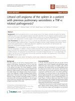
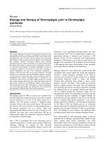
The patient underwent an upper endoscopy that showed
a prominent mass at the passage between the fundus and
the corpus of the stomach, with a hard consistency and
largely covered by fibrin; the diameter of the lesion was
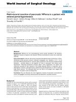
about 3 cm (Figure 1). Histological findings suggested an
undifferentiated neoplasia constituted prevalently by neo-
plastic fused cells (Figure 2). The immunohistochemical
stains for melanoma (S100, tyrosinase, Melan A and
HMB-45), carcinoma (CK), gastrointestinal stromal
tumours (CD-34, vimentin and c-Kit) and lymphoma
(LCA) were performed with negative results, except for a
weak and focal expression of vimentin. No treatment for
the gastric mass was started because of the patient's com-
promised clinical condition. Enteral nutrition was main-
Journal of Medical Case Reports 2008, 2:134 />Page 3 of 5
(page number not for citation purposes)
tained until the patient's death, a month after the
endoscopy.
Discussion
Malignant melanoma is very likely to produce regional
lymph node and distant metastasis. GIT metastases are
frequent but rarely diagnosed. In fact, only 1% to 4% of
GIT metastases are clinically diagnosed ante mortem in
patients affected by malignant melanoma, while the fre-
quency of GIT metastases is more than 60% in autopsy
series. Moreover, melanoma is the most common meta-
static tumour to the GIT; autopsy studies reported that
23% of GIT metastases derived from malignant
melanoma. These data suggest that GIT metastases are dif-
ficult to diagnose, probably because symptoms are often
absent or non-specific. Moreover, symptoms may be due
to or modified by treatment of the primary tumour, such
as chemotherapy or radiotherapy [3]. Patients with GIT
metastases are usually investigated when they present
with anaemia, gross bleeding, obstruction, abdominal
pain or weight loss; these symptoms often arise in an
advanced stage of the disease [4]. Furthermore, the diag-
nosis of GIT metastases may be difficult because they
often occur many years after the primary cutaneous
lesion. It is reported that the DFI until the onset of GIT
metastases is 43.8 ± 11.3 months [4]. Metastases confined
only to the GIT are rare; in most cases, major organs are
already involved at the time of diagnosis. GIT metastases
often occur in multiple sites: small bowel (35% to 97%),
stomach and duodenum (5% to 50%), and colon (5% to
32%) [5]. There is a significant correlation between their
occurrence with the location and nodular type of the pri-
mary lesion. Some authors also consider an ulcerated pri-
mary lesion as a risk factor for developing GIT metastases.
Risk of recurrence is directly correlated to the stage of pres-
entation. In the absence of nodal or distant metastases,
stage depends on the thickness and the depth of the pri-
mary lesion, determined by two international standard-
ized indexes, the Breslow thickness and the Clark level [6].
A primary lesion with a thickness of between 0.76 and 1.5
mm has up to a 25% chance of developing a regional
lymph node recurrence within three years. If the thickness
is between 1.5 and 4 mm the risk of nodal recurrence is
more than 60% and 15% of these patients develop distant
metastases within five years from diagnosis [7]. Moreover,
the risk of GIT metastases is higher among patients with a
primary lesion classified as Clark level III or above, which
is found in 70% to 100% of such patients, although 5% to
24% of patients present with Clark level II and 0% to 6%
with Clark level I.
In our patient the primary lesion was found on the left
sub-costal region of the trunk. Staging at diagnosis sug-
gested an advanced melanoma, nodular type, exceeding
the reticular layer of derma, corresponding to a Clark level
IV and a Breslow thickness of 8.3 mm. According to the lit-
erature, our patient presented all of the major risk factors
for developing GIT metastases: the location of the primary
melanoma on the trunk, high Clark level and Breslow
thickness, and a nodular type of lesion. Only the gastric
metastasis was diagnosed but the presence of other GIT
metastases cannot be excluded because small bowel enter-
oscopy and colonoscopy were not performed. Three types
of malignant melanoma features were described at endos-
copy: ulcerated melanocytic nodules arising on normal
rugae, sub-mucosal masses with ulcerations, and mass
lesions with necrosis and melanosis. However, the neo-
plasia may be completely amelanotic and cytomorpho-
logically variable; in such cases immunohistochemical
Endoscopic appearance of the gastric neoplasiaFigure 1
Endoscopic appearance of the gastric neoplasia.
Histological pattern of the gastric neoplasiaFigure 2
Histological pattern of the gastric neoplasia. Microscopic
aspects reveal the presence of undifferentiated cells in which
fused cells similar to the primitive melanoma can be distin-
guished.
Journal of Medical Case Reports 2008, 2:134 />Page 4 of 5
(page number not for citation purposes)
stains, regardless of the presence or not of melanin pig-
ment, are needed to diagnose malignant melanoma. The
most sensitive markers are S100 protein and HMB-45 [8];
in the literature, the sensitivity of S100 varies between
33% and 100% while HMB-45 sensitivity varies between
80% and 97%, with a high specificity (100%) [9,10].
There are other immunohistochemical markers useful in
identifying the melanocytic origin of the neoplasia.
Melanocytes contain vimentin, an intermediate filament
usually expressed in primary and metastatic melanoma
cells. However, vimentin positivity can distinguish
melanoma from undifferentiated carcinoma, but not
from lymphoma or sarcoma [11]. The Melan A protein is
a melanocytic differentiation antigen, produced by the
MART-1 gene, and it is thought to be specific to melano-
cytic cells [12]. It was found to be a useful addition to anti-
body panels when describing cutaneous melanocytic
lesions [13]. Tyrosinase is an enzyme involved in the ini-
tial stages of melanin biosynthesis in melanocytic and
melanoma cells and its hyperexpression has been pro-
posed as a biochemical marker of melanoma [14].
Thus, a broader panel of immuno-markers may be useful
in distinguishing between metastases of malignant
melanoma and other metastatic malignancies when the
lesion is morphologically undifferentiated; Gupta et al.
[15], in fact, reported four cases of morphologically undif-
ferentiated melanoma that showed a positivity for
HMB45 (two cases), S100 (one case), vimentin (three
cases), NKI/C3 (two cases), NKI/Bteb (one case) and CK
(three cases). In our patient, the gastric neoplasia was
located in the upper third of the stomach, exactly at the
passage between fundus and corpus. The gastric neoplasia
appeared as a mass covered by fibrin and was amelanotic.
These macroscopic findings are similar to the characteris-
tics described in the literature. However, histological find-
ings showed an undifferentiated neoplasm and none of
the immunohistochemical stains for melanoma, includ-
ing S100, HMB-45, Melan A, tyrosinase, CK, CD-34, c-Kit
and LCA were able to clarify its origin. The peculiarity of
this report is that neither histology nor immunohisto-
chemistry were useful in diagnosing the origin of the
lesion, although other authors have reported that tumour
markers' loss of expression is not uncommon and this has
been experienced at various degrees in other cases of met-
astatic melanoma [16-18].
Unfortunately, biopsies were only obtained from the
stomach lesion; the histological examination of one or
more lesions outside of the stomach could have permitted
a better characterization of the neoplasia. However, the
histological finding of fused cells in the gastric lesion, as
featured in the primary melanoma, suggests the diagnosis
of GIT metastases. Furthermore, this hypothesis is also
supported by the clinical history, the presence of multiple
metastases and the occurrence of a neoplasia in the stom-
ach of a patient with all of the major risk factors for devel-
oping GIT metastases. Therefore, it is very likely that the
undifferentiated gastric neoplasia is a metastasis of the
malignant melanoma. Similar cases, characterized by a
completely negative immunohistochemistry, have not
been described in the literature.
Conclusion
The present case report shows the difficulty in diagnosing
a metastatic melanoma in the GIT, due to its insidious
clinical manifestations and morphologic and immuno-
histochemical variety. This evidence suggests that in a case
of melanoma, and during the follow-up and exploration
of any gastrointestinal tract disturbances, it is necessary to
screen for possible initial or occult metastasis. Therefore,
it seems worthwhile to consider metastatic malignant
melanoma in the differential diagnosis of undifferenti-
ated neoplasia of the GIT, even in the absence of positive
immunohistochemistry.
Competing interests
The authors declare that they have no competing interests.
Authors' contributions
FA, PC, AC and AR have made substantial contributions to
the conception and design, acquisition of data, or analysis
and interpretation of data. FN, PP and DB have been
involved in drafting the manuscript or revising it critically
and have given final approval of the version to be pub-
lished.
Consent
Written informed consent was obtained from the patient's
son for publication of this case report and accompanying
images. A copy of the written consent is available for
review by the Editor-in-Chief of the journal.
References
1. Ries LAG, Eisner MP, Kosary CL: SEER Cancer Statistic Review,
1973–98. 2001 [ />CSR1973_1998]. Bethesda, MD: National Cancer Institute
2. Liang KV, Sanderson SO, Nowakowski GS, Arora AS: Metastatic
malignant melanoma of the gastrointestinal tract. Mayo Clin
Proc 2006, 81:511-516.
3. Ollila DW, Essner R, Wanek LA, Morton DL: Surgical resection
for melanoma metastatic to the gastrointestinal tract. Arch
Surg 1996, 131:975-979.
4. Kobayashi O, Murakami H, Yoshida T, Cho H, Yoshikawa T, Tsub-
uraya A, Sairenji M, Motohashi H, Sugiyama Y, Kameda Y: Clinical
diagnosis of metastatic gastric tumors: clinicopathologic
findings and prognosis of nine patients in a single cancer cen-
tre. World J Surg 2004, 28:548-551.
5. Caputy GG, Donohue JH, Goellner JR, Weaver AL: Metastatic
melanoma of the GIT. Results of surgical management. Arch
Surg 1991, 126:1353-1358.
6. Reintgen DS, Thompson W, Garbutt J, Seigler HF: Radiologic,
endoscopic, and surgical considerations of melanoma meta-
static to the gastrointestinal tract. Surgery 1984, 95:635-639.
7. Ihde JK, Coit DG: Melanoma metastatic to the stomach, small
bowel, or colon. Am J Surg 1991, 162:208-211.
Publish with BioMed Central and every
scientist can read your work free of charge
"BioMed Central will be the most significant development for
disseminating the results of biomedical research in our lifetime."
Sir Paul Nurse, Cancer Research UK
Your research papers will be:
available free of charge to the entire biomedical community
peer reviewed and published immediately upon acceptance
cited in PubMed and archived on PubMed Central
yours — you keep the copyright
Submit your manuscript here:
/>BioMedcentral
Journal of Medical Case Reports 2008, 2:134 />Page 5 of 5
(page number not for citation purposes)
8. Balch CM, Buzaid AC, Soong SJ, Atkins MB, Cascinelli N, Coit DG,
Fleming ID, Gershenwald JE, Houghton A Jr, Kirkwood JM, McMasters
KM, Mihm MF, Morton DL, Reintgen DS, Ross MI, Sober A, Thomp-
son JA, Thompson JF: Final version of the American Joint Com-
mittee on Cancer staging system for cutaneous melanoma.
J Clin Oncol 2001, 19:3635-3648.
9. Marghoob AA, Koenig K, Bittencourt FV, Kopf AW, Bart RS: Bres-
low thickness and Clark level in melanoma: support for
including level in pathology reports and in American Joint
Committee on Cancer Staging. Cancer 2000, 88:589-595.
10. Blecker D, Abraham S, Furth EE, Kochman ML: Melanoma in the
gastrointestinal tract. Am J Gastroenterol 1999, 94:3427-3433.
11. Simmons TJ, Martin SE: Fine-needle aspiration biopsy of malig-
nant melanoma. A cytologic and immunocytochemical anal-
ysis. Diagn Cytopathol 1991, 7:380-386.
12. Caselitz J, Jänner M, Breitbart E: Malignant melanomas contain
only the vimentin type of intermediate filaments. Virchows
Arch A Pathol Anat Histopathol 1983, 400:43-51.
13. Chen YT, Stockert E, Jungbluth A, Tsang S, Koplan KA, Scanlan MJ,
Old LJ: Serological analysis of Melan-A (MART1), a melano-
cyte-specific protein homogeneously expressed in human
melanomas. Proc Natl Acad Sci USA 1996, 93:5915-5919.
14. Kwon BS: Pigmentation genes: the tyrosinase gene family and
the pmel 17 gene family. J Invest Dermatol 1993, 100:134S-140S.
15. Gupta RK, Lallu S: Cytodiagnosis of amelanotic metastatic
malignant melanoma: an immunocytochemical study. Diagn
Cytopathol 1997, 16:238-241.
16. Trefzer Hofmann M, Reinke S, Guo YJ, Audring H, Spagnoli G, Sterry
W: Concordant loss of melanoma differentiation antigens in
synchronous and asynchronous melanoma metastases:
implications for immunotherapy. Melanoma Res 2006,
16:137-145.
17. Gao Z, Stanek A, Chen S: A metastatic melanoma with an unu-
sual immunophenotypic profile. Am J Dermatopathol 2007,
29:169-171.
18. Aisner DL, Maker A, Rosenberg SA, Berman DM: Loss of S100 anti-
genicity in metastatic melanoma. Hum Pathol 2005,
36:1016-1019.