báo cáo khoa học: "Littoral cell angioma of the spleen in a patient with previous pulmonary sarcoidosis: a TNF-α related pathogenesis?" pot
Bạn đang xem bản rút gọn của tài liệu. Xem và tải ngay bản đầy đủ của tài liệu tại đây (1.35 MB, 4 trang )
CAS E REP O R T Open Access
Littoral cell angioma of the spleen in a patient
with previous pulmonary sarcoidosis: a TNF-a
related pathogenesis?
Stefanie Cordesmeyer
1,2*
, Manfred Pützler
3
, Ulf Titze
4
, Harald Paulus
5
and Matthias W Hoffmann
2
Abstract
Background: Littoral cell angioma (LCA) is a rare vascular tumor of the spleen. Generally thought to be benign,
additional cases of LCA with malignant features have been described. Thus, its malignant potential seems to vary
and must be considered uncertain. The etiology remains unclear, but an immune dysregulation for the apparent
association with malignancies of visceral organs or immune-mediated diseases has been proposed.
Case Presentation: We report a case of LCA in a 43-year old male patient who presented with a loss of appetite
and intermittent upper abdominal pain. Computed tomography showed multiple hypoattenuating splenic lesions
which were hyperechogenic on abdominal ultrasound. Lymphoma was presumed and splenectomy was
performed. Pathological evaluation revealed LCA.
Conclusions: LCA is a rare, primary vascular neoplasm of the spleen that might etiologically be associated with
immune dysregulation. In addition, it shows a striking association with synchronous or prior malignancies. With
about one-third of the reported cases to date being co-existent with malignancies of visceral organs or immune-
mediated diseases, this advocates for close follow-ups in all patients diagnosed with LCA. To our knowledge, this
report is the first one of LCA associated with previous pulmonary sarcoidosis and hypothesizes a TNF-a related
pathogenesis of this splenic tumor.
Keywords: Splenic tumor, littoral cell angioma, visceral organ malignancies, sarcoidosis, TNF - α
Background
Vascular tumors are the most common primary neo-
plasms of the spleen. Among these, LCA is a very rare
tumor which arises from the littoral cells lining the sinuses
ofthesplenicredpulp.Thetumordisplaysaunique
immunohistochemical phenotype of dual endothelial and
histiocytic differentiati on but i s difficult to differentiate
from other benign and malignant splenic lesions preopera-
tively. Thus, diagn osis is usually established after electi ve
splenectomy. Currently, both etiology as well as biological
behavior remain uncertain. Increasing numbers of LCA in
associa tion with autoimmune disorders or visceral organ
tumors have been reported which hypothesizes an immu-
nological association of this entity.
Case presentation
A 43-y ear old male presented with non-specific clinical
symptoms such as loss of appetite and intermittent upper
abdominal pain which improve d slightly with antacid
medication. His medical history was unremarkable except
for pulmonal sarcoidosis in his twenties which had been
treated with corticoste roid medication. Both physic al
examination and laboratory tests were without pathologi-
cal findings. An ulcerous lesion in the duodenum was
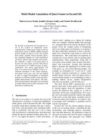
detected gastroscopically and computed tomography wa s
performed to exclude an external compressing tumor. CT
scans (Figure 1) did not de tect any tumor but revealed
mild splenomegaly with multiple hypoattenuating nodules
with a maximum diameter of 2.5 cm which were contrast-
enhancing in the late portal venous phase.
Abdominal ultrasound revealed multiple hyperecho-
genic splenic lesions without evidence of metastatic dis-
ease or infectious origin and hemangioma was assumed.
* Correspondence:
1
Department of Transplantation Medicine, University Hospital, Albert-
Schweitzer Campus 1, 48149 Münster, Germany
Full list of author information is available at the end of the article
Cordesmeyer et al. World Journal of Surgical Oncology 2011, 9:106
/>WORLD JOURNAL OF
SURGICAL ONCOLOGY
© 2011 Cordesmeyer et al; licensee BioMe d Central Ltd. This is an Open Access article distributed under the terms of the Creative
Commons Attribution License ( w hich permits unrestricted use, distribution, and
reproduction in any medium, provided the origina l work is properly cited.
Despite the absence of adenopathy, lymphoma was finally
considered the most likely diagnosis, given the quantity
of the nodules as well as their distribution within the
spleen and splenectomy was advocated. After appropriate
preoperative vaccination for Streptococcus pneumoniae,
Haemophilus influenca B and Neisseria meningitidis,
laparoscopic splenectomy was performed. The cut sur-
faceofthe12×8×4cmspecimen(Figure2)showed
multiple nodular lesions with spongy appearance, varying
from 0.5 to 2 cm in greatest dimension.
Microscopically, these lesions were composed of
cavernous sinuses which were lined by a single layer of
tall cells (Figure 3) lacking typical endothelial features.
The lacunae were filled with edemat ous fluid and blood
(Figure 4). Immunohistochemical staining (Figure 5) was
positive for both endothelial (CD 31) and histiocytic
(CD 68) markers. No cytologic atypia and mitotic figures
were found.
The combina tion of morpho logical and immunohisto-
chem ical analysis presenting this hybrid endothelial-his-
tiocytic phenotype established the diagnosis of LCA.
The postoperative course was uneventful and the patient
was discharged on day five. He will be followed-up clo-
sely for the occurrence t of visceral neoplasms.
Discussion
Littoral cell angioma (LCA) is a rare vascular tumor that
occurs exclusively in splenic tissue and was first
described by Falk et al in 1991 [1]. It ori ginates from the
specialized endothelial cells lining the sinus channels of
the splenic red pulp, called “ littoral cells”.LCAshow
neither gender nor age predilection. It might be discov-
ered incidentally i n completely asymptomatic patients or
in those presenting with non-specific clinical symptoms
like in our case. About 50% of all patients show spleno-
megaly or signs of hypersplenism li ke anemia or throm-
bocytopeni a [1]. On ultras ound, the findings vary widely
from heterogeneous echotexture without specific nodules
[2] to hyperechogenic [ 3], hypoechogenic [4] or isoecho-
genic [5] appearing lesions [6-8]. Computed tomography
typically shows multiple hypoattenuating nodules [9].
These findings are non-specific and several differential
diagnosis have to b e considered. These include benign
neoplasms like hamartoma or hemangioma but also
metastatic diseases or dissemi nated infections [2]. Since
Figure 1 Computed tomography showing multipl e
hypoattenuating lesions (arrows).
Figure 2 Sp lenectomy-specimen revea ling multiple nodular
lesions with spongy appearance (arrows).
Figure 3 Neoplastic sinuses lined by a single cell layer (20×
obj, HE-staining).
Cordesmeyer et al. World Journal of Surgical Oncology 2011, 9:106
/>Page 2 of 4
our patient did not present with adenopathy or disea se in
other organs, metastatic disease was considered an unli-
kely diagnosis. Considering disseminated infections, we
had to exclud e fungal disease, septic emb oli and granulo-
matous diseases such as sarc oidosis a nd tuberc ulosis.
Ass ociated adenopathic, pulmonary and mediastinal dis-
eases suspecting sarcoidosis or tuberculosis were not
detected. Mycoba cterium a vium-intrace llulare co mplex,
Pneumocystic carinii and disseminated Kaposi sarcoma
may also cause splenic masses but are typically seen in
immunocompromised individuals. After elaborating for
these differentials, a definite diagnosis is often still diffi-
cult to obtain and splenectomy is subsequently per-
formed for further evaluation. Gross pathology typically
shows multiple f ocal blood-f illed nodules and micro-
scopic examination reveals anastomosing vascular
channels lined with tall endothelial cells and papillary
fronds [1,7,9].
Since the littoral cells have features intermediate
between those of endothelial cells and macrophages,
they show a hybrid endothelial- histiocytic phenotype on
immunohistochemical staining. Expression of endothelial
marker factor VIII-related antigen and also of histiocytic
markers such as CD68 and lysozyme is thought to be
characteristic for LCA [1,6,7] and establishes the final
diagnosis.
Generally thought to be benign, there are additional
reports of LCA with malignant features which were
divided into a low-grade variant (littoral cell heman-
gioendothelioma [10]) and the tumor’s malignant coun-
terpart, littoral cell angiosarcoma [11]. Therefore, its
biological behavior seems to vary and must be consid-
ered uncertain [9].
The etiology of this neoplasm also remains unclear, but
for its apparent association with visceral organ tumors
and immune-mediated diseases in one-third of the
reported cases to date [12-16 ], an e tiological association
with immune dysregulation has been proposed [1,17,18].
To support this c ontention, there ar e more reports of
LCA in patients with long-term immunosupressive ther-
apy, i.e. after renal transplantation [18] or f or systemic
lupus erythematosus [19]. Reviewing literature for simila-
rities of immune-mediated diseases and LCA we found
two cases of LCA in patients with Gaucher ’ s disease
[20,21], a lipid storage disorder characterized by accumu-
lation of cerebroside in th e cytoplas m of macrophages
due to deficiency of an enzyme, glucocerebrosidase
[20,21]. Both LCA and Gaucher’ s disease involve lyso-
zymes. Since the likelihood of chance occurrence of two
rare disorders in one patient is low, Gupka et al sug-
gested a possible pathophysiologic association [20].
Since our patient had a history of pulmonal sarcoido-
sis, we concentrated on possible immunological links
between these two entities.
In sarcoidosis, a multisystemic granulomatous disorder
of unknown origin, the inflammatory response is charac-
terized by the increased p roduction of several pro-
inflammatory cytokines which mainly b elong to the
tumor necrosis factor family. Tumor necrosis factor-
alpha (TNF-a) is considered the pivotal factor in the
formation of granulomas by mediating inflammation
and cellular immune response among the cytokines
involved [22]. TNF-a is released by macrophages and
binds to two types of receptors, the 55 kDa (TNF recep-
tor I: TNF RI) and the 75 kDa receptor (TNF RII) [23].
Elevated serum levels of these receptors have been
demonstrated in a variety of diseases, e.g. rheumatic dis-
eases, malignancies and Crohn’s disease, and are thought
to reflect the disease activity. Since LCA is a neoplasm
arising from the lining cells, the spleen’ s macrophages,
Figure 4 Regul ar sple nic pare nch yma (r ight) a nd tumor (l eft)
composed of lacunae filled with oedematous fluid and blood
(4× obj, HE-staining).
Figure 5 Combined ex pression of endothelial (CD31) and
histiocytic (CD68) markers in immunohistochemical staining.
Cordesmeyer et al. World Journal of Surgical Oncology 2011, 9:106
/>Page 3 of 4
this could also be an area of increased production of
TNF-a, eventually contributing to t he pathogenesis of
LCA, since inflammatory cells including TNF-a are
known to have powerful effects on tumor development,
producing an attractive environment for tumor growth
by facilitating genomic instability and promoting angio-
genesis. The inf lammatory cells as well as the chemo-
kines and c ytokines they produce finally influence the
whole tumor organ, regulating the growth, migration
and differentiation of all cell types in the tumor micro-
environment, including neoplastic cells, fibroblasts and
endothelial cells [24]. Thus, a TNF-a related pathogen-
esis of LCA could also provide an explanation for both
the occurrence of synchronous or metachronous visceral
organtumorsaswellastheaffectionforimmune-
mediated diseases.
Conclusions
LCA is a rare, primary vascular neoplasm of the spleen.
Currently, both etiology and biological behavior remain
unclear, but an underlying immune dysregulation h as
been proposed for LCA’ s associ ation with malignancies
of visceral organs or immune-mediated disorders in
about one-third of the reported cases. Our case presenta-
tion supports the assumption of an association of LCA
and an altered immune status, hypothesizing a TNF-a-
related pathogenesis of this splenic tumor. Close follow-
upsofpatientsdiagnosedwithLCAforsubsequent
development of additional tumors is mandatory.
Consent
Written informed conse nt was obtained from the patient
for publication of this case report and any accompanying
images. A copy of the written consent is available for
review by the Editor-in-Chief of this journal.
Author details
1
Department of Transplantation Medicine, University Hospital, Albert-
Schweitzer Campus 1, 48149 Münster, Germany.
2
Department of General and
Visceral Surgery, Raphaelsklinik, Loerstraße 23, 48143 Münster, Germany.
3
Department of Radiology, Raphaelsklinik, Loerstraße 23, 48143 Münster,
Germany.
4
Department of Pathology, University Hospital, Albert-Schweitzer
Campus 1, 48149 Münster, Germany.
5
Internal Medicine, Private Practice,
Himmelreichallee 37, 48149 Münster, Germany.
Authors’ contributions
SC reviewed relevant literature and wrote the initial draft. MP reviewed the
draft and contributed the CT scans. UT contributed the histological images.
HP provided clinical expertise and reviewed the manuscript. MWH
performed the surgery and reviewed the manuscript. All authors read and
approved the final manuscript.
Competing interests
The authors declare that they have no competing interests.
Received: 14 April 2011 Accepted: 19 September 2011
Published: 19 September 2011
References
1. Falk S, Stutte HJ, Frizzera G: Littoral cell angioma: a novel splenic vascular
lesion demonstrating histiocytic differentiation. Am J Surg Pathol 1991,
15:1023-1033.
2. Kinoshita LL, Yee J, Nash SR: Littoral cell angioma of the spleen: imaging
features. AJR 2000, 174:467-469.
3. Espanol I, Lerma E, Fumanal V, et al: Littoral cell angioma with severe
thrombocytopenia. Ann Hematol 2000, 79:46.
4. Ziske C, Meyebehm M, Sauerbruch LGH, et al: Littoral cell angioma as a
rare cause of splenomegaly. Ann Hematol 2001, 80:45.
5. Oliver-Goldaracena JM, Blanco A, Miralles M, Martin-Gonzalez MA: Littoral
cell angioma of the spleen: US and MR imaging findings. Abdom Imaging
1998, 23:636-639.
6. Goldfield M, Cohen I, Loberant N, et al: Littoral cell angioma of the spleen:
appearance on sonography and CT. J Clini Ultrasound 2002, 30:510-513.
7. Levy AD, Abbott RM, Abbondanzo SL: Littoral cell angioma of the spleen:
CT features with clinicopathologic comparison. Radiology 2004,
230:485-490.
8. Bhatt S, Huang J, Dogra V: Littoral cell angioma of the spleen. AJR
American J Roentgenol 2007, 188:1365-1366.
9. Abbott RM, Levy AD, Aguilera NS, Gorospe L, Thompson WM: From the
archives of the AFIP: primary vascular neoplasms of the spleen-
radiologic-pathological correlation. RadioGraphics 2004, 24:1137-1163.
10. Ben-Izhak O, Bejar J, Ben-Eliezer S, Vlodavsky E: Splenic littoral cell
haemangioendothelioma: a new low-grade variant of malignant littoral
cell tumour. Histopathology 2001, 39:469-475.
11. Rosso R, Paulli M, Gianelli U, Boveri E, Stella G, Magrini U: Littoral cell
angiosarcoma of the spleen. Case report with immunohistochemical and
ultrastructural analysis. Am J Surg Pathol 1995, 19:1203-1208.
12. Bisceglia M, Sickel JZ, Giangaspero F, Gomes V, Amini M, Michal M: Littoral
cell angioma of the spleen: an additional report of four cases with
emphasis on the association with visceral organ cancers. Tumori 1998,
84:595-599.
13. Collins GL, Morgan MB, Taylor FM: Littoral cell angiomatosis with poorly
differentiated adenocarcinoma of the lung. Ann Diagn Pathol 2003,
7:54-59.
14. Lin CH, Yu JC, Shih ML, Peng YJ, Hsieh CB: Littoral cell angioma of the
spleen in a patient with hepatocellular carcinoma. J Formos Med Assoc
2005, 104:282-285.
15. Ercin C, Gurbuz Y, Hacihanefioglu A, Turgut Karakaya A: Multiple littoral cell
angioma of the spleen in a case of myelodysplastic syndrome.
Hematology 2005, 10:141-144.
16. Hansen T, Habekost M, Flieger D, Kirkpatrick CJ: Littoral cell angioma of
the spleen: Association with colon and hepatocellular carcinoma.
Pathologe 2010, 31(4):290-292.
17. Tholouli E, Roulson JA, Byers R, et al: Littoral cell angioma of the spleen in
a patient with severe aplastic anemia. Haematologica 2003, 88:11, ECR33.
18. Tan YM, Chuah KL, Wong WK: Littoral cell angioma of the spleen. Ann
Acad Med Singapore 2004, 33:524-526.
19. Gupka MK, Levin M, Aguilera NS, Pastores GM: Littoral cell angioma of the
spleen in a patient with Gaucher disease. Am J Hematol 2001, 68:61-62.
20. Forest F, Duband S, Clemenson A, Peoc’hM:Traumatic subcapsular
splenic hematoma revealing littoral cell angioma and Gaucher’s disease.
Ann Hematol 2010, 89(10):1061-1062.
21. Mac New HG, Fowler CL: Partial splenectomy for littoral cell angioma.
Journal of Pediatric Surgery 2008, 43:2288-2290.
22. Ziegenhagen MW, Müller-Quernheim J: The cytokine network in
sarcoidosis and its clinical relevance. Journal of Internal Medicine 2003,
253:18-30.
23. Brockhaus M, Schoenfeld HJ, Schlaeger EJ, Hunziker W, Lesslauer W,
Loetscher H: Identification of two types of tumor necrosis factor
receptors on human cell lines by monoclonal antibodies. Proc Natl Acad
Sci USA 1990, 87:3127-3131.
24. Coussens LM, Werb Z: Inflammation and cancer. Nature 2002,
420(6917):860-867.
doi:10.1186/1477-7819-9-106
Cite this article as: Cordesmeyer et al.: Littoral cell angioma of the
spleen in a patient with previous pulmonary sarcoidosis: a TNF-a
related pathogenesis? World Journal of Surgical Oncology 2011 9:106.
Cordesmeyer et al. World Journal of Surgical Oncology 2011, 9:106
/>Page 4 of 4