Báo cáo y học: " Scleroderma with crescentic glomerulonephritis: a case report" pot
Bạn đang xem bản rút gọn của tài liệu. Xem và tải ngay bản đầy đủ của tài liệu tại đây (902.21 KB, 5 trang )
BioMed Central
Page 1 of 5
(page number not for citation purposes)
Journal of Medical Case Reports
Open Access
Case report
Scleroderma with crescentic glomerulonephritis: a case report
Arunachalam Ramaswami*
1
, Thiraviam Kandaswamy
1
,
Tholappan Rajendran
1
, Kizhake Pisharam Jeyakrishnan
1
, Hla Aung
1
,
Mohammaed Iqbal
1
, Chakko K Jacob
1
, Haji Shaukat Zinna
1
and
Gazala Kafeel
2
Address:
1
Department of Nephrology, RIPAS Hospital, Bander Seri Begawan, Brunei Darussalam and
2
Department of Pathology, RIPAS Hospital,
Bander Seri Begawan, Brunei Darussalam
Email: Arunachalam Ramaswami* - ; Thiraviam Kandaswamy - ;
Tholappan Rajendran - ; Kizhake Pisharam Jeyakrishnan - ; Hla Aung - ;
Mohammaed Iqbal - ; Chakko K Jacob - ; Haji Shaukat Zinna - ;
Gazala Kafeel -
* Corresponding author
Abstract
Introduction: Systemic sclerosis or scleroderma is an autoimmune rheumatic disease
characterized by organ-based fibrosis. Renal involvement in scleroderma occurs mainly in the form
of scleroderma renal crisis, affecting 5 to 10% of patients. It remains one of the most important and
immediately life-threatening complications of scleroderma, but the prognosis improves
considerably after treatment with angiotensin-converting enzyme inhibitors. Other renal
pathologies can occur in scleroderma. These include scleroderma overlap syndromes with
associated features of lupus nephritis, myeloperoxidase anti-neutrophil cytoplasmic antibodies
(ANCA) or proteinase 3 ANCA-associated glomerulonephritis, or crescentic glomerulonephritis.
These alternative pathologies should be suspected in any individual patient with a differing clinical
picture and the patient should be appropriately investigated. Crescentic glomerulonephritis occurs
very rarely in scleroderma. This report describes a patient with scleroderma and crescentic
glomerulonephritis.
Case presentation: A 52-year-old woman with a known history of scleroderma and
hypertension on angiotensin-converting enzyme inhibitors was referred to the nephrologist
because of a rapid decline in renal function. Kidney biopsy was performed which revealed immune
complex type crescentic glomrulonephritis. Cytoplasmic-staining ANCA was negative. Despite
immunosuppressive treatment the patient rapidly went into end-stage renal failure and is still on
hemodialysis.
Conclusion: Scleroderma is a complex disease, and the best characterized renal involvement in
scleroderma is scleroderma renal crisis. However, other renal pathologies can occur in
scleroderma. These alternative pathologies should be suspected in any patient with a differing
clinical picture and the patient should be appropriately investigated, as the clinical course and
treatment are different from the more common scleroderma renal crisis.
Published: 13 May 2008
Journal of Medical Case Reports 2008, 2:151 doi:10.1186/1752-1947-2-151
Received: 6 July 2007
Accepted: 13 May 2008
This article is available from: />© 2008 Ramaswami et al; licensee BioMed Central Ltd.
This is an Open Access article distributed under the terms of the Creative Commons Attribution License ( />),
which permits unrestricted use, distribution, and reproduction in any medium, provided the original work is properly cited.
Journal of Medical Case Reports 2008, 2:151 />Page 2 of 5
(page number not for citation purposes)
Introduction
Scleroderma (systemic sclerosis) is a chronic systemic dis-
ease that targets the skin, lungs, heart, gastrointestinal
tract, kidneys and musculoskeletal system. The disorder is
characterized by three features: tissue fibrosis, small blood
vessel vasculopathy and a specific autoimmune response
associated with autoantibodies. Scleroderma is classified
into two major subsets, diffuse and limited cutaneous
sclerodermas, that are distinguished by the extent of skin
thickening. Diffuse scleroderma is characterized by wide-
spread skin thickening involving distal and proximal
body regions; rapid onset (within 1 year) of skin and
other features following appearance of Raynaud's phe-
nomenon; significant visceral involvement; high scores
on disability and organ damage indices secondary to
extensive fibrosis of tissues associated with antinuclear
antibodies; and the absence of anticentromere antibody.
Limited scleroderma shows limited skin thickening, slow
progression of disease and late visceral involvement, with
unique features of isolated pulmonary hypertension and
digital amputations associated with anticentromere anti-
body. Overlap syndromes have diffuse or limited sclero-
derma features plus features typical of one or more other
connective tissue or autoimmune diseases. Mixed connec-
tive tissue disease shows features of scleroderma, systemic
lupus erythematosus polymyositis, rheumatoid arthritis
and the presence of anti-U1 sn-RNP antibodies.
Approximately 10% of patients with scleroderma have a
renal crisis that mimics malignant hypertension, with rap-
idly progressive renal failure secondary to microvascular
disease, vasospasm and tissue ischemia. Microangiopathic
hemolytic anemia and thrombocytopenia can accompany
scleroderma renal crisis. Studies demonstrate high levels
of serum renin levels associated with vasospasm and
intrinsic renal vessel disease. A renal crisis is associated
with the use of corticosteroids or can be precipitated by
conditions compromising renal blood flow (dehydra-
tion). Any hypertension (> 140/90 mmHg) in a sclero-
derma patient should be carefully evaluated because a
renal crisis is potentially reversible with appropriate man-
agement with angiotensin converting enzyme (ACE)
inhibitors. Patients presenting with serum creatinine
above 270 μmol/l have a poor prognosis. Some patients
who progress to renal failure and dialysis can recover renal
function after months of dialysis therapy.
Variable changes may be seen in the glomeruli. In some
cases thickening of glomerular capillary walls with a dou-
ble contour appearance on silver or periodic acid-Schiff
staining may be seen. Fibrinoid necrosis may also be seen.
Crescents are very rare and those that are seen are invaria-
bly small. Interlobular arteries show intimal thickening
which is mucinous or finely fibrous. The thickening
results in a considerable reduction of the lumen.
Crescentic glomerulonephritis (GN) represents a severe
form of glomerular disease that is characterized by disrup-
tion of the glomerular basement membrane, leading to
cellular proliferation in the Bowman's space and is often
accompanied by fibrinoid necrosis. Crescentic GN is clas-
sified into three major types. Anti-glomerular basement
membrane (anti-GBM) disease is characterized by circu-
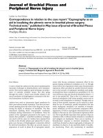
Photomicrograph of renal biopsy showing crescent formation and tuft narrowingFigure 2
Photomicrograph of renal biopsy showing crescent
formation and tuft narrowing. Periodic acid silver meth-
anamine stain.
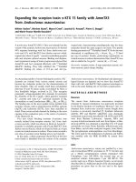
Photomicrograph of the renal biopsy showing prominent fibrocellular crescent formation and moderate mesangial proliferation in a glomerulusFigure 1
Photomicrograph of the renal biopsy showing promi-
nent fibrocellular crescent formation and moderate
mesangial proliferation in a glomerulus. Hematoxylin
and eosin stain.
Journal of Medical Case Reports 2008, 2:151 />Page 3 of 5
(page number not for citation purposes)
lating anti-GBM antibodies and linear deposition of anti-
bodies along the glomerular basement membrane. This
constitutes around 10% of cases. Pauci-immune (anti-
neutrophil cytoplasmic antibodies (ANCA)-associated
GN) is characterized by scanty glomerular deposits of
immunoglobulin and circulating ANCA, and comprises
about 60% of cases. Immune complex-mediated GN is a
heterogeneous group of diseases usually associated with
obvious granular deposits of immunoglobulins, in which
crescent formation complicates an identifiable form of
nephritis, usually proliferative in type. This constitutes
around 30% of cases. The causes of immune complex-type
crescentic GN include infection (including hepatitis C
virus (HCV) associated cryoglobulinemia), systemic
immune complex diseases (especially systemic lupus ery-
thematosus) and underlying pre-existing primary GN.
In a study of crescentic GN [1] the underlying etiology was
as follows: ANCA-associated vasculitis 37%; systemic
lupus erythematosus 23%; IgA nephropathy 12%; mesan-
giocapillary GN 6%; focal segmental GN 6%; anti-GBM
disease 6%; postinfectious GN 3%; membranous GN 2%;
focal segmental glomerulosclerosis 2%; Henoch Schon-
lein purpura 1%; others 3%.
The mainstay of therapy has remained immunosuppres-
sion with combinations including steroids, cyclophos-
phamide and azathioprine with or without
methylprednisolone, and plasma exchange [2]. In lupus
nephritis with crescents, cyclophosphamide is useful, and
mycophenolate mofetil is a useful alternative. In anti-
GBM disease and ANCA-associated nephritis with severe
renal failure plasma exchange is used. Plasma exchange
removes pathogenic autoantibodies but the process also
has an effect on inflammatory mediators and possibly
cell-mediated immune function.
Myeloperoxide ANCA-associated normotensive crescentic
GN in scleroderma has been reported from Japan. We
report a case of scleroderma with crescentic GN.
Case presentation
The patient is a 52-year-old Asian woman who presented
to the rheumatology department of our hospital in August
2003 with complaints of tightness of the skin over the face
and hands. She also had positive Raynaud's phenome-
non. She was diagnosed with scleroderma. She did not
have any difficulty swallowing. Fundus examination did
not reveal any evidence of accelerated hypertension.
Her investigations revealed ++ urine protein, ++ white
blood cell (WBC), 10 to 20 RBC/hpf (red blood cells per
high power field) and her 24-hour urine protein was 1.17
g. Her blood investigation revealed a WBC count of 7.5 ×
10
3
hemoglobin of 9.9 gm/dl, a platelet count of 378 ×
10
9
and erythrocyte sedimentation rate of 78 mm in the
first hour. Total bilirubin was 11 μmol/l, alanine transam-
inase was 10 units/l; she was HBSAg (hepatitis B surface
antigen) negative and anti-HCV antibody negative.
She was also antinuclear antibody (ANA) positive (1 in
640). Her blood urea was 7.9 mmol/l, S-Creatinine 135
μmol/l, sodium 141, and potassium 4.8. Anti-RO, -LA, -
Sm, and -RNP antibodies were negative. Anti-ds DNA
antibodies were negative. Lupus erythematosus cells were
negative. Anti-Scl70 antibodies were positive. Cytoplas-
mic-staining (C)-ANCA was negative. Perinuclear-stain-
ing (P)-ANCA was reported as 'unable to determine
because of positive ANA' (P-ANCA testing by immu-
noassay would have clarified this inconclusive result but
was unavailable to us). Rheumatoid factor was less than
8IU/ml. (reference range (RR) < 30IU/ml). Serum C3 was
0.76 g/l (RR 0.88 to 2.01 g/l). Serum C4 was 0.32 g/l (RR
0.16 to 0.58 g/l).
X-ray of the hands showed acro-osteolysis of the terminal
phalange of thumb and index finger. High-resolution
computed tomography of the lungs revealed prominent
bilateral sub-pleural honeycombing suggestive of usual
interstitial pneumonia. A Doppler study of the renal arter-
ies did not reveal any evidence of renal artery stenosis.
The patient was diagnosed with diffuse scleroderma,
treated symptomatically and given ACE inhibitors for
hypertension. In June 2004 she was referred to a nephrol-
ogist. At that time her investigations were as follows:
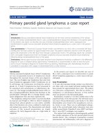
Photomicrograph of the renal biopsy showing prominent fibrocellular crescent formationFigure 3
Photomicrograph of the renal biopsy showing promi-
nent fibrocellular crescent formation. Chronic inflam-
matory cellular infiltration and fibrosis is also evident in the
interstitium. Periodic acid-Schiff stain.
Journal of Medical Case Reports 2008, 2:151 />Page 4 of 5
(page number not for citation purposes)
blood urea 22.7 mmol/l, serum sodium 137, serum potas-
sium 6.8, and serum creatinine of 555 μmol/l. Ultrasono-
gram examination of the abdomen revealed echogenic
kidneys of normal size. In view of the relatively rapid
decline in renal function and normal size kidneys a renal
biopsy was performed (Figures 1, 2 and 3).
A kidney biopsy was performed which showed 26 glomer-
uli. Most of the glomeruli (95%) showed prominent
fibrocellular and fibrous crescent formation and the
lesions were global. Capillary wall basement membrane
thickening and duplication with tram track appearance
and moderate mesangial proliferation were evident. The
glomerular hypercellularity was mainly mesangial with
occasional polymorphs with no necrotizing lesion. Five
glomeruli were completely sclerosed. The extraglomerular
renal vessels exhibited moderate thickening of the vessel
wall, particularly of its medial layer. Chronic inflamma-
tory cellular infiltration and fibrosis of interstitial tissue
were also seen. Immunoperoxide staining showed
marked positivity with ++ IgG, + IgM, ++ Focal C3 and
negative IgA. The staining pattern was granular in the
mesangium and peripheral capillary loops. The overall
impression was of crescentic GN with granular immune
deposits.
Therapy with methylprednisolone was not effective and
the patient went into end-stage renal failure and was initi-
ated on hemodialysis on 14 July 2004. She is still on
maintenance hemodialysis three times per week.
Discussion
Scleroderma is a connective tissue disorder with multi-
organ involvement. Numerous symptoms in scleroderma
such as Raynaud's phenomenon, digital tip necrosis and
angina are related to ischemic changes attributable to inti-
mal thickening of medium and small arterial vessels in the
absence of histopathologic changes of vasculitis. Renal
involvement in scleroderma is due to diminished blood
flow in the afferent arterioles and is called scleroderma
renal crisis. This includes acute deterioration of renal
function, hypertension, and hyperreninemia and absent
nephritic urinary sediment. It usually responds to ACE
inhibitors.
Histopathologically there is concentric edematous intimal
thickening of interlobular arteries and fibrinoid arteriolar
necrosis causing elevation of plasma renin level with
onset or aggravation of hypertension and rapid deteriora-
tion of renal function as a result of ischemic glomerulop-
athy [3]. There are well recognized co-occurrences of other
acute renal pathologies in scleroderma. This is especially
seen in scleroderma overlap syndromes with lupus
nephritis. In such cases there may be histological clues
such as development or rise in titer of anti-ds DNA anti-
bodies [1]. Crescentic GN is an uncommon cause of renal
failure in scleroderma but it has been observed in patients
treated with D-penicillamine. This agent is not widely
used but has been associated with antibody generation,
including ANCA.
Reports from Japan describe patients with long-standing
scleroderma and myeloperoxidase ANCA positive normo-
tensive crescentic GN [4]. The clinical features of patients
with crescentic GN are different from those with sclero-
derma renal crisis. The treatment requirements of these
two conditions also differ [5]. Those with crescentic GN
require immunosuppressive drugs such as methylpred-
nisolone and cyclophosphamide and they do not respond
to ACE inhibitors [6].
There is a recently described condition with features of
scleroderma called nephrogenic systemic fibrosis (neph-
rogenic fibrosing dermopathy) that develops in patients
with advanced kidney disease, apparently in association
with gadolinium exposure. Even though it is described
mainly in patients with end-stage renal disease (ESRD) on
dialysis, it can occur in individuals with a glomerular fil-
tration rate of <30 ml/minute. Due to this, restricted use
of gadolinium-enhanced MRI has been recommended.
Our patient does not have a history of exposure to gado-
linium.
Our patient presented with renal involvement within one
year of diagnosis of scleroderma. She did not respond to
ACE inhibitors because she did not have classical sclero-
derma renal crisis. She did not respond to immunosup-
pressive treatment with methylprednisolone. She rapidly
went into ESRD and became dialysis-dependent.
Although recovery from dialysis-dependent renal failure is
possible in crescentic nephritis, this has not occurred in
this patient. This case is presented for the rarity of crescen-
tic GN in scleroderma and the different clinical course
from scleroderma renal crisis.
Conclusion
Scleroderma is a complex disease with multisystem
involvement. The best characterized renal involvement in
scleroderma is acute or sub-acute renal hypertensive crisis
(scleroderma renal crisis), but other renal pathologies can
occur. These include scleroderma overlap syndromes with
features of lupus nephritis, myeloperoxidase ANCA, pro-
teinase 3 ANCA-associated GN or crescentic GN. These
alternative pathologies should be suspected in any indi-
vidual patient with a differing clinical picture and the
patient should be appropriately investigated because the
clinical course and treatment are different from the usual
scleroderma renal crisis.
Publish with BioMed Central and every
scientist can read your work free of charge
"BioMed Central will be the most significant development for
disseminating the results of biomedical research in our lifetime."
Sir Paul Nurse, Cancer Research UK
Your research papers will be:
available free of charge to the entire biomedical community
peer reviewed and published immediately upon acceptance
cited in PubMed and archived on PubMed Central
yours — you keep the copyright
Submit your manuscript here:
/>BioMedcentral
Journal of Medical Case Reports 2008, 2:151 />Page 5 of 5
(page number not for citation purposes)
Abbreviations
ACE: angiotensin converting enzyme; ANCA: anti-neu-
trophil cytoplasmic antibodies; anti-GBM: anti-glomeru-
lar basement membrane; ESRD: end-stage renal disease;
GN: glomerulonephritis; HCV: hepatitis C virus; RR: refer-
ence range; WBC: white blood cell.
Competing interests
The authors declare that they have no competing interests.
Authors' contributions
AR, TK, TR, KPJ, HA, MI, CKJ, HSZ wrote or contributed to
the writing of the manuscript. GK processed the kidney
biopsy specimen and wrote the biopsy report.
Consent
Written informed consent was obtained from the patient
for publication of this case report and accompanying
images. A copy of the written consent is available for
review by the Editor-in-Chief of this journal.
References
1. Crescentic glomerulonephritis. In Oxford Textbook of Clinical
Nephrology 3rd edition. Edited by: Davison AM, Jeremy Levy, Charles
D. Pusey Publisher Oxford University Press; 2005:561-570.
2. Chang A: Membranous and crescentic glomerulonephritis in
a patient with positive ANA and ANCA. Kidney Int 2007,
71:360-365.
3. Anders H-J, Wiebacke B, Haedecke C, Sanden S, Combe C, Scholen-
dorff D: MPO-ANCA-positive crescentic glomerulonephritis:
A distinct entity of scleroderma renal disease? Am J Kidney Dis
1999, 33(4):e3.
4. Kobayashi M, Saito M, Minoshima S, Arimura Y, Nagasawa T: A case
of progressive systemic sclerosis with crescentic glomeru-
lonephritis associated with MPO-ANCA and anti GBM Anti-
body. Nippon Jinzo Gakkai Shi 1995, 37:207.
5. Kamen DL, Wigley FM, Brown AN: ANCA positive crescentic
GN in scleroderma -a different kind of renal crisis. J Rheumatol
2006, 33:1886-1888.
6. Arnaud L, Huart A, Plauser F, Francois H, Mougenaut B, Tiev K, Kel-
taneh A, Ronco P, Rougier GP: ANCA-related crescentic
glomerulonephritis in systemic sclerosis: revisiting the "nor-
motensive scleroderma renal crisis". Clin Nephrol 2007,
68:165-170.