Báo cáo y học: "A step-by-step diagnosis of exclusion in a twin pregnancy with acute respiratory failure due to non-fatal amniotic fluid embolism: a case report" pps
Bạn đang xem bản rút gọn của tài liệu. Xem và tải ngay bản đầy đủ của tài liệu tại đây (226.84 KB, 4 trang )
BioMed Central
Page 1 of 4
(page number not for citation purposes)
Journal of Medical Case Reports
Open Access
Case report
A step-by-step diagnosis of exclusion in a twin pregnancy with acute
respiratory failure due to non-fatal amniotic fluid embolism: a case
report
Vasilios E Papaioannou, Christos Dragoumanis*, Vassiliki Theodorou,
Dimitrios Konstantonis and Ioannis Pneumatikos
Address: Department of Intensive Care Medicine, Alexandroupolis University Hospital, Democritus University of Thrace, Medical School, Dragana,
Alexandroupolis 68100, Greece
Email: Vasilios E Papaioannou - ; Christos Dragoumanis* - ;
Vassiliki Theodorou - ; Dimitrios Konstantonis - ; Ioannis Pneumatikos -
* Corresponding author
Abstract
Introduction: Respiratory failure may develop during the later stages of pregnancy and is usually
associated with tocolysis or other co-existing conditions such as pneumonia, sepsis, pre-eclampsia
or amniotic fluid embolism syndrome.
Case presentation: We present the case of a 34-year-old healthy woman with a twin pregnancy
at 31 weeks and 6 days who experienced acute respiratory failure, a few hours after administration
of tocolysis (ritodrine), due to preterm premature rupture of the membranes. Her chest
discomfort was significantly ameliorated after the ritodrine infusion was stopped and a Cesarean
section was performed 48 hours later under spinal anesthesia; however, 2 hours after surgery she
developed severe hypoxemia, hypotension, fever and mild coagulopathy. The patient was intubated
and transferred to the intensive care unit where she made a quick and uneventful recovery within
3 days. As there was no evidence for drug- or infection-related thromboembolic or myocardial
causes of respiratory failure, we conclude that our patient experienced a rare type of non-fatal
amniotic fluid embolism.
Conclusion: In spite of the lack of solid scientific support for our diagnosis, we conclude that our
patient suffered an uncommon type of amniotic fluid embolism syndrome and we believe that this
report highlights the need for extreme vigilance and a high index of suspicion for such a diagnosis
in any pregnant individual.
Introduction
Mild dyspnea is a common symptom during late preg-
nancy. However, some women experience severe respira-
tory distress before or immediately after labor. This could
be the result of co-existing conditions, such as asthma or
cardiovascular disease. Others may have an acute illness
such as pneumonia, pneumothorax or pulmonary embo-
lism. Finally, some pregnancies are complicated by pul-
monary edema of cardiac or non-cardiac origin. In a
previously healthy woman the first case is usually a drug-
related complication (mainly due to tocolysis). The latter
could be secondary to increased permeability of the pul-
Published: 27 May 2008
Journal of Medical Case Reports 2008, 2:177 doi:10.1186/1752-1947-2-177
Received: 9 January 2008
Accepted: 27 May 2008
This article is available from: />© 2008 Papaioannou et al; licensee BioMed Central Ltd.
This is an Open Access article distributed under the terms of the Creative Commons Attribution License ( />),
which permits unrestricted use, distribution, and reproduction in any medium, provided the original work is properly cited.
Journal of Medical Case Reports 2008, 2:177 />Page 2 of 4
(page number not for citation purposes)
monary vasculature, due to pre-eclampsia, septic shock,
placental abruption, major obstetric hemorrhage and
amniotic fluid embolism (AFE) syndrome [1].
We present a case of a previously healthy woman with a
twin pregnancy who at 31 weeks and 6 days experienced a
biphasic pattern of respiratory distress and pulmonary
edema, fever and coagulopathy, premature rupture of the
membranes (PROM) and use of tocolysis initially after
preterm labor and, subsequently, shortly after delivery. By
step-by-step exclusion of every possible cause of acute
lung injury, we concluded that this is a rare case of acute
respiratory failure due to AFE in a twin pregnancy.
Case presentation
A 34-year-old healthy woman with a twin pregnancy at 31
weeks and 6 days was admitted to our hospital with pre-
mature uterine contractions. She had no history of previ-
ous pregnancy, allergy or smoking. Vaginal examination
revealed the presence of pooled amniotic fluid on a sterile
speculum. Preterm PROM was diagnosed and a ritodrine
infusion was started at a dose of 0.10 to 0.3 mg/minute,
given in 1000 ml of normal saline, for 24 hours. At 24
hours, uterine contractions were arrested successfully, the
ritodrine infusion was tapered and oral ritodrine was
begun with 2 mg every 2 hours. She was also given dexam-
ethasone (two 12 mg doses) to improve fetal lung matu-
ration. Over the next 24 hours she became increasingly
breathless with a tachycardia of 140 beats/minute, blood
pressure of 110/70 mmHg, bilateral basal crackles and
temperature of 37.6°C. Cardiotocography (CTG) revealed
no signs of fetal distress. Pulmonary edema was diag-
nosed clinically and ritodrine administration was
stopped, while she responded to a bolus of intravenous
furosemide. Antibiotic treatment (amoxicillin/clavulanic
acid 1000 mg/100 mg four times a day intravenously and
erythromycin 1 g four times a day intravenously) was
started to prevent possible intrauterine infection and
nadroparin calcium (2850 IU once daily subcutaneously)
was added for venous thomboprophylaxis. On the suspi-
cion of an intrauterine infection an uneventful Cesarean
section was performed 48 hours later, under spinal
anesthesia, and the patient delivered healthy twins (Apgar
score: 9 and 8 at 1 minute and 10 at 5 minutes for both
neonates). As Cesarean section requires a T4 sensory level,
1.5 liters of normal saline was administered intravenously
prior to surgery and 1 liter during surgery.
A few hours after delivery the patient became acutely dys-
pnoeic with a respiratory rate of 35 breaths/minute and
bilateral rhonchi. Blood pressure was 75/45 mmHg. The
electrocardiogram showed a sinus tachycardia of 123
beats/minute. In spite of treatment with oxygen via nasal
spectacles (15 liters/minute), her arterial blood gas analy-
sis showed a severe hypoxemia with cyanosis (pH 7.46,
PaO
2
6.25 kPa, PaCO
2
3.99, bicarbonate 22 mmol/L). The
patient was intubated and transferred to the intensive care
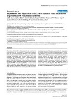
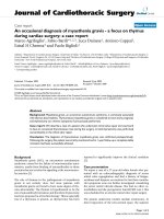
unit (ICU). A chest X-ray (Figure 1) revealed bilateral pul-
monary edema with pleural effusions while a spiral com-
puted tomography (CT) scan of the thorax supported the
above findings and excluded any case of pulmonary
embolism. A noradrenaline infusion was started at a low
rate (2 µg/minute) during initial resuscitation to support
blood pressure; noradrenaline infusion was gradually
reduced and stopped after 90 minutes as the patient's
hemodynamics stabilized. Central venous pressure was
12 mmHg under mechanical ventilatory support. An
echocardiogram showed good biventricular function with
normal chamber dimensions while there was no elevation
in cardiac enzymes. Duplex ultrasound scanning of the
lower extremities revealed no thrombosis of the femoral
and popliteal veins.
During her first day in the ICU the patient developed fever
(38.8°C), leucocytosis (17 × 10
9
/liter) and mild coagu-
lopathy (platelets 110 × 10
9
/liter, activated partial throm-
boplastin time 47 seconds, fibrinogen 140 mg/dl). A
serologic examination of pleural fluid was performed and
revealed no signs of exudate. Extensive cultures (blood,
sputum and vagina) remained negative, while C-reactive
protein (CRP) was increased (15 mg/dl). After the third
day of treatment, the patient made a quick recovery with
complete resolution of the pulmonary edema and she was
extubated 1 day later.
Discussion
Our patient matched well with the Clark criteria for AFE:
hypotension, pulmonary edema, cyanosis, coagulopathy,
dyspnea. However, the presence of PROM and prior rito-
drine toxicity complicated the clinical picture [2]. PROM
Acute bilateral pulmonary edema with pleural effusionsFigure 1
Acute bilateral pulmonary edema with pleural effusions.
Journal of Medical Case Reports 2008, 2:177 />Page 3 of 4
(page number not for citation purposes)
is defined as rupture of the chorioamniotic membranes
before the onset of labor. Maternal complications with
preterm PROM are more common, with chorioamnionitis
rates approximating 25% to 35% (see [3,4]).
Suppression of uterine contractions seems to be the obvi-
ous solution to the problem of preterm labor. Our patient
received ritodrine for tocolysis, which is a β
2
sympathom-
imetic agent and has been clearly shown to prolong preg-
nancy by 48 hours. There is a strong association between
its use and the development of maternal pulmonary
edema, especially with concomitant administration of
steroids. This complication has been reported in up to 9%
of cases and has been responsible for at least 15 maternal
deaths [5,6]. The initial tachycardia and tachypnea of the
patient was attributed to the ritodrine infusion, so the
drug was discontinued. Her clinical status was signifi-
cantly improved; however, antibiotics were administered
due to the risk of intrauterine infection.
Two days later and approximately 2 hours after the sched-
uled Cesarean section, the patient developed acute respi-
ratory distress and was transferred to the ICU. Acute
myocardial infarction was not supported by typical elec-
trocardiographic and echocardiographic changes and ele-
vated enzymes. Duplex scanning of both extremities
showed the absence of thrombosis, which made deep
venous thrombosis unlikely. The normal CT scanning
findings made pulmonary embolism less probable. Toco-
lytic therapy was also an unlikely cause because ritodrine,
with an elimination half-life of 1 to 3 hours [7], was dis-
continued approximately 48 hours before the onset of
acute respiratory failure.
High spinal anesthesia associated with vasomotor block,
profound bradycardia and respiratory insufficiency could
be responsible for the postoperative hypotension and
tachypnea. In cases such as a twin pregnancy, the gravid
uterus increases intra-abdominal pressure significantly
and decreases the epidural and subarachnoid space by the
associated engorgement of the epidural venus plexus. In
these circumstances, the usual recommended dose of spi-
nal anesthesia for non-pregnant patients will have a more
cephalad spread and may cause significant maternal
hypotension and even hypoperfusion of the medullary
respiratory center [8]. However, in our case we adminis-
tered a much lower dose of local anesthetic (8 mg of
0.75% ropivacaine), the level of sensory anesthesia never
extended above T4 and the patient was well hydrated
prior to and during surgery.
Clinical chorioamnionitis was not supported from clini-
cal examination (there was no uterine tenderness, puru-
lent vaginal discharge or fetal tachycardia), while
pneumonia, sepsis or septic shock were unlikely causes of
respiratory failure, as there was no positive culture and the
rapid resolution of pulmonary edema did not support
their diagnosis. However, alterations in cellular immunity
due to hormones prevalent during pregnancy, such as pro-
gesterone and human chorionic gonadotropine, the his-
tory of preterm PROM and the administration of tocolysis
that is associated with the development of pneumonia,
could not rule out a subclinical infection completely
[9,10]. CRP was increased but this could be due to any
inflammatory process without concomitant infection.
Radiological findings were not specific for pneumonia,
but even in patients with symptoms consistent with a
lower respiratory tract infection, radiologically proven
pneumonia is confirmed in only 39% of cases [9]. Fur-
thermore, biochemical analysis of the pleural fluid did
not reveal signs of exudate.
The final diagnosis that was made, therefore, by exclusion
of other causes of respiratory distress and pulmonary
edema, was a case of AFE syndrome. AFE occurs in about
1:40,000 to 1:60,000 deliveries and has a mortality rate of
over 85%. AFE appears to be initiated after maternal intra-
vascular exposure to fetal tissues and usually occurs dur-
ing labor, but may occur also as early as the 20th
gestational week or as late as 32 hours post-partum.
Patients present mainly with a sudden collapse associated
with dyspnea, cyanosis and hypotension [2]. Those sur-
viving the initial phase develop pulmonary edema (75%).
It has been suggested that AFE is clinically, hemodynami-
cally and hematologically indistinguishable from anaph-
ylaxis and septic shock [2]. There is no definite clinical or
laboratory diagnosis, except for necropsy that demon-
strates fetal squamous cells, mucin, hair or vernix in the
pulmonary vasculature. The diagnosis therefore is made
by exclusion of other causes with similar clinical findings
[11,12]. The hematological, pulmonary or hemodynamic
alterations can vary in presentation or can be entirely
absent. Respiratory distress is found to predominate in
51% of patients, hypotension in 27% and coagulopathy
in 12% (see [12]). The pathophysiology of AFE is not
completely understood. Although AFE in the past was
attributed to mechanical obstruction of the pulmonary
vessels by amniotic fluid, at present the endothelial injury
from the biologically active substances tissue factor,
endothelin, histamin, prostaglandins and complement
activation in the amniotic fluid seems a more likely expla-
nation for the pathogenesis of AFE [12].
The occurrence of AFE in twin pregnancy is extremely rare.
To the best of the authors' knowledge, only four cases
have been described in the literature [10,13-15]. We con-
sider this case to represent an uncommon type of AFE
with severe respiratory distress, mild hypotension, fever
and mild coagulopathy, despite the absence of any known
Journal of Medical Case Reports 2008, 2:177 />Page 4 of 4
(page number not for citation purposes)
risk factors such as tumultuous labor, use of uterine stim-
ulants, advanced maternal age or meconium in the amni-
otic fluid [12]. Although histology of the placenta was not
performed in order to definitely exclude a case of chorio-
amnionitis, and pulmonary or pleural fluid was not ana-
lyzed for the presence of lanugo or squames, because AFE
was unfortunately not considered as a possible diagnosis,
we believe that this is a case of mild acute respiratory fail-
ure due to pulmonary AFE syndrome. This highlights the
need for extreme vigilance and a high index of suspicion
for AFE by the attending physician in a case of any post-
partum individual with respiratory failure, especially in
the presence of risk factors such elderly primigravida, mul-
tipara and instrumental delivery [16].
Conclusion
Pulmonary edema may develop in pregnancy, especially
in the later stages, either as a tocolysis-related complica-
tion or due to increased permeability of the pulmonary
vasculature, due to pre-eclampsia, septic shock or AFE
syndrome. Our patient developed a biphasic pattern of
acute respiratory distress, with initial chest discomfort and
tachypnea that were attributed to, after excluding other
possible causes, ritodrine administration due to preterm
PROM, and subsequently with severe hypoxemia, hypo-
tension and mild coagulopathy following Cesarean sec-
tion that were associated with a rare type of non-fatal AFE.
Differential diagnosis of severe respiratory distress in such
patients may be extremely difficult since common symp-
toms and signs of sepsis, pneumonia, thromboembolism
and acute heart failure sometimes lack sensitivity and spe-
cificity, whereas regional anesthetic techniques that are
usually implemented for urgent Cesarean section may fur-
ther complicate the clinical picture.
AFE remains more or less a diagnosis of exclusion and
despite its severe clinical appearance it can be manifested
as a more subtle form of respiratory failure and cardiovas-
cular compromise. We believe that despite the lack of spe-
cific scientific evidence to support our diagnosis, this case
represents an uncommon type of non-fatal AFE and phy-
sicians responsible for the care of a high-risk pregnancy
should be familiar with its clinical course.
Abbreviations
AFE: amniotic fluid embolism; CRP: C-reactive protein;
CT: computed tomography; ICU: intensive care unit;
PROM: premature rupture of the membranes.
Competing interests
The authors declare that they have no competing interests.
Consent
Written informed consent was obtained from the patient
for publication of this case report and any accompanying
images. A copy of the written consent is available for
review by the Editor-in-Chief of this journal.
Authors' contributions
PV conceived of the idea for the publication, performed
the literature search and was the principal writer of the
manuscript, CD helped to draft the manuscript and
assisted with the collection of biomedical data, VT and DK
helped with the collection of biomedical data, IP critically
revised the manuscript and gave final approval of the ver-
sion to be published.
References
1. Milne JA, Howie AD, Pack AI: Dyspnoea during normal preg-
nancy. Br J Obstet Gynaecol 1978, 85:260-263.
2. Clark SL, Hankins GDV, Dudley DA, Dildy GA, Porter TF: Amniotic
fluid embolism: analysis of the national registry. Am J Obstet
Gynecol 1995, 172:1158-1169.
3. Johnson JWC, Daikoku NH, Niebyl JR, Johnson TRB, Khouzami VA,
Witter FR: Premature rupture of the membranes and pro-
longed latency. Obstet Gynecol 1981, 57:547-556.
4. Steer P, Caroline F: ABC of labour care: Preterm labour and
premature rupture of membranes. BMJ 1999, 318:1059-1062.
5. King JF, Grant A, Keirse MJN, Chalmers I: Betamimetics in pre-
term labour, an overview of the randomized controlled tri-
als. Br J Obstet Gynaecol 1988, 95:211-222.
6. The Canadian Preterm Labor Investigators Group: Treatment of
preterm labour with beta adrenergic agonist ritodrine. N
Engl J Med 1992, 327:308-312.
7. Gandar R, de Zoeten LW, Shoot JB van der: Serum level of rito-
drine in man. Eur J Clin Pharmacol 1980, 17:117-122.
8. Greene NM, Brull SJ: Physiology of Spinal Anesthesia 4th edition. Balti-
more, MD: Williams & Wilkins; 1993.
9. Lederman MM: Cell-mediated immunity in pregnancy. Chest
1984, 86:6-9.
10. Lim WS, Macfarlane JT, Colthorpe CL: Pneumonia and preg-
nancy. Thorax 2001, 56:398-405.
11. Bhatia P, Bhatia K: Pregnancy and the lungs. Postgrad Med J 2000,
76:683-689.
12. Davies S: Amniotic fluid embolus: a review of the literature.
Can J Anaesth 2001, 48:88-98.
13. Oney T, Schander K, Muller N, Fromm G, Lang N: Fruchtwasser-
embolie mit Gerinnungsstorung – ein kasuistischer Beitrag.
Geburtshilfe Frauenheilkd 1982, 42:25-28.
14. Kostamovaara PA, Ala-Kokko TI, Jouppila P: Severe maternal
hypoxemia during a twin pregnancy. Acta Obstet Gynecol Scand
2000, 79:82-83.
15. De Rooij GMN, Gelissen HPMM, Wester JPJ, Spijkstra JJ, Go ATJJ,
Girbes ARJ: Severe maternal respiratory distress due to the
amniotic fluid embolism syndrome in a twin pregnancy. Neth
J Med 2003, 61:337-340.
16. Kramer MS, Rouleau J, Baskett TF, Joseph KS, for the Maternal Health
Study Group of the Canadian Perinatal Surveillance System: Amni-
otic-fluid embolism and medical induction of labour: a retro-
spective, population-based cohort study. Lancet 2006,
21:1444-1448.